Journal List > J Stroke > v.26(3) > 1516088610

Wu, Chen, Chung, Yu, Lin, Luo, Lirng, Lee, and Chang: Early Improvement in Interstitial Fluid Flow in Patients With Severe Carotid Stenosis After Angioplasty and Stenting

Abstract

Background and Purpose

This study aimed to investigate early changes in interstitial fluid (ISF) flow in patients with severe carotid stenosis after carotid angioplasty and stenting (CAS).

Methods

We prospectively recruited participants with carotid stenosis ≥80% undergoing CAS at our institute between October 2019 and March 2023. Magnetic resonance imaging (MRI), including diffusion tensor imaging (DTI), and the Mini-Mental State Examination (MMSE) were performed 3 days before CAS. MRI with DTI and MMSE were conducted within 24 hours and 2 months after CAS, respectively. The diffusion tensor image analysis along the perivascular space (DTI-ALPS) index was calculated from the DTI data to determine the ISF status. Increments were defined as the ratio of the difference between post- and preprocedural values to preprocedural values.

Results

In total, 102 participants (age: 67.1±8.9 years; stenosis: 89.5%±5.7%) with longitudinal data were evaluated. The DTI-ALPS index increased after CAS (0.85±0.15; 0.85 [0.22] vs. 0.86±0.14; 0.86 [0.21]; P=0.022), as did the MMSE score (25.9±3.7; 24.0 [4.0] vs. 26.9±3.4; 26.0 [3.0]; P<0.001). Positive correlations between increments in the DTI-ALPS index and MMSE score were found in all patients (rs=0.468; P<0.001).

Conclusion

An increased 24-hour post-CAS DTI-ALPS index suggests early improvement in ISF flow efficiency. The positive correlation between the 24-hour DTI-ALPS index and 2-month MMSE score increments suggests that early ISF flow improvement may contribute to long-term cognitive improvement after CAS.

Introduction

Carotid angioplasty and stenting (CAS) is an interventional treatment for severe carotid stenosis [1,2]. CAS restores compromised cerebral flow [3] and decreases the potential for emboli formation after correction of the original carotid vascular turbulence [4-6]. Some previous studies have indicated that carotid revascularization may increase cognitive function in the postprocedural follow-up period [7,8] which may be attributed to the improved intracranial vascular flow [9]. However, the exact mechanisms remain enigmatic.
The glymphatic system plays a significant role in neurofluid dynamics [10,11]. Multiple physiological conditions and neurological disorders, including sleep [10,12], Alzheimer’s disease [10,13], Parkinson’s disease [14], and multiple sclerosis [15], have been linked to glymphatic dynamics. Recent studies have indicated that the glymphatic system contributes to human cognitive changes [10,16] and bridges the various pathways leading to Alzheimer’s disease [10]. Patients with severe carotid stenosis have reduced intracranial vascular flow and hypoperfusion, even without apparent neurological deficits [17]. A previous study also suggested that extracranial vascular flow is correlated with cerebrospinal fluid (CSF) flow [18]. Therefore, we hypothesized that revascularization by CAS [3] may have an impact on intracranial neurofluid dynamics and potentially further affect cognitive improvements.
Several magnetic resonance imaging (MRI) methods [19-24] have been adopted to monitor changes in intracranial neurofluid, one of which is the diffusion tensor image analysis along the perivascular space (DTI-ALPS) index [21]. By investigating water diffusivity in the perivascular space using diffusion tensor imaging (DTI), the DTI-ALPS index can be used to represent interstitial fluid (ISF) dynamics. Other contrast-enhanced MRI sequences, including fluid-attenuated T2-turbo spin echo (TSE) [19,20], T1 mapping [23] with intravenous gadolinium-based contrast agents (GBCAs), and T1-TSE with intrathecal GBCAs [24], have also been proposed. However, the DTI-ALPS index is more widely used without GBCA administration and has been found to correlate with cognition, according to the Mini-Mental State Examination (MMSE) [16,21].
The DTI-ALPS index has been used in various fields to investigate the glymphatic system hypothesis, including motor dysfunction [25], small vessel disease [26], and migraine [27]. Despite various evaluations in patients with different pathological conditions, the DTI-ALPS index was similar in terms of representing the ISF flow conditions. ISF in the perivascular space regulates the removal of intracranial CSF solutes [22], while the fluid transfer between CSF and ISF is mainly governed by the glymphatic system [28]. Although neurodegeneration seems to be a chronic change, the original DTI-ALPS index application used in Alzheimer’s disease revealed a strong correlation between the DTI-ALPS index and the MMSE cognition changes [21]. Previous studies have also revealed a strong correlation between ISF dysfunction and cognitive decline [29,30]. In addition, the DTI-ALPS index is highly reproducible under identical MRI parameters [22], which implies that the technique is suitable for tracing ISF changes longitudinally. Therefore, in this study, we adopted the DTI-ALPS index technique to longitudinally evaluate ISF differences in patients with potential cognitive changes.
The primary aim of this study was to investigate early changes in ISF flow in patients after CAS using the DTI-ALPS index. The secondary aim was to evaluate the potential correlation between ISF dynamics and cognitive changes after CAS.

Methods

Ethics

The study protocols were approved by the Institutional Review Board of the Taipei Veterans General Hospital (2022-01-011CC and 2019-07-026AC). All study participants provided written informed consent, and the investigations were conducted according to the principles of the Declaration of Helsinki.

Study participants

We prospectively enrolled study participants with carotid stenosis ≥80% (Supplementary Table 1) at neurological and interventional neuroradiological clinics of the Taipei Veterans General Hospital between October 2019 and March 2023. All patients underwent a preprocedural MRI scan within 3 days before CAS and an early postprocedural MRI scan within 24 hours after CAS. The MMSE scores were recorded at the same time as the preprocedural MRI scan and 2 months after CAS. Although the MMSE may be valid after a 1-month interval [31], we adopted a longer time interval to minimize potential confounding factors due to the patients’ memory. The final diagnosis of carotid stenosis was confirmed using digital subtraction angiography (DSA) before CAS in the same interventional session.

Imaging protocols and analysis

All magnetic resonance (MR) examinations in this study were performed using the same 3-T MR machine (MR750, GE Healthcare, Chicago, IL, USA). All MR sessions included DTI (repetition time [TR]/echo time [TE]=9,500/81.5 ms; diffusion gradient encoding in 64 directions), three-dimensional (3D) time-of-flight MR angiography (TOF-MRA; TR/TE=25.00/2.90 ms, 1 mm section thickness; multislice acquisitions), T2*-weighted imaging (susceptibility-weighted angiography [SWAN]; TR/TE=41.00/23.64 ms, 2 mm section thickness), T2-weighted imaging (T2WI; 3D-T2-TSE; TR/TE=2,500/80 ms, 1 mm section thickness), and postcontrast T1WI (T1WI+C; 3D-T1-TSE; TR/TE=600/12.98 ms, 1 mm section thickness). Since the head position and imaging angle had potential effects on DTI-ALPS reproducibility [22], all patients were positioned with the glabella facing upward and underwent scanning with imaging planes perpendicular to the z-axis. Patients with enhanced space-occupying lesions noted on T1WI+C were excluded from the study. All MR data were inspected carefully using the picture archiving and communication system (PACS) developed at our hospital (SmartIris, version 2.1.0.11; The Taiwan Electronic Data Processing Co., Taipei, Taiwan) and then transferred to the VolumeViewer (version 11.0; GE Healthcare, Chicago, IL, USA) platform, on which a complete imaging quality check was performed again.
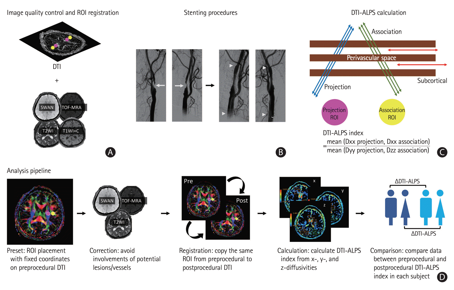
In our data analysis pipeline (Figure 1D), first, the imaging readers were asked to label the anonymized preprocedural DTI data. Initially, positions of the region of interest (ROI) centers with Montreal Neurological Institute (MNI) coordinates at (23, -13, 22), (-23, -13, 22), (34, -13, 22), and (-34, -13, 22) were applied to the right projection, left projection, right association, and left association areas, respectively (Figure 1). Spherical ROIs with fixed diameters (2.5 mm) were placed at these positions in the preprocedural DTI data. Second, the ROIs were adjusted manually to avoid the involvement of any slow-flow or arterial vessels using references from automatically synchronized SWAN and TOF-MRA [26,27]. To further minimize the potential effects of brain lesions or small regional vascular flow, ROIs were also placed on areas without apparent white matter hyperintensity on T2WI [26,27]. Therefore, the final coordinates of the ROIs between patients may not be identical. After determining the ROI positions in preprocedural DTI data, the exactly identical coordinates of the four ROIs were registered onto the postprocedural DTI to minimize the potential effects of ROI positioning on the comparisons between preprocedural and postprocedural DTI-ALPS index in each patient. The ROI positions on postprocedural DTI were confirmed again manually in all patients, to ensure correct registration. Finally, we calculated the DTI-ALPS index based on the ratio of the mean x-axis projection and association diffusivity to the mean y-axis projection and z-axis association diffusivity [21].
Imaging readers were blinded to the clinical information and randomly labeled the DTI data. Imaging analyses were performed by two experienced neuroradiologists (CHW and FCC). The diffusivities of the projection and association areas at the ventricular level on both sides were labeled and calculated. The average value for both sides was used to represent each patient’s DTI-ALPS index, which was used for further analyses. This was to avoid potentially transient glymphatic changes at the single (lesion) side [32,33], and was reasonable because cognition was dependent more on networks than on specific regional locations [34-36]. The final DTI-ALPS index was based on the consensus of both readers if a discrepancy existed.

Interventional procedures

Diagnostic DSA and interventional treatments (angioplasty and stenting) were performed during the same session. Interventional procedures were conducted under local anesthesia with femoral puncture for catheterization. Diagnostic DSA was performed using a 5-Fr angiographic catheter (Cook Medical, Bloomington, IN, USA) placed in the proximal common carotid artery. Biplanar DSA images were acquired in the frontal and lateral views. The grade of stenosis was measured according to the North American Symptomatic Carotid Endarterectomy Trial (NASCET) criteria [37] on diagnostic DSA, which is considered the gold standard for determining the stenosis grade in this study. Interventional treatments were administered if the participants met the criteria for CAS (Supplementary Table 1).
Predilatation angioplasty was performed using a noncompliant coronary or peripheral artery balloon (EMERGE PTCA Dilatation Catheter or Sterling balloon; Boston Scientific Corporation, Middlesex County, MA, USA) in approximately 80%–100% of the adjacent normal vessel segments. After adequate predilatation, carotid stents (Wallstent, Boston Scientific Corporation) were deployed meticulously. Postdilatation with a peripheral artery balloon (Sterling balloon) may be performed in selected cases. A control angiogram was acquired after CAS, and technical success was defined as successful stent placement with residual stenosis <30% [5]. In our practice, we used a distal embolic protection system (Filterwire EZ, Boston Scientific Corporation) in all cases. The filter wire basket was deployed at the level of the C1-2 vertebrae before predilatation and retrieved before the control angiogram [5]. All interventional procedures were performed by two experienced neuroradiologists (CHW and FCC).
All patients received oral premedication with dual antiplatelet therapy (300 mg aspirin and 75 mg clopidogrel daily) for at least 3 days before the interventions. The regimen (300 mg aspirin and 75 mg clopidogrel daily, same as the premedication) was continued for another 3 months if interventional procedures were performed. The medications were then shifted to single antiplatelet therapy (100 mg of aspirin daily) to be continued indefinitely 3 months after the interventional treatments [5].

Statistical analysis

Descriptive statistics are reported as percentages and means±standard deviations (SDs) or medians with interquartile ranges when appropriate. Since MMSE scores were not normally distributed, according to the Kolmogorov–Smirnov test (P<0.001), differences between preprocedural and postprocedural DTI-ALPS indices and MMSE scores were evaluated using the related-samples Wilcoxon signed-rank test. We defined increments in the DTI-ALPS index and MMSE score as the ratio of the difference between the postprocedural and preprocedural values to the preprocedural value. Correlations between increments in the DTI-ALPS index and MMSE score and between the DTI-ALPS index and DSA stenosis grade were also tested using Spearman’s rank correlation coefficients. Statistical significance was defined as P<0.05. The interrater agreement of the DTI-ALPS index was evaluated using single measurements of the intraclass correlation coefficient (ICC) [38].
The above analyses were performed using IBM SPSS Statistics software (Version 28.0; IBM Corp., Armonk, NY, USA).

Results

Study participants

In total, 109 patients with carotid stenosis ≥80% indicated for CAS were approached and recruited. All patients underwent preprocedural MRI and further interventional procedures. The preprocedural MMSE score, collected on the same day as the preprocedural MRI, was also recorded. Two patients were excluded because of procedural failure. A total of 107 study patients underwent early postprocedural MRI within 24 hours after CAS. Another three patients were excluded because of the presence of metallic artifacts on DTI. Since it was not reasonable for MMSE scores to increase dramatically within 2 months solely by CAS, patients with MMSE increments ≥50% were excluded (n=2; MMSE increments of 71.4% and 66.7%). Finally, the remaining 102 patients completed the postprocedural MMSE 2 months after CAS, and their data were used for further analysis (Table 1 and Supplementary Figure 1).

DTI-ALPS index and MMSE score before and after CAS

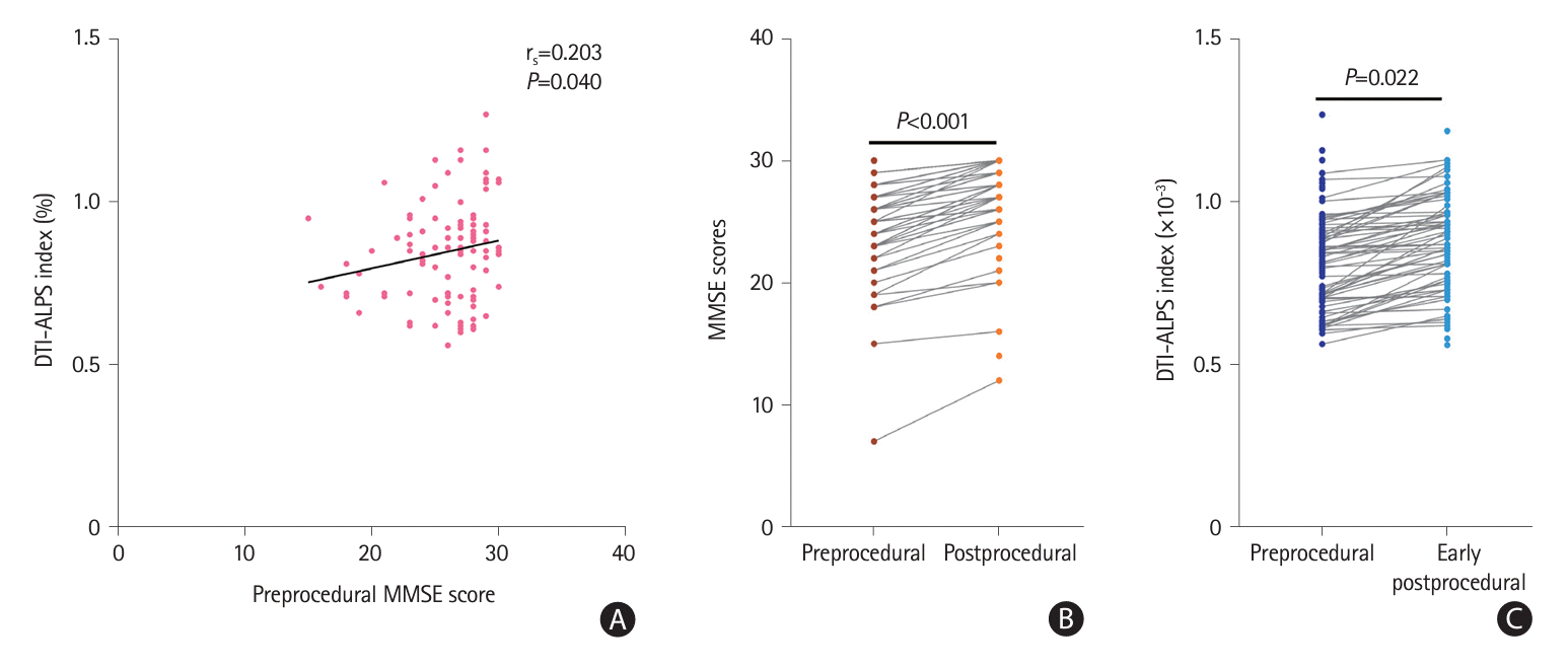
Positive correlations between the preprocedural DTI-ALPS index and preprocedural MMSE scores were observed in all participants (n=102; rs=0.203; P=0.040) (Figure 2A). The postprocedural MMSE scores (26.9±3.4; 26.0 [3.0]) were higher than the preprocedural MMSE scores (25.9±3.7; 24.0 [4.0]; P<0.001) (Figure 2B and Table 2). The DTI-ALPS index also increased after CAS (0.86±0.14; 0.86 [0.21]) compared to the values before the procedure (0.85±0.15; 0.85 [0.22]; P=0.022) (Figure 2C).

Correlations between MMSE score and DTI-ALPS index increments

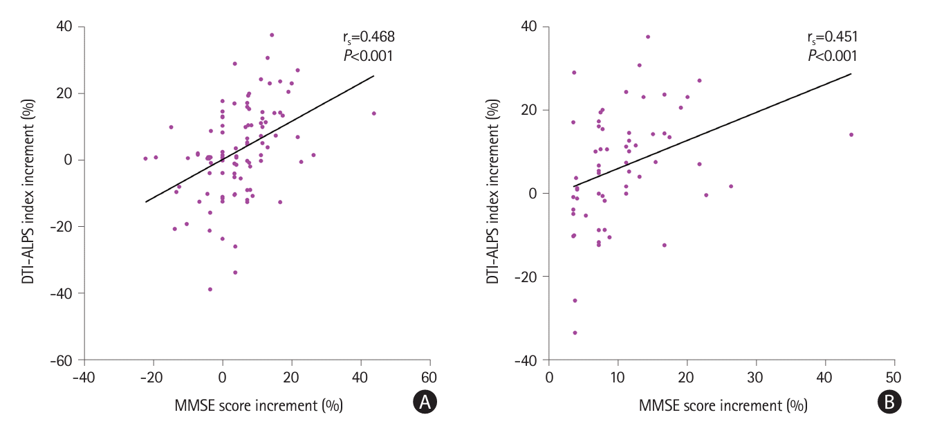
A positive correlation between increments in the DTI-ALPS index and increments in MMSE was observed (n=102; rs=0.468; P<0.001) (Figure 3A). We further analyzed the potential correlations between MMSE scores and DTI-ALPS index increments in selected patients with increased postprocedural MMSE scores (n=61). A positive correlation was found between the MMSE score and DTI-ALPS index increments in 61 patients (rs=0.451; P<0.001) (Figure 3B).

Correlation between stenosis grade and DTI-ALPS index increments

The correlation between the DSA stenosis grade and DTI-ALPS index increments was not significant in all patients (rs=-0.023; P=0.819) or in patients with increased MMSE scores (n=61; rs=-0.044; P=0.738).

Interrater agreement

The ICC between the two readers for the preprocedural and postprocedural DTI-ALPS index was 0.860 and 0.864, respectively.

Discussion

This is the first large-scale study investigating changes in ISF status after CAS. We successfully demonstrated an increased ISF flow within 24 hours after CAS using an increased DTI-ALPS index in each patient. Furthermore, increments in the DTI-ALPS index were positively correlated with increments in the MMSE scores in all patients and in those with improved MMSE scores 2 months after CAS. Although the increments in the MMSE scores at 2 months were small, their correlations with the DTI-ALPS index increments within 24 hours may indicate that the potential cognitive improvements can be attributed to the early improvement in ISF flow after CAS.
The DTI-ALPS index increased within 24 hours after CAS in each patient. The DTI-ALPS index represents perivascular flow and indicates diffusivity of ISF flow [21,22,25]. The glymphatic system plays a significant role in fluid transfer between the CSF and ISF [28]. The ISF travels along the perivascular spaces and regulates CSF waste removal [22]. Failure to remove CSF wastes may exacerbate cognition decline [10] and other neurological disorders [26,27]. One of the proposed driving forces of ISF flow is pulsation of the cerebral arterial wall [39,40]. A previous study suggested that extracranial blood flow is correlated with CSF flow [18]. In fact, ligation of the internal carotid artery may decrease cerebral arterial pulsation and further result in a reduction in CSF-ISF transfer and ISF flow [41]. We speculated that the early improvement in ISF flow after CAS in our study may be at least partially attributed to improved cerebral arterial pulsation after the restoration of the extracranial carotid blood flow. However, the correlation between early DTI-ALPS index changes within 24 hours and MMSE changes at 2 months after CAS may not be solely due to the theory of CSF waste accumulation. Previous studies have revealed that perfusion changes may occur within 3–5 days [42] and persist for 3–24 months after CAS [34,43]. Early improved functional connectivity was also observed after CAS [34,44]. Since the compromised vascular flow is highly correlated with functional MRI output [45], and the extracranial vascular flow may have impacts on the intracranial blood and CSF flow [18], future research is warranted to elucidate the relative contributions of these factors to cognitive changes.
The MMSE scores were higher at the 2-month follow-up than before the procedure. Although the differences between the mean scores were small, we evaluated the MMSE score improvements in each patient longitudinally. Therefore, the specific increase in the MMSE score in each patient may be parallel to that reported in previous studies, which suggests improved cognition in patient during the long-term follow-up after CAS [46-48]. In selected patients with increased MMSE scores, we demonstrated that increments in the MMSE score and DTI-ALPS index were positively correlated. Although the relatively small differences between the preprocedural and postprocedural MMSE scores may not be clinically apparent, the positive correlation between increments in the DTI-ALPS index within 24 hours and the MMSE score at 2 months after CAS may indicate the potential impact of early improvement in ISF flow on future cognitive improvement.
Although the data in this study indicate potential correlations between improved ISF flow and future cognitive improvement after CAS in patients with severe (≥80%) carotid stenosis, the DSA stenosis grades were not correlated with DTI-ALPS index increments. Since we only recruited patients with carotid stenosis ≥80%, with a relatively narrow stenosis grade sample distribution (SD=5.7), and with similar improvements in vessel diameter (all patients had residual stenosis <30% on the control angiogram, considered technical success), it was difficult to demonstrate potential correlations between the stenosis grade and ISF status using the current data. Therefore, a sophisticated study protocol that enrolls patients with a broader stenosis grade distribution may be required in the future.
This study has several strengths. First, all MR data were acquired using the same 3-T MRI machine with a standardized protocol. Although the DTI-ALPS index is reproducible for different MR machines, its values may differ for different TEs [22]. Therefore, conducting all MRI scans on the same MR machine using the same protocol avoids potential errors stemming from MR settings. This is also the first study to demonstrate early postprocedural ISF status changes within 24 hours after CAS. Furthermore, the DTI-ALPS index calculation showed excellent ICCs for both the preprocedural and early postprocedural values.
This study also has some limitations. First, we did not recruit completely healthy controls in this study. As recruitment of study participants was conducted during the COVID-19 pandemic, it was against our facility’s regulations to conduct healthy control recruitment. Furthermore, as per our study protocol, we acquired T1WI+C data to exclude patients with potential intracranial space-occupying lesions, and it was not ethical to impose unnecessary risks on healthy participants during GBCA administration [49]. In fact, comparisons were performed between the preprocedural and postprocedural values in each patient to demonstrate ISF changes after the intervention. With significant DTI-ALPS index improvements, positive correlations between the preprocedural DTI-ALPS index and MMSE score in all patients, which are similar to those in previous studies [21,50], and excellent interrater agreement, we believe our results are robust. Second, we did not exclude patients with vascular variants, including those with incomplete circle of Willis. However, changes in ISF flow are considered a global phenomenon, and the DTI-ALPS index is significantly correlated with global functional changes [51]. In fact, an incomplete circle of Willis does not contribute to lateral differences in brain perfusion [52]; therefore, this factor may have had a limited impact on the final results of this study. Third, although the longitudinal improvement in the MMSE score at 2 months after CAS was significant for each patient, the average improvement was relatively small. Therefore, to carefully inspect the correlations between increments in the MMSE score and DTI-ALPS index, we excluded patients with relatively high MMSE increments (≥50%; n=2) before the analysis. Although a sophisticated study is still needed to evaluate the exact causative effects of improved ISF flow efficiency on cognition, the data from our study suggest that ISF flow may improve early within 24 hours after CAS and may contribute to future changes in cognition. Finally, this study adopted the DTI-ALPS index to elaborate on the ISF function. Although we adopted several attempts to enhance the DTI-ALPS index reproducibility, including identical MRI machine/parameters, standardized scanning protocol (including head positioning and scanning plane angles), software-based synchronization and identical coordinates between preprocedural and postprocedural DTI analysis in each patient, and we compared the differences between preprocedural and postprocedural data in each patient instead of between patients (coordinates may not be identical between patients due to ROI avoidance of brain lesion or vessels), potential differences between imaging readers may not be completely excluded. Therefore, future studies with advanced MR techniques, including the combined use of DTI and dynamic imaging [27,53], may be needed to delineate the detailed ISF dynamics in patients after CAS.

Conclusions

The DTI-ALPS index improved within 24 hours after CAS. Increments in the DTI-ALPS index and MMSE scores at 2 months were positive. These results indicate that early improvements in ISF status may contribute to future cognitive improvements after CAS.

Supplementary materials

Supplementary materials related to this article can be found online at https://doi.org/10.5853/jos.2023.04203.
Supplementary Table 1.
Criteria for carotid stenting indications by Taiwan National Health Insurance (code: A220-1)
jos-2023-04203-Supplementary-Table-1.pdf
Supplementary Figure 1.
Flow diagram of the study subject recruitment. CAS, carotid angioplasty and stenting; MMSE, Mini-Mental State Examination; MRI, magnetic resonance imaging; DSA, digital subtraction angiography; DTI, diffusion tensor image; ROIs, regions of interest.
jos-2023-04203-Supplementary-Fig-1.pdf

Notes

Funding statement

This study was funded by the Taipei Veterans General Hospital, Taiwan (V111B-032, V112B-007 [to CHW]; V110C-037, V111C-028, V112C-059, V112D67-002-MY3-1 [to FCC]), Veterans General Hospitals and University System of Taiwan Joint Research Program (VGHUST 109V1-5-2 and VGHUST 110-G1-5-2 [to FCC]), National Science and Technology Council, Taiwan (NSTC 110-2314-B-075-005- and 111-2314-B-075-025-MY3 [to CHW] and 109-2314-B-075-036, 110-2314-B-075-032, 112-2314-B-075-066-, and 113-2314-B-075-037- [to FCC]), Yen Tjing Ling Medical Foundation, Taiwan (CI-109-3, CI-111-2, and CI-112-2 [to CHW]), Professor Tsuen CHANG’s Scholarship Program from Medical Scholarship Foundation In Memory Of Professor Albert Ly-Young Shen (to CHW and TML), and Vivian W. Yen Neurological Foundation (to CHW and FCC).

Conflicts of interest

The authors have no financial conflicts of interest.

Author contribution

Conceptualization: FCC, CHW. Study design: FCC, CHW, IHL. Methodology: CHW, SPC. Data collection: CHW, CPC. Investigation: CHW, KWY, TML. Statistical analysis: CHW, CBL. Writing—original draft: CHW, SPC, CPC. Writing—review & editing: CHW, JFL, IHL, FCC. Funding acquisition: CHW, TML, FCC. Approval of final manuscript: all authors.

References

1. Yadav JS, Wholey MH, Kuntz RE, Fayad P, Katzen BT, Mishkel GJ, et al. Protected carotid-artery stenting versus endarterectomy in high-risk patients. N Engl J Med. 2004; 351:1493–1501.
crossref
2. Ederle J, Bonati LH, Dobson J, Featherstone RL, Gaines PA, Beard JD, et al. Endovascular treatment with angioplasty or stenting versus endarterectomy in patients with carotid artery stenosis in the carotid and vertebral artery transluminal angioplasty study (CAVATAS): long-term follow-up of a randomised trial. Lancet Neurol. 2009; 8:898–907.
crossref
3. Haubrich C, Kruska W, Diehl RR, Möller-Hartmann W, Klötzsch C. Recovery of the blood pressure – cerebral flow relation after carotid stenting in elderly patients. Acta Neurochir (Wien). 2007; 149:131–136. discussion 137.
crossref
4. Lu CJ, Kao HL, Sun Y, Liu HM, Jeng JS, Yip PK, et al. The hemodynamic effects of internal carotid artery stenting: a study with color-coded duplex sonography. Cerebrovasc Dis. 2003; 15:264–269.
crossref
5. Wu CH, Lin TM, Chung CP, Yu KW, Tai WA, Luo CB, et al. Prevention of in-stent restenosis with drug-eluting balloons in patients with postirradiated carotid stenosis accepting percutaneous angioplasty and stenting. J Neurointerv Surg. 2023; 16:73–80.
6. Hsu YJ, Wu CH, Tai WA, Yu KW, Luo CB, Lin TM, et al. Early recurrent ischemic stroke involving the independent carotid territory after intravenous thrombolysis: a case report and literature review. J Radiol Sci. 2023; 48:e00017.
crossref
7. Chen YH, Lin MS, Lee JK, Chao CL, Tang SC, Chao CC, et al. Carotid stenting improves cognitive function in asymptomatic cerebral ischemia. Int J Cardiol. 2012; 157:104–107.
crossref
8. Kwok CHR, Park JC, Joseph SZ, Foster JK, Green DJ, Jansen SJ. Cognition and cerebral blood flow after extracranial carotid revascularization for carotid atherosclerosis: a systematic review. Clin Ther. 2023; 45:1069–1076.
crossref
9. Turowicz A, Czapiga A, Malinowski M, Majcherek J, Litarski A, Janczak D. Carotid revascularization improves cognition in patients with asymptomatic carotid artery stenosis and cognitive decline. Greater improvement in younger patients with more disordered neuropsychological performance. J Stroke Cerebrovasc Dis. 2021; 30:105608.
crossref
10. Nedergaard M, Goldman SA. Glymphatic failure as a final common pathway to dementia. Science. 2020; 370:50–56.
11. Wang DJ, Hua J, Cao D, Ho ML. Neurofluids and the glymphatic system: anatomy, physiology, and imaging. Br J Radiol. 2023; 96:20230016.
crossref
12. Siow TY, Toh CH, Hsu JL, Liu GH, Lee SH, Chen NH, et al. Association of sleep, neuropsychological performance, and gray matter volume with glymphatic function in community-dwelling older adults. Neurology. 2022; 98:e829–e838.
crossref
13. Kamagata K, Andica C, Takabayashi K, Saito Y, Taoka T, Nozaki H, et al. Association of MRI indices of glymphatic system with amyloid deposition and cognition in mild cognitive impairment and Alzheimer disease. Neurology. 2022; 99:e2648–e2660.
crossref
14. Ding XB, Wang XX, Xia DH, Liu H, Tian HY, Fu Y, et al. Impaired meningeal lymphatic drainage in patients with idiopathic Parkinson’s disease. Nat Med. 2021; 27:411–418.
crossref
15. Carotenuto A, Cacciaguerra L, Pagani E, Preziosa P, Filippi M, Rocca MA. Glymphatic system impairment in multiple sclerosis: relation with brain damage and disability. Brain. 2022; 145:2785–2795.
16. Hsiao WC, Chang HI, Hsu SW, Lee CC, Huang SH, Cheng CH, et al. Association of cognition and brain reserve in aging and glymphatic function using diffusion tensor image-along the perivascular space (DTI-ALPS). Neuroscience. 2023; 524:11–20.
crossref
17. Khan AA, Patel J, Desikan S, Chrencik M, Martinez-Delcid J, Caraballo B, et al. Asymptomatic carotid artery stenosis is associated with cerebral hypoperfusion. J Vasc Surg. 2021; 73:1611–1621.e2.
crossref
18. Lokossou A, Metanbou S, Gondry-Jouet C, Balédent O. Extracranial versus intracranial hydro-hemodynamics during aging: a PC-MRI pilot cross-sectional study. Fluids Barriers CNS. 2020; 17:1.
crossref
19. Wu CH, Lirng JF, Ling YH, Wang YF, Wu HM, Fuh JL, et al. Noninvasive characterization of human glymphatics and meningeal lymphatics in an in vivo model of blood–brain barrier leakage. Ann Neurol. 2021; 89:111–124.
crossref
20. Wu CH, Kuo Y, Chang FC, Lirng JF, Ling YH, Wang YF, et al. Noninvasive investigations of human glymphatic dynamics in a diseased model. Eur Radiol. 2023; 33:9087–9098.
21. Taoka T, Masutani Y, Kawai H, Nakane T, Matsuoka K, Yasuno F, et al. Evaluation of glymphatic system activity with the diffusion MR technique: diffusion tensor image analysis along the perivascular space (DTI-ALPS) in Alzheimer’s disease cases. Jpn J Radiol. 2017; 35:172–178.
crossref
22. Taoka T, Ito R, Nakamichi R, Kamagata K, Sakai M, Kawai H, et al. Reproducibility of diffusion tensor image analysis along the perivascular space (DTI-ALPS) for evaluating interstitial fluid diffusivity and glymphatic function: CHanges in Alps index on Multiple conditiON acquIsition eXperiment (CHAMONIX) study. Jpn J Radiol. 2022; 40:147–158.
23. Lee S, Yoo RE, Choi SH, Oh SH, Ji S, Lee J, et al. Contrast-enhanced MRI T1 mapping for quantitative evaluation of putative dynamic glymphatic activity in the human brain in sleepwake states. Radiology. 2021; 300:661–668.
crossref
24. Eide PK, Ringstad G. MRI with intrathecal MRI gadolinium contrast medium administration: a possible method to assess glymphatic function in human brain. Acta Radiol Open. 2015; 4:2058460115609635.
crossref
25. Qin Y, Li X, Qiao Y, Zou H, Qian Y, Li X, et al. DTI-ALPS: an MR biomarker for motor dysfunction in patients with subacute ischemic stroke. Front Neurosci. 2023; 17:1132393.
26. Hsu SL, Liao YC, Wu CH, Chang FC, Chen YL, Lai KL, et al. Impaired cerebral interstitial fluid dynamics in cerebral autosomal dominant arteriopathy with subcortical infarcts and leucoencephalopathy. Brain Commun. 2023; 6:fcad349.
crossref
27. Wu CH, Chang FC, Wang YF, Lirng JF, Wu HM, Pan LH, et al. Impaired glymphatic and meningeal lymphatic functions in patients with chronic migraine. Ann Neurol. 2024; 95:583–595.
crossref
28. Klostranec JM, Vucevic D, Bhatia KD, Kortman HGJ, Krings T, Murphy KP, et al. Current concepts in intracranial interstitial fluid transport and the glymphatic system: part I—anatomy and physiology. Radiology. 2021; 301:502–514.
crossref
29. Han F, Chen J, Belkin-Rosen A, Gu Y, Luo L, Buxton OM, et al. Reduced coupling between cerebrospinal fluid flow and global brain activity is linked to Alzheimer disease-related pathology. PLoS Biol. 2021; 19:e3001233.
crossref
30. van der Thiel MM, Freeze WM, Verheggen ICM, Wong SM, de Jong JJA, Postma AA, et al. Associations of increased interstitial fluid with vascular and neurodegenerative abnormalities in a memory clinic sample. Neurobiol Aging. 2021; 106:257–267.
31. Clark CM, Sheppard L, Fillenbaum GG, Galasko D, Morris JC, Koss E, et al. Variability in annual Mini-Mental State Examination score in patients with probable Alzheimer disease: a clinical perspective of data from the Consortium to Establish a Registry for Alzheimer’s Disease. Arch Neurol. 1999; 56:857–862.
crossref
32. Gaberel T, Gakuba C, Goulay R, Martinez De Lizarrondo S, Hanouz JL, Emery E, et al. Impaired glymphatic perfusion after strokes revealed by contrast-enhanced MRI: a new target for fibrinolysis? Stroke. 2014; 45:3092–3096.
crossref
33. Li C, Lin L, Sun C, Hao X, Yin L, Zhang X, et al. Glymphatic system in the thalamus, secondary degeneration area was severely impaired at 2nd week after transient occlusion of the middle cerebral artery in rats. Front Neurosci. 2022; 16:997743.
crossref
34. Wang T, Sun D, Liu Y, Mei B, Li H, Zhang S, et al. The impact of carotid artery stenting on cerebral perfusion, functional connectivity, and cognition in severe asymptomatic carotid stenosis patients. Front Neurol. 2017; 8:403.
crossref
35. Mesulam M. Defining neurocognitive networks in the BOLD new world of computed connectivity. Neuron. 2009; 62:1–3.
36. Eijlers AJC, Wink AM, Meijer KA, Douw L, Geurts JJG, Schoonheim MM. Reduced network dynamics on functional MRI signals cognitive impairment in multiple sclerosis. Radiology. 2019; 292:449–457.
crossref
37. Ferguson GG, Eliasziw M, Barr HW, Clagett GP, Barnes RW, Wallace MC, et al. The North American symptomatic carotid endarterectomy trial: surgical results in 1415 patients. Stroke. 1999; 30:1751–1758.
crossref
38. Koo TK, Li MY. A guideline of selecting and reporting intraclass correlation coefficients for reliability research. J Chiropr Med. 2016; 15:155–163.
crossref
39. Schley D, Carare-Nnadi R, Please CP, Perry VH, Weller RO. Mechanisms to explain the reverse perivascular transport of solutes out of the brain. J Theor Biol. 2006; 238:962–974.
crossref
40. Mestre H, Tithof J, Du T, Song W, Peng W, Sweeney AM, et al. Flow of cerebrospinal fluid is driven by arterial pulsations and is reduced in hypertension. Nat Commun. 2018; 9:4878.
41. Iliff JJ, Wang M, Zeppenfeld DM, Venkataraman A, Plog BA, Liao Y, et al. Cerebral arterial pulsation drives paravascular CSF-interstitial fluid exchange in the murine brain. J Neurosci. 2013; 33:18190–18199.
crossref
42. Chang TY, Liu HL, Lee TH, Kuan WC, Chang CH, Wu HC, et al. Change in cerebral perfusion after carotid angioplasty with stenting is related to cerebral vasoreactivity: a study using dynamic susceptibility-weighted contrast-enhanced MR imaging and functional MR imaging with a breath-holding paradigm. AJNR Am J Neuroradiol. 2009; 30:1330–1336.
crossref
43. Lin CJ, Chang FC, Lin CJ, Liaw YC, Tu PC, Wang PN, et al. Long-term cognitive and multimodal imaging outcomes after carotid artery stenting vs intensive medication alone for severe asymptomatic carotid stenosis. J Formos Med Assoc. 2022; 121(1 Pt 1):134–143.
crossref
44. Tani N, Yaegaki T, Nishino A, Fujimoto K, Hashimoto H, Horiuchi K, et al. Functional connectivity analysis and prediction of cognitive change after carotid artery stenting. J Neurosurg. 2018; 131:1709–1715.
crossref
45. Wu CH, Hsu TW, Lai KL, Wang YF, Fuh JL, Wu HM, et al. Disrupted brain functional status in patients with reversible cerebral vasoconstriction syndrome. Ann Neurol. 2023; 94:772–784.
46. Gupta AN, Bhatti AA, Shah MM, Mahajan NP, Sadana DK, Huded V. Carotid artery stenting and its impact on cognitive function: a prospective observational study. Neurointervention. 2020; 15:74–78.
crossref
47. Lin CJ, Tu PC, Chern CM, Hsiao FJ, Chang FC, Cheng HL, et al. Connectivity features for identifying cognitive impairment in presymptomatic carotid stenosis. PLoS One. 2014; 9:e85441.
crossref
48. Turk AS, Chaudry I, Haughton VM, Hermann BP, Rowley HA, Pulfer K, et al. Effect of carotid artery stenting on cognitive function in patients with carotid artery stenosis: preliminary results. AJNR Am J Neuroradiol. 2008; 29:265–268.
crossref
49. Jost G, Frenzel T, Boyken J, Lohrke J, Nischwitz V, Pietsch H. Long-term excretion of gadolinium-based contrast agents: linear versus macrocyclic agents in an experimental rat model. Radiology. 2019; 290:340–348.
crossref
50. Steward CE, Venkatraman VK, Lui E, Malpas CB, Ellis KA, Cyarto EV, et al. Assessment of the DTI-ALPS parameter along the perivascular space in older adults at risk of dementia. J Neuroimaging. 2021; 31:569–578.
51. Lee DA, Park BS, Ko J, Park SH, Park JH, Kim IH, et al. Glymphatic system function in patients with newly diagnosed focal epilepsy. Brain Behav. 2022; 12:e2504.
crossref
52. Urbanski PP, Lenos A, Blume JC, Ziegler V, Griewing B, Schmitt R, et al. Does anatomical completeness of the circle of Willis correlate with sufficient cross-perfusion during unilateral cerebral perfusion? Eur J Cardiothorac Surg. 2008; 33:402–408.
crossref
53. Wu CH, Lirng JF, Wu HM, Ling YH, Wang YF, Fuh JL, et al. Blood-brain barrier permeability in patients with reversible cerebral vasoconstriction syndrome assessed with dynamic contrast-enhanced MRI. Neurology. 2021; 97:e1847–e1859.
crossref

Figure 1.
Imaging process in this study. (A) Diffusion tensor imaging (DTI) was performed on a 3-T magnetic resonance imaging (MRI) machine along with susceptibility-weighted angiography (SWAN), time-of-flight MR angiography (TOF-MRA), T2-weighted imaging (T2WI), and postcontrast T1-weighted imaging (T1WI+C) in the same session. Any patients with visible intracranial space-occupying lesions depicted on T1WI+C were excluded. T2WI data were then used as a reference to avoid placing regions of interest (ROIs) on areas of white matter hyperintensity. SWAN and TOF-MRA data were used as references to avoid placing ROIs on slow-flow and arterial vessels. The pink and yellow circles indicate ROI registration onto the projection and association area, respectively, as on DTI. (B) Diagnostic digital angiography (DSA) was performed to calculate the stenosis grade based on the North American Symptomatic Carotid Endarterectomy Trial (NASCET) criteria. Arrows on the diagnostic DSA image indicate carotid stenosis. A control angiogram was obtained after carotid stenting. Technical success was defined as residual stenosis <30%. Arrowheads on the control angiogram depict the proximal and distal ends of the carotid stent. (C) The diffusion tensor image analysis along the perivascular space (DTI-ALPS) index was calculated as the ratio of the mean diffusivity of projection and association areas on the x-axis to the mean diffusivity of projection on the y-axis and association on the z-axis. (D) Detailed analysis pipeline for the DTI-ALPS analysis was demonstrated. We first applied the coordinates of (23, -13, 22), (-23, -13, 22), (34, -13, 22), and (-34, -13, 22) to the right projection, left projection, right association, and left association areas, respectively, on the preprocedural DTI. Corrections were made to avoid these ROIs being placed on areas with slow-flow, arterial vessels, or apparent white matter intensities on SWAN, TOF-MRA, or T2WI. The ROIs with the same locations were copied onto the postprocedural DTI in the same patients, and the locations were confirmed manually again on the postprocedural DTI. The DTI-ALPS index of both preprocedural and postprocedural imaging was then calculated and compared for each patient.
jos-2023-04203f1.tif
Figure 2.
Correlations of and longitudinal changes in the MMSE score and DTI-ALPS index before and after CAS. (A) A positive correlation between the preprocedural MMSE score and DTI-ALPS index is depicted. (B) The MMSE score increased 2 months after carotid stenting. (C) The DTI-ALPS index increased within 24 hours after carotid stenting. The gray lines in (B) and (C) connect the preprocedural and postprocedural values in patients with increased postprocedural values. MMSE, Mini-Mental State Examination; DTI-ALPS, diffusion tensor image analysis along the perivascular space; CAS, carotid angioplasty and stenting.
jos-2023-04203f2.tif
Figure 3.
Increments in the DTI-ALPS index and MMSE score. Increments in the DTI-ALPS index and MMSE score were positively correlated in (A) all patients and (B) patients with increased MMSE scores after CAS. The increment was defined as the ratio of differences between postprocedural and preprocedural values (DTI-ALPS index or MMSE scores) to preprocedural values. DTI-ALPS, diffusion tensor image analysis along the perivascular space; MMSE, Mini-Mental State Examination; CAS, carotid angioplasty and stenting.
jos-2023-04203f3.tif
Table 1.
Demographics of all subjects with severe carotid stenosis
Characteristic Value (n=102)
Total number of MRI scans 204
Age (yr) 67.1±8.9
Number of females 16 (15.7)
NASCET stenosis grade (%) 89.5±5.7
Laterality of the carotid stenosis (right side) 44 (43.1)
CCA involvement (≥50% stenosis in CCA) 71 (69.6)
Timing intervals
 Preprocedural MRI/MMSE to stenting (day) 1.7±0.6
 Stenting to early postprocedural MRI (h) 20.2±1.8
 Stenting to postprocedural MMSE (day) 61.2±1.5

Values are presented as mean±standard deviation or n (%) unless otherwise indicated.

MRI, magnetic resonance imaging; NASCET, North American Symptomatic Carotid Endarterectomy Trial; CCA, common carotid artery; MMSE, Mini-Mental State Examination.

Table 2.
Increased values of MMSE scores and DTI-ALPS index after CAS
Preprocedure Postprocedure P
All subjects (n=102)
 MMSE score 25.9±3.7; 24.0 [4.0] 26.9±3.4; 26.0 [3.0] <0.001
 DTI-ALPS index 0.85±0.15; 0.85 [0.22] 0.86±0.14; 0.86 [0.21] 0.022
Subjects with increased MMSE scores after CAS (n=61)
 DTI-ALPS index 0.84±0.17; 0.85 [0.22] 0.88±0.15; 0.89 [0.23] 0.001

Values are presented as mean±standard deviation; and median [interquartile range].

MMSE, Mini-Mental State Examination; DTI-ALPS, diffusion tensor image analysis along the perivascular space; CAS, carotid angioplasty and stenting.

TOOLS
Similar articles