Journal List > Intest Res > v.22(4) > 1516088600

Jo, Kim, Lee, Im, Kim, and Koh: Predictors of histologic remission in patients with biologic-naïve, moderate-to-severe ulcerative colitis treated with first-line biologic agents and small-molecule drugs: a single-center, retrospective cohort study

Abstract

Background/Aims

The prevalence and incidence of ulcerative colitis (UC) in Korea is increasing. Each patient has a different disease course and treatment response. Recently, with the development of biologic agents, histological remission has become a treatment goal. In this study, we aimed to identify the predictors of histological remission after first-line biologic agent treatment in patients with biologic agent-naïve UC.

Methods

We retrospectively analyzed the medical records of 92 patients who had been diagnosed with UC and treated with first-line biologic agent treatment at our center, between 2015 and 2022. The clinical characteristics, laboratory test results, and endoscopic and biopsy findings were analyzed. Histological remission was defined as the absence of cryptitis, crypt abscesses, and inflammatory cells on histology. Univariate and multivariate logistic regression analyses were performed to identify the predictors of histological remission after first-line treatment.

Results

Of the total 92 patients, 25 (27.2%) achieved histological remission. Each cohort had a varied body mass index (BMI) distribution, with a statistically significant overweight ratio, as defined by the Asian-Pacific BMI category of 23–25 kg/m2, of 48.0% in the histological remission cohort (P= 0.026). A causal correlation between the overweight category and histological remission was confirmed (odds ratio, 3.883; 95% confidence interval, 1.141–13.212; P= 0.030).

Conclusions

We confirmed that the overweight category was a predictor of histological remission after first-line treatment with a biological agent. However, as BMI does not account for skeletal muscle mass, future studies are required to confirm the correlation between skeletal muscle mass and histological remission.

INTRODUCTION

Ulcerative colitis (UC) is a type of inflammatory bowel disease (IBD) that is limited to the colon and is characterized by continuous mucosal inflammation and subsequent symptoms, such as diarrhea and hematochezia [1,2]. The prevalence has been increasing globally over the past 2 decades, particularly in East Asian countries, including Korea; due to Westernization and urbanization in this region [3-5]. The exact pathophysiological mechanisms have not been established; however, they are believed to be caused by a combination of various factors, including genetic disposition of the host, mucosal immunity, and an imbalance of the intestinal microbiome [1,6-8].
Several molecules involved in the inflammatory cascade, such as tumor necrosis factor-α, interleukin (IL)-12, and their receptors have been identified. Moreover, a number of biologic agents and small-molecule drugs have been developed to target them, revealing excellent therapeutic responses; and they are used as first-line treatment [1,9-12]. New drugs targeting other mechanisms are additionally in research and development [13,14].
In the interim, the Selecting Therapeutic Targets in Inflammatory Bowel Disease-II Initiative has expanded the goal of treatment for IBD from conventional, symptomatic clinical remission to endoscopic remission. This has resulted in the use of objective markers, absence of disability, and restoration of quality of life with prognostic considerations [15].
More recently, targeted treatment has been expanded to include histological mucosal healing [16,17]. Findings of meta-analysis by Yoon et al. [18] have revealed that patients who achieve histological remission have a lower risk of relapse and better prognosis than that of those who do not. Furthermore, Chateau et al. [19] have suggested expanding the treatment goal of UC to histological remission. However, each patient possesses a different course of disease and responds differently to drugs, with some patients not responding at all or their response regressing quickly, making it challenging for clinicians to select the appropriate biologic agents [7,20]. In addition, due to the high cost of biologic agents, the economic burden on patients and overall healthcare costs have tended to increase, intensifying the demand for predicting the response to biologic agents, in order to be administered to the appropriate patients [21,22]. Several studies have reported predictors of response and remission regarding biologic agents; however, these studies were conducted primarily in Western populations. Thus, these predictors are difficult to apply to the Korean clinical setting, due to racial differences [7,23,24].
Therefore, in this study, we identified the predictors of histological remission after first-line biologic agent and small-molecule drug treatment in patients with biologic agent-naïve UC. We hypothesized that biological agents can appropriately be administered to patients, to improve prognosis and reduce the associated healthcare costs.

METHODS

1. Ethical Approval

This study was approved by the Institutional Review Board (IRB) of the Seoul National University of Korea (IRB No. H2105-055-1218). Written informed consent was waived.

2. Study Design and Definition of Variables

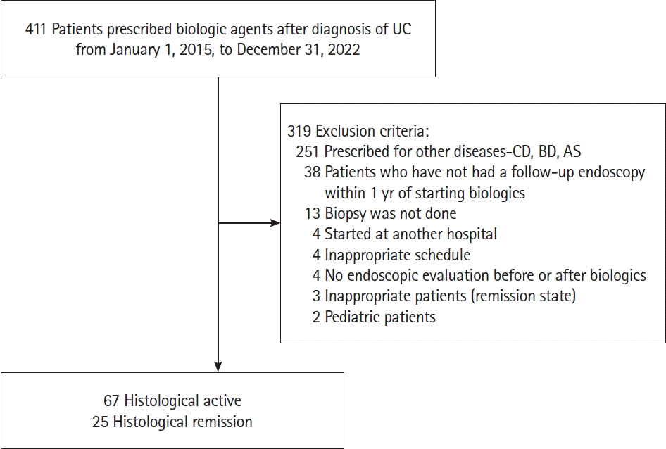
We conducted a single-center, retrospective cohort study and collected data on patients with at least 1 diagnosis related to UC (K51 code in the Korean Standard Classification of Diseases and Causes of Death) and recorded minimal 1 dose of infliximab, adalimumab, golimumab, vedolizumab, ustekinumab, or tofacitinib, between January 1, 2015 and December 31, 2022 (Fig. 1). Of the 411 patients, 92 patients were included in the study.
The exclusion criteria were as follows: (1) patients who were prescribed biological agents and small-molecule drugs for other immune-related diseases; however, the diagnosis was incorrectly recorded; (2) patients transferred from other hospitals; thus, no data were available at our center; (3) patients without baseline biopsies; (4) patients who were administered an inappropriate regimen; (5) patients who were administered drugs, without proper endoscopic evaluation; and (6) pediatric patients.
Subsequently, the clinical information, medical history, laboratory test results at the time of diagnosis, and endoscopic and histological findings pre- and post-treatment with biologic agents and small-molecule drugs were retrospectively reviewed.
Medical history included age, sex, body mass index (BMI), date of diagnosis, smoking history, underlying medical conditions, disease extent at diagnosis, and endoscopic severity. Laboratory tests included the complete blood count, albumin level, erythrocyte sedimentation rate, C-reactive protein, fecal calprotectin, antineutrophil cytoplasmic antibody, and anti-Saccharomyces cerevisiae antibody. The full Mayo score and endoscopic and histological findings at the time of the initiation of the biological agent were reviewed. Additionally, the 1-year post-treatment follow-up endoscopic and histological findings were assessed. Colonofiberscopy (CFS) was performed by an experienced endoscopist and evaluated using the Mayo endoscopic score (MES). Tissue samples were obtained from active lesions, and random biopsies were performed for patients in remission.
The Elderly onset of UC was defined as being diagnosed at the age of ≥ 65 years, as per a previous definition [25]. Smoking status was defined as, “ex-smoker,” “current smoker,” or “never smoked,” according to the patient’s in-clinic response. The BMI was measured in kg/m2. The Asia-Pacific BMI classification was used to categorize underweight, normal, overweight, and obese as < 18.5, 18.5–22.9, 23.0–24.9, and > 25.0 kg/m2 respectively [26].
Endoscopic remission was defined as 0–1 on the MES. An improvement in the MES was defined as a decrease of ≥ 1, from the baseline to the follow-up CFS.
Histological remission was defined as the absence of active cryptitis, crypt abscess, and inflammatory cell infiltration on microscopic examination by an experienced histopathologist [17,27]. A comparative analysis of the clinical characteristics between patients with and without histological remission on follow-up endoscopic biopsies was conducted.

3. Statistical Analysis

A logistic regression analysis was performed to identify the statistically significant predictors of histological remission. The primary outcome was histological remission after first-line treatment with a biological agent, and the secondary outcomes were endoscopic improvement and remission.
Statistical analyses were performed using the SPSS 25 software (IBM Corp., Armonk, NY, USA). Categorical variables were presented as total counts and percentages. For continuous variables that were normally distributed, the mean values and standard deviations were presented. For continuous variables that were not normally distributed, the medians and ranges were presented. Demographic, clinical, therapeutic, and endoscopic factors in patients with and without histological remission were compared, using chi-square or t-tests. A univariate logistic regression analysis was performed to identify potential clinical predictors, with a cutoff P-value of 0.2. Factors of potential statistical significance were included in a multiple logistic regression analysis, to identify independent associations. Moreover, multiple logistic regression analysis was used to calculate the odds ratios (ORs) and 95% confidence intervals (CIs), for the endoscopic and histological remission and non-remission cohorts, to identify predictors of remission. Statistical significance was set at P< 0.05.

RESULTS

1. Baseline Demographics and Patient Characteristics

Of the 92 patients included in the study, 25 (27.2%) achieved histological remission, after first-line biological agent treatment. The baseline characteristics of the 2 cohorts are presented in Table 1. The median age at diagnosis and sex ratio did not differ between the 2 cohorts. The mean BMI did not differ statistically significantly between the 2 cohorts; however, the proportion of patients who were overweight was statistically significantly higher in the remission cohort than that in the non-remission cohort (48.0% and 16.4%, respectively, P= 0.026). Statistically significant differences in the comorbidities were not observed between the 2 cohorts.
At the time of diagnosis, statistically significant differences were not observed in the disease extent and MES between the 2 cohorts, and most patients had a MES of 2–3. Regarding the laboratory test results, the hemoglobin and albumin level were statistically significantly higher in the remission cohort than that in the non-remission cohort (P= 0.022 and P= 0.002, respectively). Laboratory results revealed no other statistically significant differences between the 2 cohorts. Before first-line treatment with biologic agents, the Mayo score and MES did not differ statistically significantly between the 2 cohorts; and all patients had moderate-to-severe disease and a MES of 2–3. In addition, histological activity on pretreatment biopsy was observed in 92% and 97% of the patients in the histological remission and non-remission cohorts, respectively.

2. Trends in First-Line Biologic Agents and Small-Molecule Drugs

Trends regarding first-line treatment with biologic agents are shown in Table 2. No statistically significant differences in the time taken from diagnosis to first use of the biological agent were observed between the remission and non-remission cohorts (665 days and 1,118 days, respectively, P= 0.109). Infliximab, vedolizumab, adalimumab, ustekinumab, tofacitinib, and golimumab were used as first-line biologic agents and small-molecule drugs in the order listed, with no statistically significant differences between the 2 cohorts.

3. Results of Follow-up Studies and Endoscopic Improvement

The results of the first-line treatment with biologic agents are summarized in Table 3. At follow-up, complete remission was revealed in 16 (64.0%) and 2 (93.0%) patients in the histological remission and non-remission cohorts, respectively. Moreover, 8 (32.0%) and 23 (34.3%) patients showed endoscopic improvement, in the histological remission and non-remission cohorts, respectively (P< 0.001). Compared to pretreatment CFS, all patients in the histological remission cohort showed an improvement in the MES; nonetheless, in the non-remission cohort, an improvement was only observed in 49.3% of the patients (P< 0.001).

4. Univariate and Multivariate Analysis for Predictors of Histological Remission

Results of the binomial logistic regression analysis, identifying statistically significant predictors of histological remission are presented in Table 4. In the logistic regression analysis to identify predictors of histological remission, the Asian-Pacific BMI classification, albumin, azathioprine usage, and class of firstline biologic agents were found to be statistically significant predictors in the univariate logistic regression analysis. In the multivariate logistic regression analysis, the overweight category of the BMI was found to be a statistically significantly stronger predictor of histological remission than normal weight (OR, 3.883; 95% CI, 1.141–13.212; P= 0.030).

DISCUSSION

This study identified predictors of endoscopic and histological remission after first-line therapy with biologic agents and small-molecule drugs, in patients with biologic agent-naïve UC, in real-world clinical practice.
Previous studies have shown endoscopic remission rates ranging from 29% to 60%, depending on the agent, and this study revealed an endoscopic remission rate of 57.2% [13,28,29]. Histological remission rates for current UC treatments range from 15.0% to 44.9%, according to drug class and patient population [30]. The histological remission rate was 23.8% in this study. Although the endoscopic and histological examinations were not performed according to uniform protocols due to retrospective nature; nevertheless, they were evaluated by trained endoscopists and histopathologists. Moreover, the results are consistent with those of previous studies.
In our study, on follow-up endoscopic biopsies, we found that patients who were overweight were statistically significantly more likely to achieve histological remission than patients of a normal weight. This can be explained by obesity being a poor prognostic factor in the course of immune-related diseases, including UC [31,32]. The distribution of the drug in patients who are obese is different from that in patients who are of a normal weight. This is due to the increase in body fat composition, which results in different activation rates in the body [33]. Additionally, pro-inflammatory adipokines secreted by the immune cells of adipose tissue and an imbalance in the gut microbiota have been found to influence the pathophysiology of UC, by exacerbating the overall inflammatory cascades [34-36].
In addition to the increased proportion of adipose tissue in obesity, sarcopenia, a decrease in skeletal muscle mass, is theorized to play a role in the inflammatory response [37-39]. A systematic review by Fatani et al. [39] has confirmed that 17% of patients with IBD have sarcopenia, which is associated with the risk of surgical and medical treatment failure. Findings of a prospective cohort study by Liu et al. [40] have revealed that sarcopenia in patients with IBD is associated with poor clinical outcomes, such as increased rates of surgery (OR, 6.651; P< 0.001), re-hospitalization (OR, 6.344; P< 0.001), and mortality (P= 0.003) in IBD. Cushing et al. [41] have confirmed that sarcopenia as determined by abdominal computed tomography is a poor prognostic factor (OR, 3.98; P= 0.033) for acute, severe UC, requiring further treatment. This is due to the secretion of myokines by skeletal muscles, such as IL-6 and IL-15, which exert direct anti-inflammatory effects and inhibit the release of pro-inflammatory adipokines from visceral fat [42]. In a murine model of colitis with increased physical activity, symptoms of colitis symptoms were significantly reduced by diarrheal episodes. Immunological changes, such as decreased regulatory T-cells and tumor necrosis factor-α secretion; as well as enhanced secretion of a well-known anti-inflammatory cytokine, IL-10 have been identified [43-45].
Past research has shown that the BMI does not accurately reflect actual skeletal muscle and fat mass. Possessing a large amount of skeletal muscle mass can distort the BMI into that of the overweight category, which is a numerical limitation of BMI [46]. In the present study, accurate body composition was not measured by a body composition analyzer, such as InBody® (InBody Co.) or abdominal computed tomography; however, the inference was that the patients who were overweight had relatively more skeletal muscle mass than did the other patients. Patients with UC may have a poor oral intake, due to chronic abdominal symptoms. Furthermore, they are relatively susceptible to sarcopenia, due to repeated steroid use and reduced physical activity. Thus, maintenance of the skeletal muscle mass may predict a better disease course and good response to biologic agents. Lifestyle modifications, such as nutritional education, weight management, and encouragement of physical activity in patients with sarcopenia or sarcopenic obesity may improve the treatment response to biological agents [42,45,47,48]. However, previous studies have not confirmed a direct correlation between sarcopenia and biologic agent treatment failure, and further studies with precise designs are required [49].
Our study had several limitations. First, due to the retrospective nature of the study, the examination protocols were not standardized; and the timing of blood tests, endoscopies, and histological examinations were inconsistent. Selection bias may have been induced by excluding patients who did not have the biopsies required for this study. Second, many patients did not undergo laboratory tests, such as fecal calprotectin, at the discretion of the attending clinician; therefore, data was missing. Third, due to the small population size, limited to a single-center, we included multiple biologic agents and small-molecule drugs with different pharmacological actions. This made it difficult to identify the predictors of remission for each agent. Still, the choice of biologic agents and small-molecule drugs as first-line treatment for patients with biologic agent-naïve UC should be based on the insurance policy of the region, unique characteristics of the drug, route of administration, patient preference, cost, and consideration of existing head-to-head trials.
Despite these limitations, this real-world study identified predictors of histological remission after first-line treatment with biologic agents and small-molecule drugs in patients with biologic agent-naïve UC. This is the first study conducted in Korea that has identified predictors of histological remission in patients with biologic agent-naïve. The predictors identified in this study can be used to improve patient outcomes and reduce healthcare costs, by appropriately administering biologic agents to patients expected to achieve histological remission.
In conclusion, this retrospective cohort study confirmed that the overweight category of BMI is a statistically significant predictor of histological remission. Further prospective and translational studies with a molecular basis are required to confirm these findings, and to improve the prognosis.

Notes

Funding Source

This work was supported by a National Research Foundation of Korea (NRF) grant by the Korean government (The Ministry of Science and ICT; No. NRF-2020R1F1A1066491 and No. RS-2023-00227939). This work was supported by the National Research Foundation of Korea (NRF) grant funded by the Korean government (MSIT) (NRF-2022R1F1A1076019). This work was granted by general clinical research grant-in-aid from the Seoul Metropolitan Government Seoul National University (SMG-SNU) Boramae Medical Center (04-2023-0012).

Conflict of Interest

Kim KW and Im JP are editorial board members of the journal but were not involved in the peer reviewer selection, evaluation, or decision process of this article. No other potential conflicts of interest relevant to this article were reported.

Data Availability Statement

The datasets generated during and/or analyzed during the current study are available from the corresponding author on reasonable request.

Author Contributions

Conceptualization: Jo K, Kim KW, Koh SJ, Lee HJ, Im JP, Kim JS. Data curation: Jo K. Formal analysis: Jo K. Funding acquisition: Kim KW, Koh SJ. Investigation: Jo K, Kim KW. Methodology: Jo K. Project administration: Kim KW. Resources: Jo K. Software: Jo K. Supervision: Kim KW, Koh SJ, Lee HJ, Im JP, Kim JS. Validation: Jo K. Visualization: Jo K. Writing - original draft: Jo K. Writing - review & editing: Kim KW, Koh SJ, Lee HJ, Im JP, Kim JS. Approval of final manuscript: all authors.

REFERENCES

1. Ungaro R, Mehandru S, Allen PB, Peyrin-Biroulet L, Colombel JF. Ulcerative colitis. Lancet. 2017; 389:1756–1770.
crossref
2. Fumery M, Singh S, Dulai PS, Gower-Rousseau C, Peyrin-Biroulet L, Sandborn WJ. Natural history of adult ulcerative colitis in population-based cohorts: a systematic review. Clin Gastroenterol Hepatol. 2018; 16:343–356.
3. Cha JM, Park SH, Rhee KH, et al. Long-term prognosis of ulcerative colitis and its temporal changes between 1986 and 2015 in a population-based cohort in the Songpa-Kangdong district of Seoul, Korea. Gut. 2020; 69:1432–1440.
crossref
4. Lee JW, Eun CS. Inflammatory bowel disease in Korea: epidemiology and pathophysiology. Korean J Intern Med. 2022; 37:885–894.
crossref
5. Cosnes J, Gower-Rousseau C, Seksik P, Cortot A. Epidemiology and natural history of inflammatory bowel diseases. Gastroenterology. 2011; 140:1785–1794.
crossref
6. Xu XR, Liu CQ, Feng BS, Liu ZJ. Dysregulation of mucosal immune response in pathogenesis of inflammatory bowel disease. World J Gastroenterol. 2014; 20:3255–3264.
crossref
7. Quetglas EG, Mujagic Z, Wigge S, et al. Update on pathogenesis and predictors of response of therapeutic strategies used in inflammatory bowel disease. World J Gastroenterol. 2015; 21:12519–12543.
8. Lee CH, Koh SJ, Radi ZA, Habtezion A. Animal models of inflammatory bowel disease: novel experiments for revealing pathogenesis of colitis, fibrosis, and colitis-associated colon cancer. Intest Res. 2023; 21:295–305.
crossref
9. Raine T, Bonovas S, Burisch J, et al. ECCO guidelines on therapeutics in ulcerative colitis: medical treatment. J Crohns Colitis. 2022; 16:2–17.
crossref
10. Rubin DT, Ananthakrishnan AN, Siegel CA, Sauer BG, Long MD. ACG clinical guideline: ulcerative colitis in adults. Am J Gastroenterol. 2019; 114:384–413.
crossref
11. Nakase H, Sato N, Mizuno N, Ikawa Y. The influence of cytokines on the complex pathology of ulcerative colitis. Autoimmun Rev. 2022; 21:103017.
crossref
12. Na SY, Choi CH, Song EM, et al. Korean clinical practice guidelines on biologics and small molecules for moderate-to-severe ulcerative colitis. Intest Res. 2023; 21:61–87.
13. Al-Bawardy B, Shivashankar R, Proctor DD. Novel and emerging therapies for inflammatory bowel disease. Front Pharmacol. 2021; 12:651415.
crossref
14. Song HY, Seo GS. Treatment of inflammatory bowel diseases: focusing on biologic agents and new therapies. J Korean Med Assoc. 2021; 64:605–613.
crossref
15. Turner D, Ricciuto A, Lewis A, et al. STRIDE-II: an update on the Selecting Therapeutic Targets in Inflammatory Bowel Disease (STRIDE) Initiative of the International Organization for the Study of IBD (IOIBD): determining therapeutic goals for treat-to-target strategies in IBD. Gastroenterology. 2021; 160:1570–1583.
crossref
16. Magro F, Lopes SI, Lopes J, et al. Histological outcomes and predictive value of faecal markers in moderately to severely active ulcerative colitis patients receiving infliximab. J Crohns Colitis. 2016; 10:1407–1416.
crossref
17. Peyrin-Biroulet L, Bressenot A, Kampman W. Histologic remission: the ultimate therapeutic goal in ulcerative colitis? Clin Gastroenterol Hepatol. 2014; 12:929–934.
18. Yoon H, Jangi S, Dulai PS, et al. Incremental benefit of achieving endoscopic and histologic remission in patients with ulcerative colitis: a systematic review and meta-analysis. Gastroenterology. 2020; 159:1262–1275.
crossref
19. Chateau T, Feakins R, Marchal-Bressenot A, Magro F, Danese S, Peyrin-Biroulet L. Histological remission in ulcerative colitis: under the microscope is the cure. Am J Gastroenterol. 2020; 115:179–189.
crossref
20. O’Toole A, Moss AC. Optimizing biologic agents in ulcerative colitis and Crohn’s disease. Curr Gastroenterol Rep. 2015; 17:32.
crossref
21. Zhao M, Gönczi L, Lakatos PL, Burisch J. The burden of inflammatory bowel disease in Europe in 2020. J Crohns Colitis. 2021; 15:1573–1587.
22. Kim JW, Lee CK, Lee JK, et al. Long-term evolution of direct healthcare costs for inflammatory bowel diseases: a population-based study (2006-2015). Scand J Gastroenterol. 2019; 54:419–426.
23. Song EM, Yang SK. Natural history of inflammatory bowel disease: a comparison between the East and the West. Intest Res. 2022; 20:418–430.
crossref
24. Gisbert JP, Chaparro M. Predictors of primary response to biologic treatment [anti-TNF, vedolizumab, and ustekinumab] in patients with inflammatory bowel disease: from basic science to clinical practice. J Crohns Colitis. 2020; 14:694–709.
crossref
25. Zammarchi I, Lanzarotto F, Cannatelli R, et al. Elderly-onset vs adult-onset ulcerative colitis: a different natural history? BMC Gastroenterol. 2020; 20:147.
crossref
26. World Health Organization. The Asia-Pacific perspective: redefining obesity and its treatment. Sydney: Health Communications Australia;2000.
27. Gheorghe C, Cotruta B, Iacob R, Becheanu G, Dumbrava M, Gheorghe L. Endomicroscopy for assessing mucosal healing in patients with ulcerative colitis. J Gastrointestin Liver Dis. 2011; 20:423–426.
28. Singh S, Murad MH, Fumery M, Dulai PS, Sandborn WJ. Firstand second-line pharmacotherapies for patients with moderate to severely active ulcerative colitis: an updated network meta-analysis. Clin Gastroenterol Hepatol. 2020; 18:2179–2191.
29. Lu X, Jarrett J, Sadler S, Tan M, Dennis J, Jairath V. Comparative efficacy of advanced treatments in biologic-naïve or biologic-experienced patients with ulcerative colitis: a systematic review and network meta-analysis. Int J Clin Pharm. 2023; 45:330–341.
crossref
30. Battat R, Duijvestein M, Guizzetti L, et al. Histologic healing rates of medical therapies for ulcerative colitis: a systematic review and meta-analysis of randomized controlled trials. Am J Gastroenterol. 2019; 114:733–745.
crossref
31. Guerbau L, Gerard R, Duveau N, et al. Patients with Crohn’s disease with high body mass index present more frequent and rapid loss of response to infliximab. Inflamm Bowel Dis. 2017; 23:1853–1859.
crossref
32. Kim JH, Oh CM, Yoo JH. Obesity and novel management of inflammatory bowel disease. World J Gastroenterol. 2023; 29:1779–1794.
crossref
33. Knibbe CA, Brill MJ, van Rongen A, Diepstraten J, van der Graaf PH, Danhof M. Drug disposition in obesity: toward evidence-based dosing. Annu Rev Pharmacol Toxicol. 2015; 55:149–167.
34. Karaskova E, Velganova-Veghova M, Geryk M, Foltenova H, Kucerova V, Karasek D. Role of adipose tissue in inflammatory bowel disease. Int J Mol Sci. 2021; 22:4226.
35. Teixeira LG, Leonel AJ, Aguilar EC, et al. The combination of high-fat diet-induced obesity and chronic ulcerative colitis reciprocally exacerbates adipose tissue and colon inflammation. Lipids Health Dis. 2011; 10:204.
36. Harper JW, Zisman TL. Interaction of obesity and inflammatory bowel disease. World J Gastroenterol. 2016; 22:7868–7881.
37. Nishikawa H, Nakamura S, Miyazaki T, et al. Inflammatory bowel disease and sarcopenia: its mechanism and clinical importance. J Clin Med. 2021; 10:4214.
38. Bano G, Trevisan C, Carraro S, et al. Inflammation and sarcopenia: a systematic review and meta-analysis. Maturitas. 2017; 96:10–15.
39. Fatani H, Olaru A, Stevenson R, et al. Systematic review of sarcopenia in inflammatory bowel disease. Clin Nutr. 2023; 42:1276–1291.
40. Liu S, Ding X, Maggiore G, et al. Sarcopenia is associated with poor clinical outcomes in patients with inflammatory bowel disease: a prospective cohort study. Ann Transl Med. 2022; 10:367.
crossref
41. Cushing KC, Kordbacheh H, Gee MS, Kambadakone A, Ananthakrishnan AN. Sarcopenia is a novel predictor of the need for rescue therapy in hospitalized ulcerative colitis patients. J Crohns Colitis. 2018; 12:1036–1041.
42. Bilski J, Brzozowski B, Mazur-Bialy A, Sliwowski Z, Brzozowski T. The role of physical exercise in inflammatory bowel disease. Biomed Res Int. 2014; 2014:429031.
43. Cook MD, Martin SA, Williams C, et al. Forced treadmill exercise training exacerbates inflammation and causes mortality while voluntary wheel training is protective in a mouse model of colitis. Brain Behav Immun. 2013; 33:46–56.
44. Hoffman-Goetz L, Pervaiz N, Packer N, Guan J. Freewheel training decreases pro- and increases anti-inflammatory cytokine expression in mouse intestinal lymphocytes. Brain Behav Immun. 2010; 24:1105–1115.
crossref
45. Sharif K, Watad A, Bragazzi NL, Lichtbroun M, Amital H, Shoenfeld Y. Physical activity and autoimmune diseases: get moving and manage the disease. Autoimmun Rev. 2018; 17:53–72.
46. Groothof D, Post A, Polinder-Bos HA, Hazenberg BP, Gans RO, Bakker SJ. Muscle mass versus body mass index as predictor of adverse outcome. J Cachexia Sarcopenia Muscle. 2021; 12:517–518.
crossref
47. Pan L, Xie W, Fu X, et al. Inflammation and sarcopenia: a focus on circulating inflammatory cytokines. Exp Gerontol. 2021; 154:111544.
48. Rozich JJ, Holmer A, Singh S. Effect of lifestyle factors on outcomes in patients with inflammatory bowel diseases. Am J Gastroenterol. 2020; 115:832–840.
49. Feng Y, Feng W, Xu M, et al. Sarcopenia and treatment failure in inflammatory bowel disease: a systematic review and meta-analysis. Rev Esp Enferm Dig. 2024; 116:68–76.

Fig. 1.
Flowchart of subject selection. UC, ulcerative colitis; CD, Crohn’s disease; BD, Behçet’s disease; AS, ankylosing spondylitis.
ir-2024-00044f1.tif
Table 1.
Baseline Clinical Characteristics of the Histological Remission and Non-Remission Cohorts Are Depicted
Characteristic Histological non-remission (n = 67) Histological remission (n = 25) P-value
Male sex 50 (74.6) 16 (64.0) 0.455
Age at diagnosis 39.0 (26.0–51.5) 38.0 (24.0–51.0) 0.739
Smoking status 0.983
 Current 6 (9.0) 2 (8.0)
 Ex-smoker 13 (19.4) 4 (16.0)
 Never smoked 31 (46.3) 13 (52.0)
 Unknown 17 (25.4) 6 (24.0)
BMI (kg/m2) 23.6 ± 4.2 24.8 ± 3.5 0.221
Asian-Pacific BMI classificationa,b 0.026
 Normal 41 (61.2) 10 (40.0)
 Obese 6 (9.0) 2 (8.0)
 Overweight 11 (16.4) 12 (48.0)
 Underweight 3 (4.5) 1 (4.0)
Disease extent (Montreal classification) 0.674
 E1 (proctitis) 13 (19.4) 3 (12.0)
 E2 (left-sided colitis) 24 (35.8) 11 (44.0)
 E3 (extensive colitis) 30 (44.8) 11 (44.0)
MES at diagnosis 0.368
 MES 1 3 (4.5) 1 (4.0)
 MES 2 30 (44.8) 15 (60.0)
 MES 3 34 (50.7) 9 (36.0)
Underlying disease
 Hypertension 3 (4.5) 0 0.560
 Diabetes mellitus 4 (6.0) 0 0.571
 Tuberculosis 2 (3.0) 1 (4.0) 1.000
 Chronic kidney disease 2 (3.0) 0 1.000
Medicationc
 5-ASA 63 (94.0) 25 (100.0) 0.571
 AZA (entire duration) 56 (83.6) 16 (64.0) 0.082
 AZA (before biologic agents) 52 (77.6) 16 (64.0) 0.291
 Steroid (entire duration) 0.679
  High-dose 63 (94.0) 25 (100.0)
  Low-dose 1 (1.5) 0
 Steroid (before biologic agents) 0.759
  High-dose 60 (89.6) 24 (96.0)
  Low-dose 1 (1.5) 0
Baseline laboratory test results
 White blood cell (103/μL) 8.2 ± 3.1 8.9 ± 2.8 0.312
 Hemoglobin (g/dL) 12.5 ± 2.4 13.7 ± 1.8 0.022
 Albumin (g/dL) 4.0 ± 0.5 4.2 ± 0.3 0.002
 Hypoalbuminemia 12 (17.9) 0 0.032
 ESR (mm/hr) 31.3 ± 24.7 25.2 ± 15.8 0.175
 CRP (mg/dL) 1.2 ± 2.2 1.2 ± 2.4 0.992
 Fecal calprotectin (μg/g) 951 ± 731 1,024 ± 653 0.745
 ANCA-positive 2 (3.0) 1 (4.0) 0.027
 ASCA-positive 5 (7.5) 2 (8.0) 0.025
Mayo score 9 (8–10) 9 (8–10) 0.529
Mayo severity 0.276
 Moderate 58 (86.6) 24 (96.0)
 Severe 9 (13.4) 1 (4.0)
Baseline endoscopy 0.551
 MES 2 31 (46.3) 14 (56.0)
 MES 3 36 (53.7) 11 (44.0)
Baseline histologic activity 0.297
 Active 65 (97.0) 23 (92.0)
 Inactive 2 (3.0) 2 (8.0)
Duration to FU CFS from 1st line biologics start (day) 96 (65–120) 89 (79–99) 0.341

Values are presented as a number (%), median (interquartile range), or mean±standard deviation.

a Asian-Pacific BMI classification: underweight (<18.5 kg/m2), normal weight (18.5–22.9 kg/m2), overweight (23.0–24.9 kg/m2), obesity class I (>25.0 kg/m2).

b The histological non-remission group contains 6 (8.9) of "unknown." It was included in statistical processing but omitted for the readability of the table.

c Steroid (entire duration) contains 3 (4.5) of "no use" in the histological non-remission group. Steroids (before biologic agents) contain 6 (8.9) of "no use" in the histological non-remission group and 1 (4.0) of "no use" in the histological remission group. Each was included in the statistical process but omitted for the readability of the table.

BMI, body mass index; MES, Mayo endoscopic score; 5-ASA, 5-aminosalicylate; AZA, azathioprine; ESR, erythrocyte sedimentation rate; CRP, C-reactive protein; ANCA, anti-neutrophil cytoplasmic antibody; ASCA, anti-Saccharomyces cerevisiae antibody; FU, follow-up; CFS, colonofiberscopy.

Table 2.
Trends Regarding the First-Line Biologic Agents and Small-Molecule Drugs in the Remission and Non-Remission Cohorts Are Depicted
Variable Histological non-remission (n = 67) Histological remission (n = 25) P-value
Time from diagnosis to first biologics treatment (day) 665 (355–1,697) 1,118 (470–2,655) 0.109
First-line biologic agents 0.056
 IFX 32 (47.8) 6 (24.0)
 ADA 10 (14.9) 1 (4.0)
 GOL 2 (3.0) 1 (4.0)
 VDZ 18 (26.9) 13 (52.0)
 UST 3 (4.5) 2 (8.0)
 TOF 2 (3.0) 2 (8.0)

Values are presented as median (interquartile range) or number (%).

IFX, infliximab; ADA, adalimumab; GOL, golimumab; VDZ, vedolizumab; UST, ustekinumab; TOF, tofacitinib.

Table 3.
Results of the Follow-up Studies and Endoscopic Improvement in the Remission and Non-Remission Cohorts Are Depicted
Variable Histological non-remission (n = 67) Histological remission (n = 25) P-value
Results of follow-up endoscopy < 0.001
 Complete remission (MES 0) 2 (3.0) 16 (64.0)
 Endoscopic healing (MES 1) 23 (34.3) 8 (32.0)
 No response (MES 2) 25 (37.3) 1 (4.0)
 No response (MES 3) 17 (25.4) 0
Improvement in MES < 0.001
 Non-response 34 (50.7) 0
 Response 33 (49.3) 25 (100.0)

Values are presented as number (%).

MES, Mayo endoscopic score.

Table 4.
Univariate and Multivariate Logistic Regression Analysis for Predictors of Histological Remission
Variable Univariate
Multivariate
OR (95% CI) P-value OR (95% CI) P-value
Sex
 Male (ref.) 1.000
 Female 1.654 (0.617–4.429) 0.316
Smoking status
 Never smoked (ref.) 1.000
 Ex-smoker 0.733 (0.201–2.677) 0.639
 Current 0.794 (0.141–4.467) 0.794
 Unknown 0.841 (0.270–2.615) 0.765
Asian-Pacific BMI classification
 Normal (ref.) 1.000 1.000
 Underweight 1.366 (0.128–14.567) 0.795 3.096 (0.249–38.541) 0.380
 Overweight 4.472 (1.532–13.053) 0.006 3.883 (1.141–13.212) 0.030
 Obese 1.366 (0.239 –7.811) 0.725 0.835 (0.116–5.997) 0.858
Disease extent (Montreal classification)
 E1 (ref.) 1.000
 E2 1.986 (0.468–8.416) 0.351
 E3 1.588 (0.379–6.658) 0.526
Mayo severity
 Moderate (ref.) 1.000
 Severe 0.268 (0.032–2.237) 0.224
Medication-AZA 0.513 (0.189–1.392) 0.190 0.586 (0.170–2.018) 0.397
CRP 1.001 (0.811–1.234) 0.991
Albumin 4.827 (1.316–17.698) 0.017 2.633 (0.574–12.066) 0.213
First-line biologic agents
 IFX (ref.) 1.000 1.000
 ADA 0.533 (0.057–4.974) 0.581 0.817 (0.076–8.797) 0.868
 GOL 2.666 (0.207–34.286) 0.451 7.643 (0.330–177.120) 0.205
 VDZ 3.851 (1.248–11.883) 0.018 2.739 (0.677–11.073) 0.157
 UST 3.555 (0.485–26.019) 0.211 2.834 (0.291–27.629) 0.370
 TOF 5.333 (0.624–45.565) 0.126 5.620 (0.512–61.642) 0.158

OR, odds ratio; CI, confidence interval; ref., reference; BMI, body mass index; AZA, azathioprine; CRP, C-reactive protein; IFX, infliximab; ADA, adalimumab; GOL, golimumab; VDZ, vedolizumab; UST, ustekinumab; TOF, tofacitinib.

TOOLS
Similar articles