This article has been
cited by other articles in ScienceCentral.
Abstract
Central nervous system involvement of multiple myeloma (MM) is rare and may be overlooked. Here, we report a case of MM presenting as a spheno-orbital plasmacytoma and improper serum separation, initially mistaken for spheno-orbital meningioma. A 39-year-old man presented with exophthalmos and left eye pain. Magnetic resonance imaging revealed a spheno-orbital mass, raising suspicion of meningioma. Preoperative testing of the specimen showed improper serum separation, which was suspected to be caused by high viscosity attributed to hypergammaglobulinemia. Routine chemistry tests showed a reversed albumin to globulin ratio, requiring further evaluation for monoclonal gammopathy. Subsequent findings, including a monoclonal peak in serum protein electrophoresis and increased plasma cells in bone marrow analysis, ultimately led to a diagnosis of MM. This case highlights the importance of suspecting monoclonal gammopathy in cases of improper serum separation, especially when extramedullary masses are present.
Go to :

초록
다발성 골수종의 중추신경계 침범은 드물기 때문에 의심이 없으면 간과될 수 있다. 초기에 접형-안와 수막종으로 의심되었으나 부적절한 혈청 분리 현상을 보여서 다발성 골수종으로 진단된 증례를 보고하고자 한다. 39세 남자가 왼쪽 안구 돌출 및 안구 통증으로 내원하였다. 자기공명영상 검사 결과 접형-안와 종괴가 발견되어 수막종이 의심되었다. 수술 전 검사를 위한 검체에서 부적절한 혈청분리 현상이 나타났으며 고감마글로불린혈증으로 인한 고점도를 의심할 수 있었다. 화학 검사 결과 역전된 알부민 글로불린 비율이 확인되어 단클론성 감마글로불린병증이 의심되었으며, 혈청 단백질 전기영동에서 나타난 단클론성 피크와 골수에서의 형질 세포 증가를 통해 다발성 골수종으로 최종 진단되었다. 이 사례는 부적절한 혈청 분리를 보이는 경우, 특히 골수 외 종괴가 있다면 단클론성 감마글로불린병증을 의심하는 것이 중요하다는 것을 시사한다.
Go to :

Keywords: Multiple myeloma, Plasmacytoma, Hyperviscosity syndrome
INTRODUCTION
Multiple myeloma (MM) is a mature B-cell neoplasm characterized by a clonal bone marrow plasma cell percentage of more than 10% or biopsy-proven plasmacytoma, along with one or more myeloma-defining events [
1]. Extramedullary plasmacytomas are rare at diagnosis, with the skin being the most common site and central nervous system (CNS) involvement being infrequent [
2,
3]. Hyperviscosity syndrome is an uncommon complication observed in about 3% to 4% of MM cases [
4]. Elevated serum viscosity can influence the positioning of the separation gel, leading to improper serum separation and complicating the blood tests.
Here, we report a case of MM presenting as a spheno-orbital plasmacytoma and improper serum separation, initially misdiagnosed as spheno-orbital meningioma. This study was approved by the Institutional Review Board of Kangbuk Samsung Hospital (IRB No. 2022-11-052).
Go to :

CASE REPORT
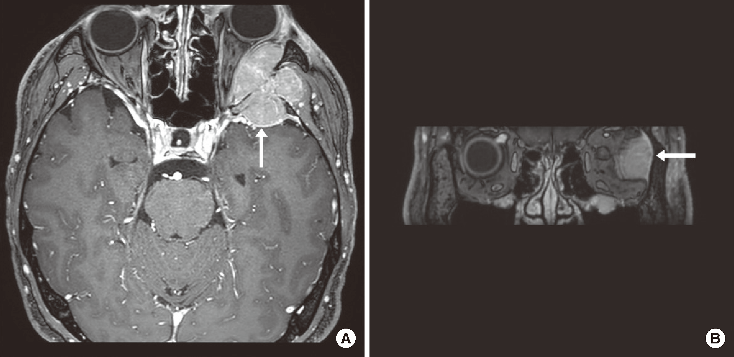
A 39-year-old male presented with exophthalmos and left eye pain, along with headache in the left V1 region, diplopia, and numbness in the left temporal region. Head and neck magnetic resonance imaging (MRI) showed a 4.1×3.5×5.4 cm homogeneous enhancing intracranial mass involving the extraconal space of the lateral aspect of the left orbit, left middle cranial fossa, left temporalis muscle, and greater wing of the sphenoid bone (
Fig. 1). Spheno-orbital meningioma was suspected, and surgical intervention was planned.
 | Fig. 1T1-weighted post-gadolinium head and neck MRI images at the time of diagnosis shows homogenous enhancing mass involving extraconal space of the lateral aspect of the left orbit, left middle cranial fossa, left temporalis muscle, and greater wing of the sphenoid bone. (A) Axial view. (B) Coronal view. 
|
Chemistry tests were requested, but the serum was barely separated. The separator gel was positioned at the top, blocking the serum layer (
Fig. 2). After redrawing the blood, the same phenomenon occurred, and only a tiny amount of serum could be obtained. The test could be conducted after dilution with normal saline at a 1:1 ratio. The results were as follows: increased total protein at 18.2 g/dL (reference interval: 6.7–8.1 g/dL), decreased albumin at 2.8 g/dL (reference interval: 4.1–5.1 g/dL), increased globulin at 15.4 g/dL, increased creatinine at 3.66 mg/dL (reference interval: 0.7–1.2 mg/dL), and hypercalcemia at 14.6 mg/dL (reference interval: 8.6–10.2 mg/dL). The complete blood count (CBC) revealed anemia with hemoglobin at 8.5 g/dL (reference interval: 14.0–17.6 mg/dL) and 5% of plasmacytoid lymphocytes. Rouleaux formation was observed in peripheral blood smears.
 | Fig. 2Improper gel barrier formation in serum separator tube. 
|
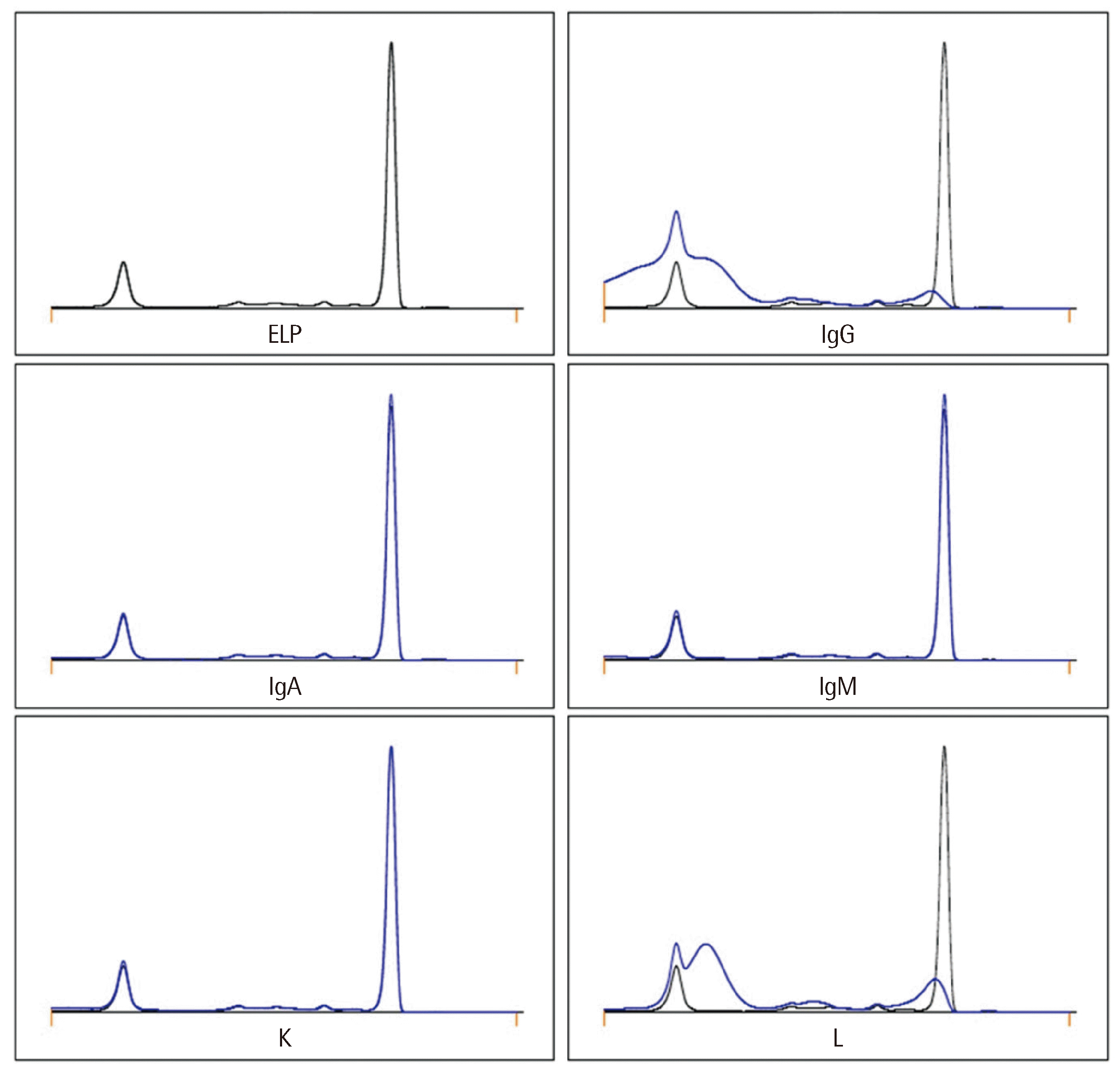
Monoclonal gammopathy was suspected, and a diagnostic workup was conducted. Free light chain analysis showed a decreased kappa of 6.35 mg/L and increased lambda of 1,038.00 mg/L, resulting in a decreased kappa/lambda ratio of 0.01. Serum protein electrophoresis (EP) revealed a peak of 8.01 g/dL in the gamma globulin region, confirmed as IgG-lambda restriction by the serum immunotyping with immunosubtraction by capillary electrophoresis (
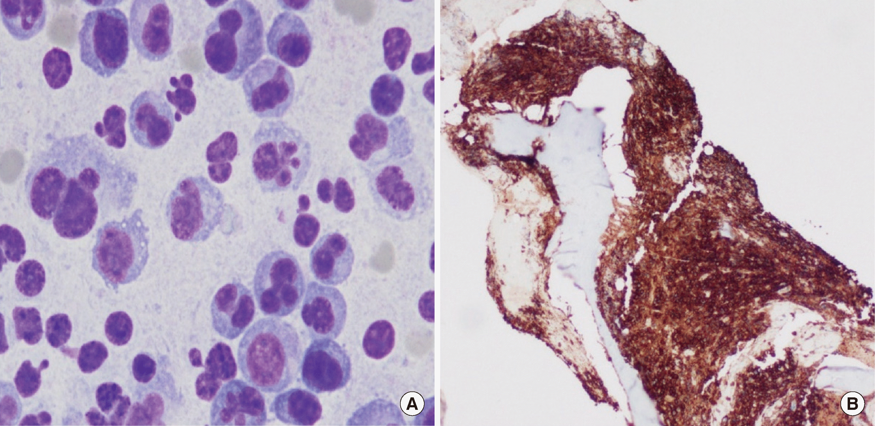
Fig. 3). The bone marrow aspiration smear showed an increased proportion of plasma cells, constituting 61.8% of the total bone marrow cells. Immunohistochemical staining of the bone marrow biopsy revealed diffuse positivity for CD138, indicating the diffuse infiltration of plasma cells (
Fig. 4).
 | Fig. 3Results of serum protein electrophoresis (EP) and immunotyping (IT) by capillary electrophoresis. An IgG-lambda monoclonal peak observed in the gamma globulin region. 
|
 | Fig. 4Dysplastic plasma cells are observed in the bone marrow aspiration (×1,000). (B) Immunohistochemical staining of the bone marrow biopsy reveals diffuse positivity for CD138 (×200). 
|
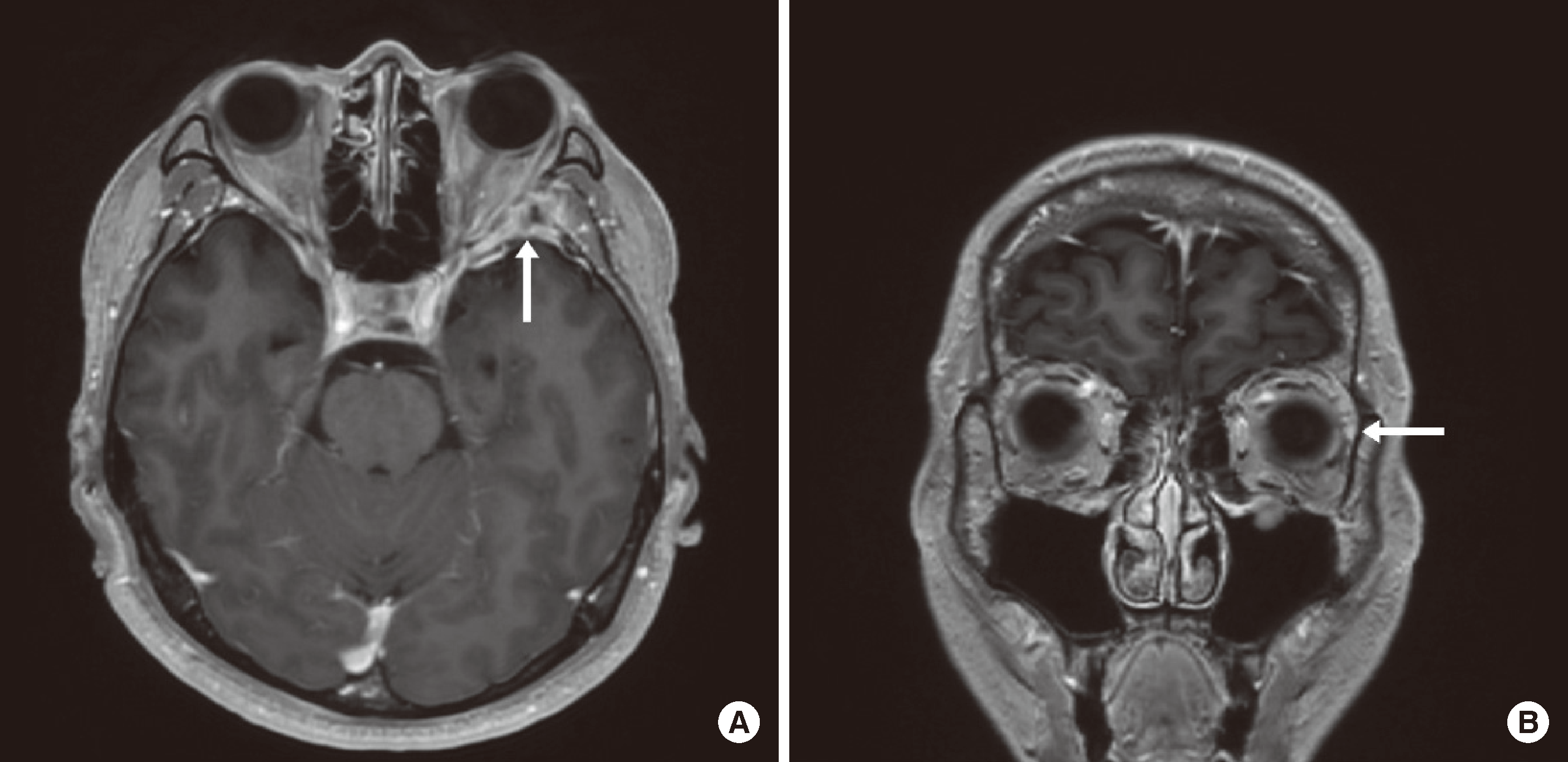
Finally, MM was diagnosed, and the surgery was canceled. Therapeutic plasma exchange (TPE) was performed to manage hyperviscosity in hypergammaglobulinemia. After two sessions of TPE, total protein, globulin, and calcium levels decreased to 7.4 g/dL, 3.0 g/dL, and 8.1 mg/dL, respectively. Following chemotherapy and radiotherapy, there was an improvement in exophthalmos and eye pain. Subsequent MRI revealed a reduction in the intracranial mass (
Fig. 5).
 | Fig. 5T1-weighted post-gadolinium head and neck MRI images after receiving chemotherapy and radiotherapy showed decreased size of enhancing mass. (A) Axial view. (B) Coronal view. 
|
Go to :

DISCUSSION
MM with CNS involvement is extremely rare, accounting for approximately 1% of MM cases [
5]. Although previous studies have been contradictory [
6], CNS involvement predominantly occurs during disease relapse [
7,
8]. Typical ocular symptoms such as blurred vision, proptosis, and eye movement disturbances suggest orbital involvement of plasmacytoma [
9]. However, neuroradiological findings in intracranial plasmacytomas typically lack distinctive features, making it challenging to distinguish them from other malignancies such as meningioma, dural sarcoma, and leptomeningeal carcinomatosis [
10]. In the present case, the MRI findings also suggested a spheno-orbital meningioma and required surgical preparations. Routine chemistry tests and CBC performed as part of preoperative preparations indicated the possibility of monoclonal gammopathy.
Hyperviscosity syndrome is a rare complication of MM. While monoclonal gammopathies such as MM, Waldenström macroglobulinemia, and cryoglobulinemia are common causes, polyclonal gammopathy resulting from autoimmune diseases such as rheumatoid arthritis, systemic lupus erythematosus, and Sjögren’s syndrome can also lead to hyperviscosity syndrome [
11]. The traditional triad of hyperviscosity syndrome consists of mucosal or skin bleeding, visual changes, and focal neurologic deficits [
12]. In the present case, visual changes and neurologic deficits were observed. Although these symptoms could also be attributed to the mass effects of the plasmacytoma, TPE led to improvements.
Improper serum separation is a rare phenomenon, with a prevalence of 0.05 %, but it is highly associated with monoclonal gammopathy [
13-
15]. Chakraborty et al [
13]. reported that over a 2-year period, 16 samples exhibited improper serum separation despite redraws, all of which were associated with monoclonal gammopathy. In monoclonal gammopathy, elevated serum viscosity resulting from hypergammaglobulinemia can influence the positioning of the separation gel and potentially lead to improper serum separation [
14]. In the present case, improper serum separation and a reversed A/G ratio raised suspicion of monoclonal gammopathy. Consequently, MM was diagnosed, and the planned surgery for meningioma was canceled. Therefore, monoclonal gammopathy should be suspected when samples show improper serum separation, and tests for total protein, albumin, and globulin, as well as, if possible, immunoglobulin and free light chain, should be performed. This approach can aid in diagnosing unexpected monoclonal gammopathy and guide patients toward proper management.
This study had some limitations. First, histological confirmation of plasmacytoma was not conducted due to the unavailability of tissue. Second, serum viscosity at the time of diagnosis was not assessed.
In conclusion, we report a case of MM presenting as a sphenoorbital plasmacytoma with improper serum separation, initially misdiagnosed as spheno-orbital meningioma. When encountering improper serum separation in a blood sample, it is essential to conduct further evaluation for monoclonal gammopathy. Especially when an extramedullary mass is present, suspicion of plasmacytoma is warranted for appropriate patient management.
Go to :


 PDF
PDF Citation
 Print
Print


 XML Download