Journal List > Lab Med Online > v.14(4) > 1516088575

Synovial Calprotectin as a Biomarker for Distinguishing Septic Arthritis from Non-Septic Arthritis: A Preliminary Study

Abstract

Background

Delayed septic arthritis treatment exacerbates cartilage degradation and causes persistent functional impairments. Hence, the treatment timing profoundly affects the prognosis, although diagnosing specific cases may prove challenging. This study aimed to investigate whether calprotectin can be used as a biomarker to differentiate between septic and non-septic arthritis.

Methods

Twenty patients with suspected septic arthritis between February and April 2024 were included in this prospective study. The calprotectin levels in the synovial fluid and serum were measured and compared between the patients with septic and non-septic arthritis. Receiver operating characteristic (ROC) curve analyses were performed to differentiate between the two groups.

Results

Synovial fluid calprotectin levels were significantly higher in the septic arthritis group than in the non-septic arthritis group (15,291.5 vs. 6,465.5 mg/L, P <0.001). The area under the ROC curve for synovial fluid calprotectin level in detecting septic arthritis was 0.881 (sensitivity 100%, specificity 78.6%, positive predictive value 66.7%, and negative predictive value 100%).

Conclusions

Synovial calprotectin level is a reliable diagnostic parameter for the differential diagnosis of septic arthritis from non-septic arthritis.

초록

배경

Calprotectin은 백혈구에서 분비되는 단백으로 다양한 염증성 질환에서 염증 정도를 확인하는 지표로 이용되고 있다. 이 연구에서는 관절액과 혈청에서의 calprotectin이 화농성 및 비화농성 관절염을 구별하는 지표로 이용될 수 있는지 확인하였다.

방법

2024년 2월부터 4월까지 화농성 관절염이 의심되는 20명 환자의 관절액과 혈청에 대하여 calprotectin 검사를 시행하였다. 대상자들을 화농성 관절염군과 비화농성 관절염군으로 나누어 관절액과 혈청에서의 calprotectin값을 서로 비교하였다.

결과

화농성 관절염군은 6명(30%)이었다. 화농성 관절염군의 관절액 calprotectin값은 비화농성 관절염군보다 유의하게 높았다(15,291.5 mg/L vs. 6,465.5 mg/L, P<0.001). 화농성 관절염을 진단하는 관절액 calprotectin의 area under the curve (AUC)값은 0.881이었다(민감도 100%, 특이도 78.6%, 양성예측도 66.7%, 음성예측도 100%).

결론

관절액에서 측정한 calprotectin은 화농성 관절염과 비화농성 관절염을 구별하는 신뢰할 만한 진단 지표로서 임상적 유용성이 높다.

INTRODUCTION

Prompt identification and treatment are crucial for acute septic arthritis because of its associated elevated morbidity and mortality rates, particularly in the elderly population [1]. Delayed diagnosis and treatment of septic arthritis can cause subchondral bone loss and permanent joint dysfunction [2]. Although antibiotic development has made treatment easier than in the past, antibiotic overuse has increased bacterial resistance, increasing septic arthritis prevalence, which is further compounded by increased life expectancy and frequent intraarticular injections. The incidence of septic arthritis has significantly increased. A recent study reported that the incidence of septic arthritis per 100,000 people in Korea increased from 2005 to 2018 (4.072 and 15.298, respectively) [3]. An accurate and prompt septic arthritis diagnosis is crucial for improving patient outcomes and minimizing the required resource use.
Septic arthritis is diagnosed by combining clinical findings, laboratory markers, and microbiological analyses. Traditional criteria to diagnose septic arthritis is synovial fluid culture, with >25,000 or >50,000 white blood cell (WBC)/μL or at least 90% polymorphonuclear leukocyte (PMN) count in the synovial fluid; however, they are not particularly sensitive and specific for diagnosing septic arthritis [4, 5]. Early recognition of septic arthritis is often challenging when direct Gram-stained synovial fluid smears are negative [6].
Calprotectin is a cytosolic calcium-binding protein released as S100A8/S100A9-heterodimers predominantly from phagocytes such as neutrophils and monocytes [7]. Human calprotectin functions as a host defense protein, inhibiting microbial colonization by sequestering manganese and zinc [8]. Therefore, calprotectin may form a distinctive biomarker of infections, including septic arthritis. Recent studies have demonstrated its diagnostic precision in distinguishing between inflammatory and bacterial infections [6, 9, 10]. Till date, few studies have focused on synovial calprotectin levels for diagnosing septic arthritis in South Korea. This study evaluated the discriminatory ability of synovial and serum calprotectin levels in septic arthritis diagnosis, compared with synovial WBC count and PMN and serum C-reactive protein (CRP) levels in patients with suspected septic arthritis.

MATERIALS AND METHODS

1. Patients

We prospectively included patients with suspected septic arthritis who requested synovial fluid culture at our laboratory. Samples were collected between February and April 2024. The differences in synovial WBC count, synovial PMN count, and synovial and serum calprotectin levels between patients with septic and non-septic arthritis were assessed. Additionally, ideal cut-off values were derived using the area under the curve (AUC) analysis.
The diagnosis of septic arthritis was confirmed if bacteria were present in synovial fluid culture without crystals in synovial fluid [6]. Gram staining and matrix-assisted desorption/ionization-time of flight mass spectrometry (MALDI-TOF MS; Bruker Daltonics, Billerica, MA, USA) identified the microbial etiology. Since the presence or absence of inflammatory bowel disease (IBD) may affect calprotectin levels, we investigated this through medical records. This cohort did not have any patients with IBD.

2. Biomarkers

Synovial fluids were centrifuged at 3,000 rpm for 7 min at 4℃. The supernatant was transferred to a new tube and stored at −80℃ until use. Calprotectin was measured in the synovial fluid and serum using the ELIATM Calprotectin 2 assay, a sandwichprinciple-based fluoro-enzyme-immunoassay (FEIA) on PhadiaTM 200 (Thermo Fisher Scientific, Freiburg, Germany). This assay, performed according to the manufacturer’s instructions, utilizes monoclonal mouse anti-calprotectin antibodies, which are highly specific for heterodimeric calprotectin complexes [11]. The measured calprotectin range in fecal specimens is 3.8–6,000 mg/kg. In this study, the samples were diluted 1/200. Other synovialbased biomarkers (WBC (cells/μL) and PMN (%)) and serum CRP were retrieved from the medical records. In our hospital, the cells in body fluids are counted manually using a counting chamber and light microscopy. The study protocol was approved by the Institutional Review Board and Ethics Committee of Daejeon St. Mary’s Hospital (No. DC24TASI0009).

3. Statistics

Categorical variables are presented as numerical values (percentages), while continuous variables are presented as medians and interquartile ranges (IQRs) for non-normally distributed data. Univariate analyses were conducted utilizing the chi-square and Mann–Whitney U tests for categorical and continuous variables, respectively. Suggestions for the most effective threshold values were obtained by analyzing the AUC. The sensitivity, specificity, positive predictive value (PPV), and negative predictive value (NPV) were calculated using optimal threshold values. Statistical analyses were performed using MedCalc (version 20.111; MedCalc Software Ltd., Ostend, Belgium) and two-tailed P<0.05 was considered statistically significant.

RESULTS

Among the 20 patients with a median age of 82 years (range, 56–96 years) included, 13 were female (65%). The final diagnoses were septic arthritis in six patients (30%) and non-septic arthritis in 14 patients (70%). The baseline patient characteristics are shown in Table 1. The difference in the parameters between the patients with septic and non-septic arthritis was not significant. The causative bacteria of septic arthritis were Staphylococcus aureus (four cases), Escherichia coli (one case), and Streptococcus dysgalactiae (one case).
The median synovial calprotectin concentration in patients with septic arthritis was significantly higher than that in those with non-septic arthritis (15,291.5 [IQR 14,597.0–15,935.0] vs. 6,465.5 [IQR 3,038.0–13,940.0] mg/L, P=0.006; Fig. 1, Table 2). The patients with septic arthritis had a significantly higher median synovial PMN% than those with non-septic arthritis (94.5% [IQR 85.0–98.0] vs. 77.5% [IQR 65.0–83.0], P=0.0148). The median synovial WBC count in the patients with septic and non-septic arthritis was 48,800 (IQR 8,000–132,500) and 24,600 (IQR 3,000–34,080) cells/μL, respectively. Synovial WBC count did not differ significantly between the two groups. The median serum calprotectin concentration was 2,722.5 (IQR 946.0–3,397.0) mg/L in the patients with septic arthritis and 764.5 (IQR 376.0–1,502.0) mg/L in the patients with non-septic arthritis. Serum calprotectin levels tended to be higher in the patients with septic arthritis than those in the patients with non-septic arthritis group, although the difference was not statistically significant. The median serum CRP concentration in the patients with septic and non-septic arthritis were 18.7 (IQR 11.6–22.4) and 2.9 (IQR 0.8–8.3) mg/dL, respectively.
In the ROC curve analysis, the synovial calprotectin (AUC, 0.881 [95% CI 0.659–0.981]) was more accurate for septic arthritis diagnosis than >50,000 synovial WBC/μL (AUC, 0.714 [95% CI, 0.472–0.891]) and synovial PMN >90% (AUC, 0.726 [95% CI, 0.484–0.898]) (Table 3). The overall sensitivity, specificity, PPV, and NPV of >13,940 mg/L synovial calprotectin for septic arthritis were 100.0%, 78.6%, 66.7%, and 100.0%, respectively. The overall sensitivity, specificity, PPV, and NPV of >50,000 synovial WBC/μL for septic arthritis were 50.0%, 92.9%, 75.0%, and 81.3%, respectively. The overall sensitivity, specificity, PPV, and NPV of >90% synovial PMNs for septic arthritis were 66.7%, 78.6%, 57.1%, and 84.6%, respectively. Among serum biomarkers, the AUC of CRP was higher than that of serum calprotectin (0.869 vs. 0.786, respectively).

DISCUSSION

Timely diagnosis of septic arthritis is crucial because as any delay or insufficient treatment may cause irreversible joint destruction, permanent loss of function, and a life-threatening situation [12]. However, the markedly sensitive and specific tests for the early differentiation between septic and non-septic arthritis are lacking. This study demonstrated that synovial calprotectin levels were significantly higher in the patients with septic arthritis than those in the patients with non-septic arthritis. With an AUC of 0.881 and a cut-off value of 13,940 mg/L, it showed excellent potential for septic arthritis diagnosis.
Calprotectin is a calcium-binding antimicrobial protein found exclusively in neutrophils and monocytes and has been thoroughly investigated under various inflammatory conditions, including IBD. Synovial fluid calprotectin has recently been recognized as a promising diagnostic test [6, 9, 13, 14]. Wouthuyzen-Bakker et al. reported that synovial calprotectin demonstrates high sensitivity (86.7%) and specificity (91.7%) when employing the lateral flow assay (BÜHLMANN Laboratories AG, Sch€onenbuch, Switzerland) for diagnosing chronic prosthetic joint infection [9]. Lazic et al. demonstrated that a synovial calprotectin lateral flow assay yielded 88% sensitivity (95% CI 64–99), 81% specificity (95% CI 54–96), 83% PPV (95% CI 59–96), and 87% NPV (95% CI 60–98) in diagnosing a periprosthetic joint infection [13]. Couderc et al. noted that synovial calprotectin exhibited 73% sensitivity and 67% specificity, with a threshold of 854 mg/L for diagnosing septic arthritis. However, the commercial ELISA kits used were unspecified [14]. Baillet et al. investigated the ability of synovial calprotectin to discriminate between septic arthritis, rheumatoid arthritis, and pseudogout [6]. They reported 76% sensitivity and 94% specificity at a threshold of 150 mg/L, using the HK325 kit (Hycult Biotech, Uden, the Netherlands). These findings suggest that the exact cutoff value and concentration range of synovial calprotectin for determining joint infections remain ambiguous. Therefore, it necessitates the integration of research utilizing various calprotectin testing methods with population groups in the future.
Traditional diagnosis of septic arthritis uses synovial fluid culture, a synovial fluid with >50,000 WBC/μL and >90% PMN, but they are not highly sensitive or specific for diagnosing septic arthritis [15, 16]. Carpenter et al. discovered that the pooled sensitivity for synovial fluid WBC exceeding 50,000 cells/μL and PMN% in diagnosing septic arthritis was 56% and 60%, respectively [15]. The outcomes correspond closely with our results, where >50,000 synovial WBC/μL displayed 50.0% sensitivity and 92.9% specificity. Similarly, >90% synovial PMN exhibited 66.7% sensitivity and 78.6% specificity. Importantly, our research suggests that synovial calprotectin may offer a new alternative, significantly surpassing both the synovial WBC count and PMN% regarding accuracy.
As this was a preliminary study, it had some limitations. First, the sample size was small, thus a larger patient cohort is required to validate the utility of synovial calprotectin as a biomarker for septic arthritis. Nevertheless, this study is valuable as a preliminary pilot study owing to the low prevalence of septic arthritis. Second, we excluded patients with periprosthetic joint infections because of their predominantly chronic nature. We primarily aimed to investigate the role of synovial calprotectin in acute joint infections. Further studies should aim to explore the utility of calprotectin in other conditions that pose clinical challenges in differentiation, such as crystal arthritis, rheumatoid arthritis, or periprosthetic joint infection. Third, fecal and/or serum calprotectin is a widely applied biomarker for diagnosis and monitoring in patients with IBD [17]; therefore, calprotectin level may increase if the patient also suffers from IBD. This study did not recruit any patient with IBD. Further studies are needed to include various inflammatory diseases such as IBD.
In conclusion, the result of this study suggests that synovial calprotectin is better than synovial WBC count and PMN% for differentiating between septic and non-septic arthritis.

Acknowledgement

This study was funded by a grant from the Korean Society of Diagnostic Immunology (Grant No. KSDI2022-6).

Notes

Conflicts of Interest

None declared.

REFERENCES

1. Choi JY, Lee EY, Oh SY, Seong MK, Lee OJ. 2017; Causative factors regarding the clinical outcomes after arthroscopic treatment for pyogenic knee arthritis. J Korean Orthop Assoc. 52:257–63. DOI: 10.4055/jkoa.2017.52.3.257.
2. Earwood JS, Walker TR, Sue GJC. 2021; Septic arthritis: diagnosis and treatment. Am Fam Physician. 104:589–97. PMID: 34913662.
3. Choi HJ, Yoon HK, Oh HC, Hong JH, Choi T, Park SH. 2022; Mortality of septic knee arthritis in Korea: risk factors analysis of a large national database. Sci Rep. 12:14008. DOI: 10.1038/s41598-022-18420-4. PMID: 35978108. PMCID: PMC9385853. PMID: 64e87cc2a9d740c4a9d42443c02fc8d0.
4. Margaretten ME, Kohlwes J, Moore D, Bent S. 2007; Does this adult patient have septic arthritis? JAMA. 297:1478–88. DOI: 10.1001/jama.297.13.1478. PMID: 17405973.
5. Varady NH, Schwab PE, Kheir MM, Dilley JE, Bedair H, Chen AF. 2022; Synovial fluid and serum neutrophil-to-lymphocyte ratio: novel biomarkers for the diagnosis and prognosis of native septic arthritis in adults. J Bone Joint Surg Am. 104:1516–22. DOI: 10.2106/JBJS.21.01279. PMID: 35726876.
6. Baillet A, Trocmé C, Romand X, Nguyen CMV, Courtier A, Toussaint B, et al. 2019; Calprotectin discriminates septic arthritis from pseudogout and rheumatoid arthritis. Rheumatology (Oxford). 58:1644–8. DOI: 10.1093/rheumatology/kez098. PMID: 30919904.
7. Odink K, Cerletti N, Brüggen J, Clerc RG, Tarcsay L, Zwadlo G, et al. 1987; Two calcium-binding proteins in infiltrate macrophages of rheumatoid arthritis. Nature. 330:80–2. DOI: 10.1038/330080a0. PMID: 3313057.
8. Brophy MB, Hayden JA, Nolan EM. 2012; Calcium ion gradients modulate the zinc affinity and antibacterial activity of human calprotectin. J Am Chem Soc. 134:18089–100. DOI: 10.1021/ja307974e. PMID: 23082970. PMCID: PMC3579771.
9. Wouthuyzen-Bakker M, Ploegmakers JJW, Ottink K, Kampinga GA, Wagenmakers-Huizenga L, Jutte PC, et al. 2018; Synovial calprotectin: an inexpensive biomarker to exclude a chronic prosthetic joint infection. J Arthroplasty. 33:1149–53. DOI: 10.1016/j.arth.2017.11.006. PMID: 29224989.
10. Zhang Z, Cai Y, Bai G, Zhang C, Li W, Yang B, et al. 2020; The value of calprotectin in synovial fluid for the diagnosis of chronic prosthetic joint infection. Bone Joint Res. 9:450–7. DOI: 10.1302/2046-3758.98.BJR-2019-0329.R2. PMID: 32832073. PMCID: PMC7418777.
11. Mylemans M, Nevejan L, Van Den Bremt S, Stubbe M, Cruyssen BV, Moulakakis C, et al. 2021; Circulating calprotectin as biomarker in neutrophil-related inflammation: pre-analytical recommendations and reference values according to sample type. Clin Chim Acta. 517:149–55. DOI: 10.1016/j.cca.2021.02.022. PMID: 33689693.
12. Weston VC, Jones AC, Bradbury N, Fawthrop F, Doherty M. 1999; Clinical features and outcome of septic arthritis in a single UK Health District 1982-1991. Ann Rheum Dis. 58:214–9. DOI: 10.1136/ard.58.4.214.
13. Lazic I, Prodinger P, Stephan M, Haug AT, Pohlig F, Langer S, et al. 2022; Synovial calprotectin is a reliable biomarker for periprosthetic joint infections in acute-phase inflammation - a prospective cohort study. Int Orthop. 46:1473–9. DOI: 10.1007/s00264-022-05421-1. PMID: 35524793. PMCID: PMC9166865.
14. Couderc M, Peyrode C, Pereira B, Miot-Noirault E, Mathieu S, Soubrier M, et al. 2019; Comparison of several biomarkers (MMP-2, MMP-9, the MMP-9 inhibitor TIMP-1, CTX-II, calprotectin, and COMP) in the synovial fluid and serum of patients with and without septic arthritis. Joint Bone Spine. 86:261–2. DOI: 10.1016/j.jbspin.2018.04.008.
15. Carpenter CR, Schuur JD, Everett WW, Pines JM. 2011; Evidence-based diagnostics: adult septic arthritis. Acad Emerg Med. 18:781–96. DOI: 10.1111/j.1553-2712.2011.01121.x. PMID: 21843213. PMCID: PMC3229263.
16. Horowitz DL, Katzap E, Horowitz S, Barilla-LaBarca ML. 2011; Approach to septic arthritis. Am Fam Physician. 84:653–60. DOI: 10.1093/med/9780199550647.003.011005.
17. Khaki-Khatibi F, Qujeq D, Kashifard M, Moein S, Maniati M, Vaghari-Tabari M. 2020; Calprotectin in inflammatory bowel disease. Clin Chim Acta. 510:556–65. DOI: 10.1016/j.cca.2020.08.025. PMID: 32818491. PMCID: PMC7431395.
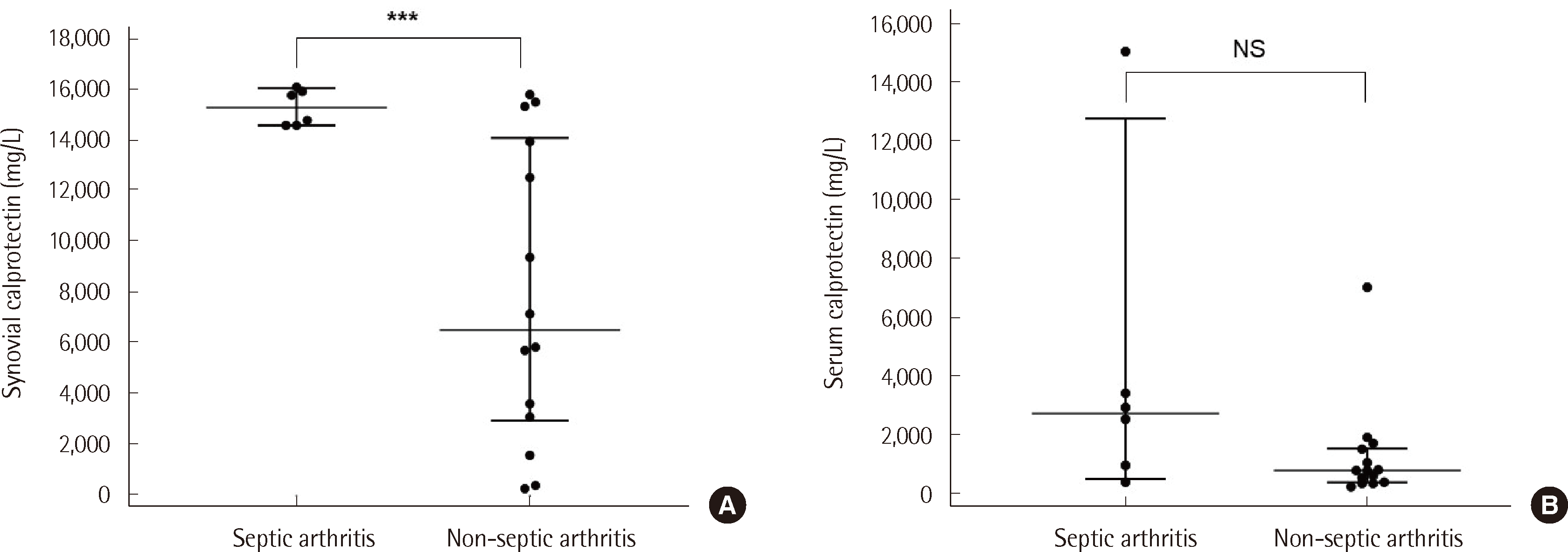
Fig. 1
Comparison of calprotectin concentrations between patients with septic arthritis and non-septic arthritis. The (A) synovial fluid and (B) serum calprotectin concentrations in patients with septic arthritis and non-septic arthritis. The line indicates the median and the bars show the interquartile range. The Mann–Whitney U-test was used for the statistical comparison between the groups.
Abbreviation: NS, no significant difference.
***P<0.001.
lmo-14-4-358-f1.tif
Table 1
Baseline clinical characteristic and biomarkers of patients with septic arthritis and non-septic arthritis
Septic arthritis (N=6) Non-septic arthritis (N=14) P value
Age (years), median (IQR) 77.5 (69.0–82.0) 82.5 (71.0–87.0) 0.5359*
Female, N (%) 3 (50.0) 10 (71.4) 0.3695
Medical history
Diabetes, N (%) 1 (16.7) 7 (50) 0.1741
Dyslipidemia, N (%) 1 (16.7) 2 (14.3) 0.8940
Hypertension, N (%) 4 (66.7) 11 (78.6) 0.5829

*Mann–Whitney U-test; Chi-square test.

Abbreviation: IQR, interquartile range.

Table 2
Intergroup differences between synovial WBC count, synovial PMN, synovial calprotectin, and serum calprotectin
Biomarker median (IQR) Septic arthritis (N=6) Non-septic arthritis (N=14) P value*
Synovial WBC (cells/μL) 48,800 (8,000–132,500) 24,650 (3,000–34,080) 0.1786
Synovial PMN (%) 94.5 (85.0–98.0) 77.5 (65.0–83.0) 0.0148
Synovial calprotectin (mg/L) 15,291.5 (14,597.0–15,935.0) 6,465.5 (3,038.0–13,940.0) 0.0064
Serum CRP (mg/dL) 18.7 (11.6-22.4) 2.9 (0.8-8.3) 0.0087
Serum calprotectin (mg/L) 2,722.5 (946.0–3,397.0) 764.5 (376.0–1,502.0) 0.0507

*Mann–Whitney U-test.

Abbreviations: IQR, interquartile range; WBC, white blood cell; PMN, polymorphonuclear leukocyte; CRP, C-reactive protein.

Table 3
Performance of synovial WBC count, synovial PMN, and synovial and serum calprotectin levels in differentiating septic arthritis and nonseptic arthritis
Biomarkers AUC (95% CI) P value Cut-off Sensitivity% (95% CI) Specificity% (95% CI) PPV (95% CI) NPV (95% CI)
Synovial WBC (cells/μL) 0.714 (0.472–0.891) 0.068 >50000 50.0 (11.8–88.2) 92.9 (66.1–99.8) 75.0 (27.8–95.9) 81.3 (65.8–90.7)
Synovial PMN (%) 0.726 (0.484–0.898) 0.059 >90 66.7 (22.3–95.7) 78.6 (49.2–95.3) 57.1 (29.7–80.8) 84.6 (63.2–94.6)
Synovial calprotectin (mg/L) 0.881 (0.659–0.981) <0.001 >13,940* 100.0 (54.1–100.0) 78.6 (49.2–95.3) 66.7 (42.3–95.5) 100.0 (71.5–100.0)
Serum CRP (mg/dL) 0.869 (0.644–0.976) 0.001 >11.5* 83.3 (35.9–99.6) 92.9 (66.1–99.8) 83.3 (42.2–97.2) 92.9 (68.3–98.7)
Serum calprotectin (mg/L) 0.786 (0.548–0.935) 0.024 >1896* 66.7 (22.3–95.7) 92.9 (66.1–99.8) 80.0 (35.8–96.6) 86.7 (67.5–95.3)

The optimal threshold for the diagnosis of septic diagnosis was calculated using the Youden index, which is the point of maximum potential effectiveness of a biomarker (the maximum sum of sensitivity and specificity).

Abbreviations: AUC, area under the curve; CI, confidence interval; PPV, positive predictive value; NPV, negative predictive value; PMN, polymorphonuclear leukocyte; CRP, Creactive protein.

TOOLS
Similar articles