Journal List > Lab Med Online > v.14(4) > 1516088569

Improvement of Clinical Diagnosis Accuracy of Alzheimer’s Disease Using Multiple Blood Biomarkers

Abstract

Background

Blood biomarkers significantly change the diagnostic and prognostic approaches to Alzheimer’s disease (AD). There have also been many reports of enhanced accuracy achieved by combining multiple biomarkers. Our study aimed to assess the efficacy of the QPLEX™ Alz plus assay kit (QM-Alz; Quantamatrix Inc., Korea), which includes multiple blood biomarkers, in clinical AD diagnosis. After adding the apolipoprotein E genotype (ApoE) and galectin-3 (Gal-3) to QM-Alz, we tested whether the diagnostic accuracy increased.

Methods

A total of 128 clinically diagnosed participants were included. We identified the optimal algorithm combining QM-Alz, ApoE, and Gal-3 for diagnosing clinical AD dementia and cerebral amyloid-β deposition. The area under the curve (AUC), sensitivity (Sen), and specificity (Spe) of each biomarker combination were calculated.

Results

The AUC for the QM-Alz algorithm to differentiate between cognitively normal patients and patients with AD was 0.758 (Sen=71.3%, Spe=72.9%). When the QM-Alz algorithm was enriched with the addition of ApoE and Gal-3, their individual AUC values improved to 0.804 (Sen=73.8%, Spe=75.0%) and 0.842 (Sen=77.5%, Spe=79.2%), respectively. Furthermore, when all biomarkers were integrated into the algorithm, the resulting AUC was elevated to 0.860 (Sen=77.5%, Spe=77.1%).

Conclusions

The combination of various blood biomarkers, especially when integrating QM-Alz, ApoE, and Gal-3, significantly improves diagnostic accuracy for a more precise AD diagnosis.

초록

배경

혈액 바이오마커는 알츠하이머병(Alzheimer’s disease, AD)의 진단 및 예후에 대한 접근 방식을 크게 변화시켰으며, 여러 바이오마커를 조합하여 정확도가 향상되었다는 보고가 많이 나오고 있다. 이 논문에서는 여러 혈액 바이오마커를 활용한 QPLEX™ Alz plus assay kit (QM-Alz; Quantamatrix Inc., Korea)가 AD의 임상 진단에 도움이 될 수 있는지 살펴보고자 하였다. 또한, QM-Alz에 아포지단백 E 유전자형(ApoE)과 galectin-3 (Gal-3)을 추가했을 때 진단 정확도를 확인하였다.

방법

임상적 진단을 통해 정상으로 분류된 48명과 AD로 분류된 80명을 대상으로 QM-Alz, ApoE, Gal-3 측정을 수행하였다. 임상적 AD 진단 결과 및 대뇌 아밀로이드 베타 침착 결과를 종속변수로 한 회귀분석 결과를 토대로 각 바이오마커 조합별 알고리즘을 도출하고, 각 바이오마커 조합의 곡선하면적(area under the curve, AUC), 민감도, 특이도를 계산하였다.

결과

인지기능이 정상인 환자와 AD 환자를 구별하는 QM-Alz 알고리즘의 AUC는 0.758 (민감도 71.3%, 특이도 72.9%)이었다. QMAlz에 ApoE 또는 Gal-3 결과를 추가하였을 때, 각각의 AUC 값은 0.804 (민감도 73.8%, 특이도 75.0%)와 0.842 (민감도 77.5%, 특이도 79.2%)로 향상되었다. 모든 바이오마커를 알고리즘에 통합했을 때 AUC는 0.860 (민감도 77.5%, 특이도 77.1%)이 되었다.

결론

QM-Alz는 AD의 임상 진단에 AUC 0.758의 정확도를 보여주었으며, 다양한 혈액 바이오마커를 조합함으로써 진단 정확도를 더욱 향상시킬 수 있었다.

INTRODUCTION

Alzheimer’s disease (AD) is a neurodegenerative disorder related to aging and is the most widespread form of dementia. The global prevalence of dementia including AD is predicted to rise from 57.4 million cases in 2019 to 152.8 million cases by 2050 [1]. With the aging global population, AD is becoming a major healthcare issue worldwide, with an estimated financial burden of $2 trillion by the year 2030 [2]. Hence, it is crucial to develop effective strategies such as early diagnosis and treatment to improve patient outcomes and reduce costs for patients, caregivers, and the healthcare system.
Because cerebrospinal fluid (CSF) proteins and positron emission tomography (PET) tracers specific to amyloid-β (Aβ) and phosphorylated tau (p-tau) are directly connected to AD, both candidates are excellent sources of information for the detection of biochemical abnormalities within the brain for an AD diagnosis. Nevertheless, the broad application of CSF and imaging-based biomarkers faces limitations due to the invasive nature of lumbar punctures and the high cost and limited availability of PET imaging [3, 4]. Using blood-based biomarkers for AD diagnosis has been challenging despite the convenience of blood sampling compared to CSF sampling and PET imaging. One reason for this is that only a small proportion of brain proteins are found in the blood, making it difficult to detect AD biomarkers. Additionally, the presence of high levels of plasma proteins, such as albumin and immunoglobulin G in blood samples, can cause analytical interference when measuring AD biomarkers [5]. The release of brain proteins into the bloodstream may also cause degradation by proteases, leading to metabolism by the liver or elimination by the kidneys. This results in unpredictable changes that are not related to brain function, making it hard to find consistent blood markers for AD [6]. Despite these challenges, technical advancements such as ultrasensitive immunoassays, mass spectrometrybased proteomic analyses, and bead-based multiplex assays offer renewed optimism [7, 8]. In particular, blood biomarkers have recently demonstrated an ability to significantly change the diagnostic and prognostic approaches for AD and improve the planning of interventional trials [9-11].
We previously developed a bead-based multiple biomarker diagnostic tool named the QPLEX™ Alz plus assay kit (QM-Alz), which can predict the existence of cerebral Aβ deposition. The kit incorporates four blood biomarkers consisting of Aβ40, a galectin-3-binding protein (LGALS3BP), an angiotensin-converting enzyme (ACE), and periostin (POSTN), which have been employed in a previous study [9-11] and can distinguish between individuals who test positive or negative for PET imaging [8, 12]. These four biomarkers are unique to this platform and have shown promising results in identifying cerebral Aβ deposition, and amyloid plaques and neurofibrillary tangles are two diagnostic candidates for AD. Moreover, efforts have been made to improve the performance of QM-Alz by using additional markers.
This study aimed to demonstrate the predictive effectiveness of QM-Alz, a composite of four markers, in differentiating between clinical groups—cognitively normal (CN) and those with AD— using an independent cohort from South Korea. We also aimed to compare the predictive capabilities of Aβ40 alone against those of QM-Alz, both with and without Aβ40 because the diagnostic performance of Aβ for AD may be compared with that of LGALS3BP, ACE, and POSTN, suggested previously as new AD biomarkers. Additionally, we intended to explore the potential improvement in the performance of QM-Alz in terms of clinical diagnosis and PET positivity by incorporating novel factors such as the apolipoprotein E genotype (ApoE) and galectin-3 (Gal-3). We propose a combination of biomarkers that offers the best potential for accurately diagnosing AD, which could be particularly useful in clinical assessments such as the Mini-Mental State Examination (MMSE) and the Clinical Dementia Rating (CDR).

MATERIALS AND METHODS

1. Participants

We recruited participants over the age of 40 who voluntarily decided to participate in the study after hearing the detailed explanation and fully understanding it from 15 referral hospitals in South Korea. We excluded participants who had other psychiatric diseases or specific surgical history. Detailed exclusion criteria are described in the Supplementary File. Finally, a QM-Alz assay was performed on 128 participants. Most participants were recruited from the Samsung Medical Center (N=47) and Soonchunhyang University Bucheon Hospital (N=45). This project was part of a nationwide multicenter consortium named the Precision Medicine Platform for Mild Cognitive Impairment, based on Multiomics, Imaging, Evidence-based R&BD [11]. This study was conducted following the Declaration of Helsinki, and approved by the Institutional Review Board of the Samsung Medical Center (IRB No. 2020-01-024[M1]) and each center. Informed consent was obtained from all subjects involved in the study. Written informed consent has been obtained from the patients for the publication of this paper.

2. Clinical diagnosis

Experienced neurologists diagnosed the participants based on general diagnostic criteria. The criteria for AD dementia are based on the proposal by the National Institute on Aging-Alzheimer’s Association Research Framework. The patients with AD exhibited CDR scores ranging from 0.5 to 3, and their MMSE scores were 10 or higher, indicating their suitability for Seoul neuropsychological screening battery-II (SNSB-II) testing. The SNSB-II evaluates many cognitive factors including verbal and visual memory, visuo-constructive function, language, praxis, components of Gerstmann syndrome (acalculia, agraphia, right/left disorientation, finger agnosia), and frontal/executive functions [13].

3. Amyloid PET imaging and analysis

All participants underwent either F-florbetaben or F-flutemetamol PET scanning at each center using a Discovery Ste. PET/computed tomography scanner (GE Medical Systems, Milwaukee, USA). Briefly, mean doses of 311.5 MBq F-florbetaben or 185 MBq F-flutemetamol were injected into each individual and, 90 minutes later, an emission PET scan with dynamic mode (4×5-minute frames) was performed. Three-dimensional (3D) PET images were reconstructed using the ordered-subsets expectation maximization algorithm (iterations=4, subset=20). We performed image processing as in the previous studies to obtain a direct comparison Centiloid. The cut-off value of the direct comparison Centiloid was derived from receiver operating characteristic (ROC) curve analysis, which was previously described and calculated as 25.11 [11].

4. Blood sampling and storage

Blood samples were collected in dipotassium ethylene-diamine-tetraacetic acid (K2 EDTA) tubes (BD Vacutainer Systems, Plymouth, UK) and centrifuged at 700×g for 5 minutes at 20°C. The plasma supernatants were stored at -80°C [8, 12].

5. QM-Alz assay protocol

Aβ40, LGALS3BP, ACE, and POSTN were quantified using QM-Alz as it is. Gal-3 (R&D Systems, Minneapolis, USA) was coupled to Quantamatrix microdisks, the raw material used to manufacture QM-Alz. The coupling protocol was as follows: we chose a microdisk with a different code from the codes already used in QM-Alz, washed the microdisk with 50 mM MES, pH 5.0 (Sigma-Aldrich, Milwaukee, USA), activated the carboxylic acid on the surface of the microdisk with 1-Ethyl-3-(3-Dimethylaminopropyl) Carbodiimide Hydrochloride (ThermoFisher, Waltham, USA) and N-Hydroxysulfosuccinimide sodium salt (ThermoFisher), incubated the microdisk for two hours with Gal-3 in MES, and washed it thrice with 1% bovine serum albumin (BSA).
Microdisks were placed in 96-well plates and 35 μL of diluted human plasma samples and equal amounts of biotin-conjugated detection antibodies were incubated for 90 minutes at room temperature. The microdisks were washed with a 0.1% BSA buffer and incubated with 2 μg/mL of R-phycoerythrin-conjugated streptavidin for 15 minutes at room temperature. The microdisks were washed, re-suspended in 0.1% BSA buffer, and analyzed using the Quantamatrix multiplex assay platform (Quantamatrix, Seoul, Korea). All reagents, microdisks, and 96-well plates that were used were included in QM-Alz.

6. ApoE genotyping

ApoE genotyping was performed as previously described. Briefly, the DNA of the patients was extracted from the blood samples using the QIAamp DNA Blood Mini Kit (QIAGEN GmbH, Hilden, Germany). The genotypes of rs7412 and rs429358 were analyzed by allele-specific real-time polymerase chain reaction (PCR). Depending on the genotypes of rs7412 and rs429358, variants of the ApoE gene were classified as ε1, ε2, ε3, or ε4. The detailed genotype results are included in the supplementary data (Supplementry Table 1), and participants with the ε4 gene were classified as ApoE-positive [13].

7. Data analysis

Statistical analyses were performed using MedCalc 20.115 (Ostend, Belgium). Several algorithms were generated using logistic regression to discriminate between CN and AD groups for each biomarker combination. The algorithms for distinguishing PET positivity were also separately generated through logistic regression. The basic equation of algorithm value was as follows:
Pi=E1+EE=expa1×m1+a2×m2++an×mn+C
Pi, algorithm values; an, coefficient values for each biomarker; mn, measured concentration for each biomarker; C, constant. Each biomarker concentration of the samples obtained with QM-Alz was multiplied by the coefficient values and Pi was calculated. To confirm the discrimination performance, area under the curve (AUC), ROC curve analysis, independent t-test, sensitivity, specificity, and correlation were calculated to evaluate the discriminatory power of the algorithms. The cut-off value was set to the value where the sum of sensitivity and specificity is maximized. Analysis of covariance (ANCOVA) was performed to investigate the effect of age on the overall analysis.

RESULTS

1. Demographic characteristics of participants

This study included 128 participants (aged 45–88 years). The demographic data are shown in Table 1. There were statistically significant differences between the CN and AD groups in age, ApoE, MMSE, CDR, and PET positivity. There were no significant differences between the two groups in sex and total lifetime education years.

2. Performance of QM-Alz for clinical diagnosis of AD

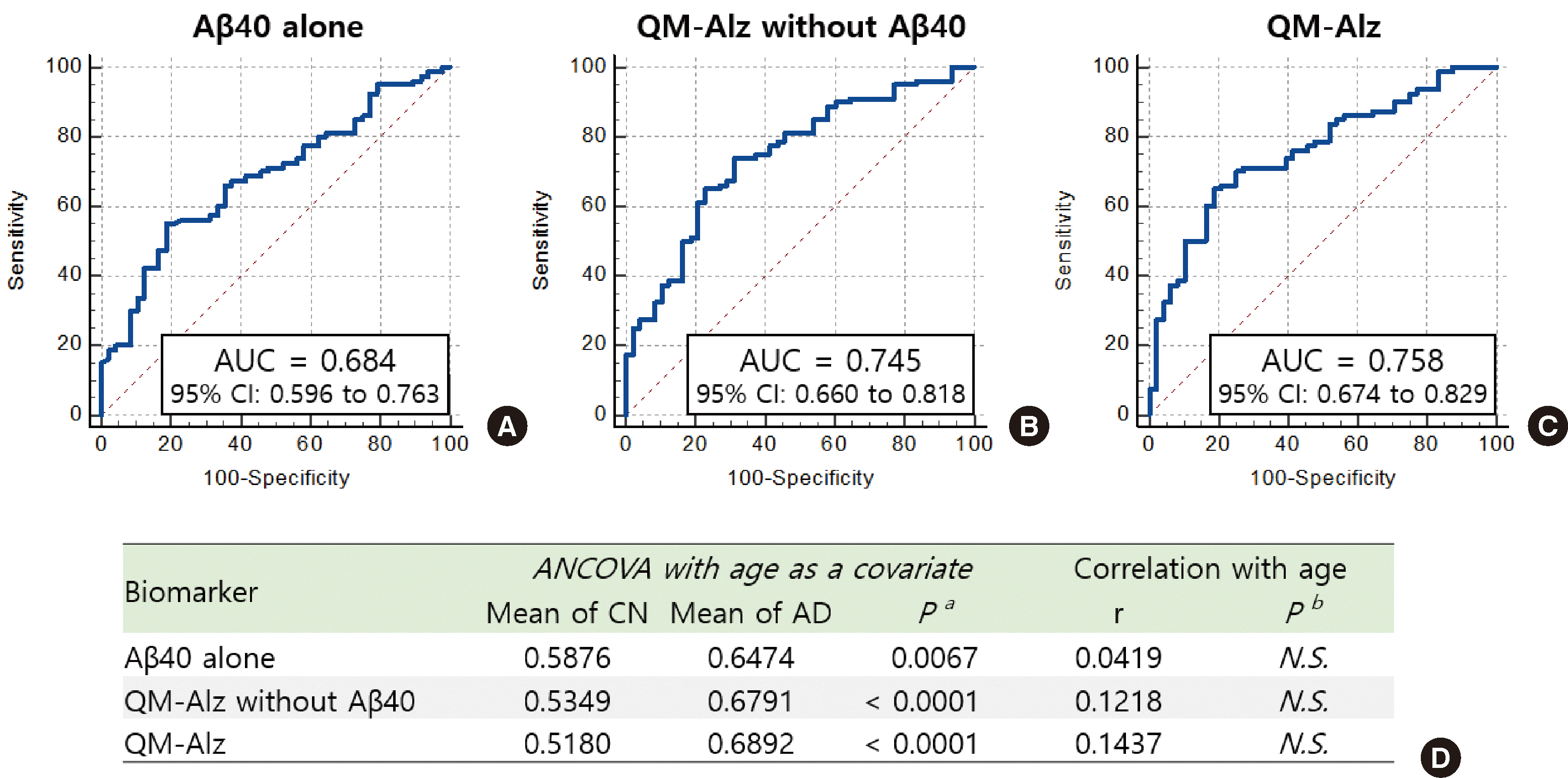
The ROC curves and statistical data for algorithms derived from each combination are shown in Fig. 1. The AUC in the case of Aβ40 alone was 0.684. The AUC of QM-Alz without Aβ40 was 0.745, which indicates higher discriminatory accuracy than that of Aβ40 alone. The AUC of the full set of QM-Alz including Aβ40 was 0.758. As described in Table 1, the CN group is significantly younger than the AD group. To investigate the effect of age on the algorithm results, ANCOVA and correlation analysis were performed. The ANCOVA result with age as a covariate shows that there was a significant difference in the mean of algorithm values between CN and AD even after removing the effect of age. Furthermore, the algorithm values of each combination do not significantly correlate with age. ANCOVA was performed with several clinical histories such as hypertension and diabetes as covariates, and the differences in algorithm values between CN and AD were significant for the factors we checked (Supplementry Table 2).

3. Evaluation of clinical diagnosis accuracy for AD of algorithms derived from various biomarker combinations

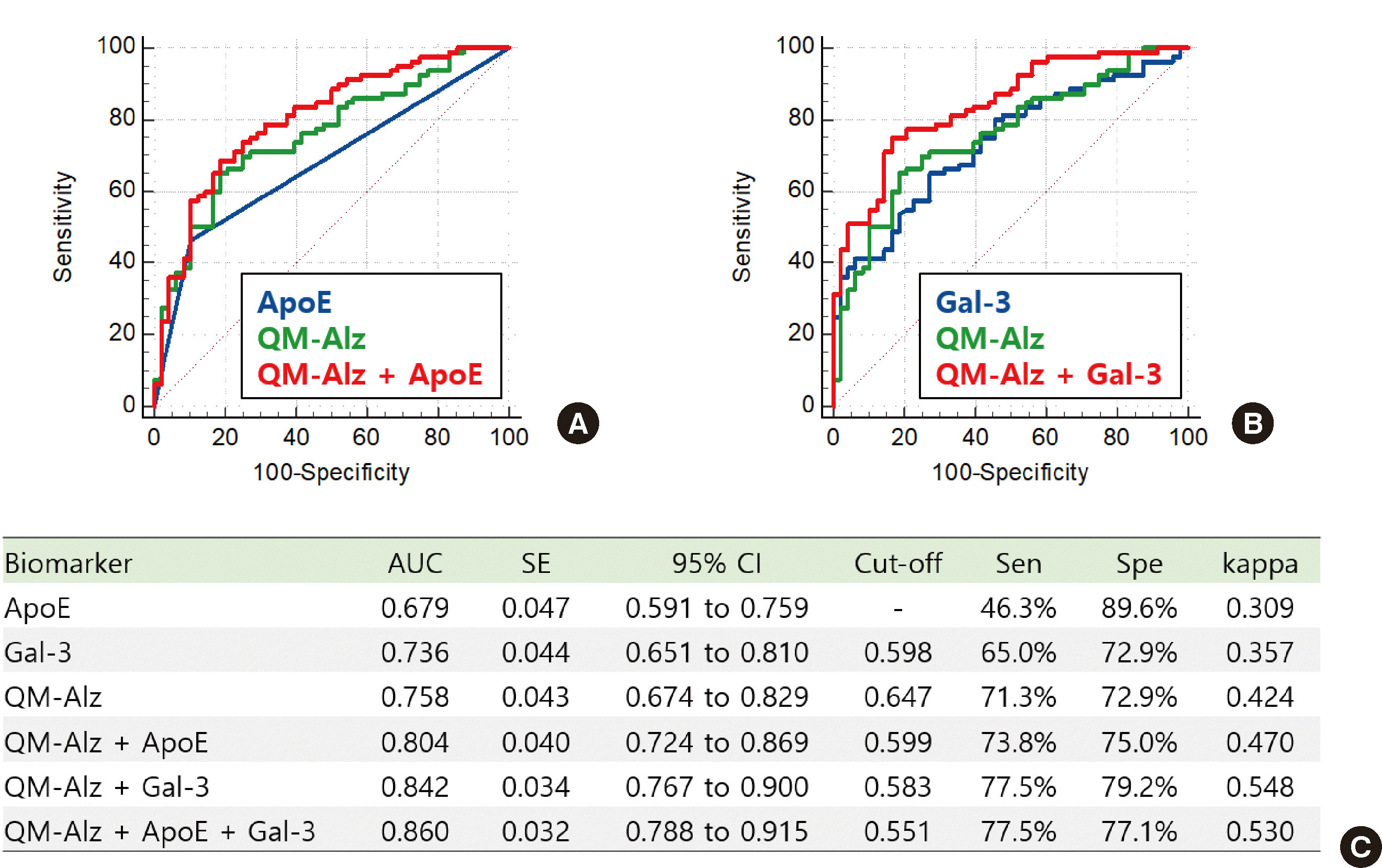
We derived algorithms based on logistic regression aimed at distinguishing CN and AD individually and in combinations of QM-Alz, ApoE, and Gal-3 (Fig. 2). In the case of ApoE, as only the presence or absence of the ε4 gene is checked, there is no cut-off setting, and thus sensitivity and specificity are fixed. ApoE provides information on potential risk based on the genomic characteristics, but dementia can occur due to various factors, so the sensitivity is low in diagnosis based on ApoE only. Gal-3, a novel biomarker that has shown potential for use in AD diagnosis, exhibited a higher AUC than ApoE, which indicates higher diagnostic accuracy in distinguishing CN from AD. QM-Alz, which creates an algorithm by combining four biomarkers, exhibited higher accuracy than when using individual biomarkers alone. Adding additional biomarkers to QM-Alz increases diagnostic accuracy. Combining QM-Alz with ApoE yielded an AUC of 0.804, combining QM-Alz with Gal-3 yielded 0.842, and combining all biomarkers increased the AUC up to 0.860.

4. Correlation between clinical tests and each algorithm derived from various biomarker combinations

We checked the correlation between the clinical tests and the algorithms derived from each biomarker combination (Table 2). The MMSE is a screening test that provides information about global cognition, where a low score indicates proximity to dementia. Meanwhile, in the algorithms we derived, a high score indicates proximity to dementia. Therefore, algorithm values and MMSE scores negatively correlate. The CDR is a composite evaluation used to determine functional impairment, and a high score indicates proximity to dementia. Therefore, algorithm values and CDR scores positively correlate. In terms of correlation with MMSE, ApoE and Gal-3 exhibited correlation coefficients of -0.2799 and -0.3081, respectively. QM-Alz exhibited a higher correlation of -0.3263 than the previous three cases using only a single biomarker. When QM-Alz was combined with ApoE, Gal-3, and both, the correlation coefficient increased to -0.3959, -0.4537, and -0.4734 respectively. In terms of correlation with CDR, ApoE, and Gal-3 exhibited correlation coefficients of 0.3156, and 0.2292, respectively. The correlation coefficient between QM-Alz and CDR was 0.2627, which was lower than that of ApoE alone. When QM-Alz was combined with ApoE, Gal-3, and both, the correlation coefficient increased to 0.3723, 0.3924, and 0.4423, respectively.

5. Performance of biomarkers accuracy test for PET positivity

As with previous studies using QM-Alz, the association of each biomarker combination with PET positivity, which is highly related to cerebral Aβ deposition, was also analyzed (Table 3). When the model using only ApoE was evaluated based on the PET results, AUC, sensitivity, and specificity were 0.698, 52.3%, and 87.3%, respectively. As the ApoE test is an on/off test, it is impossible to balance sensitivity and specificity by adjusting the cutoff value. Gal-3 also exhibited a similar AUC of 0.660. When using QM-Alz to measure four biomarkers, the AUC was 0.759, exhibiting higher accuracy than using only ApoE positivity or Gal-3. When combining QM-Alz and ApoE, their AUC exhibited the highest value of 0.823. Combining QM-Alz with Gal-3 was not as favorable as combining it with ApoE, but combining all biomarkers resulted in an AUC, sensitivity, and specificity of 0.852, 78.5%, and 79.4%, respectively, indicating the highest accuracy.

DISCUSSION

We aimed to evaluate the diagnostic ability of QM-Alz in detecting AD using blood samples. The QPLEX™ platform is a system with strengths in multiplexing, capable of measuring multiple biomarkers simultaneously. Furthermore, it is a bead-based 3D suspension array system that enhances reactivity and sensitivity. This advanced system enables the analysis of rare or volume-limited samples; it requires only 20 μL of undiluted human plasma to detect multiple biomarkers. QM-Alz simultaneously measures four blood biomarkers, Aβ40, ACE, POSTN, and LGALS3BP, and integrates the measurement results into an algorithmic framework. Previous studies have established the association of these four biomarkers with AD [9-11]. Notably, Aβ40 emerges as a prominent biomarker for AD, reflecting its significance in disease pathology [9]. ACE, primarily recognized for its involvement in blood pressure regulation [14], has also exhibited inhibitory effects on Aβ aggregation [15]. Reduced ACE activity levels have been observed in patients with AD compared to cognitively normal individuals [16]. POSTN, implicated in inflammatory diseases, has been identified within the cerebral cortex of patients with AD [8], suggesting its potential role in the activated inflammatory response during AD pathogenesis. LGALS3BP functions as a receptor for Gal-3 and its interaction with ligands contributes to the inhibition of neutrophil activation [17]. Our primary findings indicate that QM-Alz can differentiate not only PET positivity but also clinical AD diagnosis. To validate the results of previous studies, we confirmed the reliability and efficacy of QM-Alz to identify individuals with AD symptoms. This was consistent with our previous studies that demonstrated the efficacy of the kit as a bloodbased diagnostic tool for AD in an alternative cohort [8, 12].
Furthermore, we demonstrated that the application of a multiple-marker approach may yield superior diagnostic results for AD compared to relying on Aβ alone. In agreement with our results, earlier studies reported that the AUC for blood Aβ in isolation was not as high as that for CSF Aβ, particularly for Aβ40, with an AUC ranging from 0.51 to 0.69 [18, 19]. Our data suggest that QM-Alz with and without Aβ40 results in a higher AUC compared to Aβ40 alone (Fig. 1), endorsing the use of multiple markers to enhance diagnostic precision.
Using biomarker combinations has advantages as a diagnostic tool. First, due to the heterogeneous nature of AD, a combination of markers appears to perform better than individual biomarkers in diagnosing and predicting the disease. For instance, a diagnostic prediction model based on a combination of plasma Aβ42/Aβ40 ratio, p-tau181, and neurofilament light chain (NFL) exhibited higher diagnostic value than each factor alone [20]. Based on the complexity of AD pathogenesis, multivariate biomarker panels associated with various biological pathways may provide a more accurate diagnosis than individual markers. Second, bloodbased biomarker panels have been designed to estimate diseaserelated phenotypes such as cognitive decline, brain atrophy, and neocortical Aβ deposition beyond case-control studies [21]. The combination of plasma biomarkers, namely P-tau217, the Aβ42/Aβ40 ratio, and NFL, provided the most robust results to predict cognitive decline in individuals with normal cognition [22]. Furthermore, the addition of plasma P-tau181 to factors such as demographics, genetics, and clinical information significantly improved the prediction of memory decline in individuals with normal cognition and mild cognitive impairment [4].
In our secondary analysis, we observed that the performance of QM-Alz could be improved in clinical diagnosis (Fig. 2) and PET positivity (Table 3) by including ApoE and an additional blood protein, Gal-3. The ε4 allele of ApoE remains the most potent genetic risk factor for sporadic AD [23]. ApoE, a 34 kDa glycoprotein consisting of 299 amino acids, is co-deposited with Aβ in amyloid plaques. The interaction between ApoE and pathological Aβ deposition appears to be the central mechanism through which ApoE influences the risk [24]. Recently, a combination of ApoE and biomarkers has been employed in the development of diagnostic tools for AD. For instance, combining the plasma Aβ42/40 ratio with ApoE and age improved the accuracy of identifying amyloid positivity compared to using ApoE and age alone [25]. Moreover, Gal-3 is a member of the galectin protein family known for its binding affinity toward β-galactoside molecules. Through its carbohydrate recognition domain, it identifies proteins with β-galactoside modifications, initiating a range of biological responses [26]. Elevated Gal-3 levels in the bloodstream could be linked to AD, potentially serving as an early biomarker for disease detection [27]. This connection may be attributed to the activation of pathways promoting apoptosis, inflammation, and compromised neurodegeneration, which are commonly observed in individuals with AD. Additionally, there is evidence suggesting a relationship between serum Gal-3 levels and cognitive status, observed both in patients with AD and those with normal cognitive function [28]. We believe that the diagnostic accuracy of
QM-Alz can be improved by including ApoE and Gal-3. QM-Alz, either alone or in combination with ApoE and Gal-3, exhibited some correlation with clinical tests, such as the MMSE and CDR (Table 2). The MMSE is a well-known and frequently employed cognitive screening tool to detect AD that offers a concise method for the evaluation of overall cognitive function in clinical, research, and community environments [29]. The CDR is a widely used clinical scale with proven diagnostic and severity ranking values and has been extensively applied in international epidemiological studies, case series, and clinical trials [30]. Therefore, these new combinations, as well as our kit, likely have the potential to serve as useful tools for cognitive impairment without a long survey time.
This study had some limitations. First, although the cohort used here was new and independent from the cohort that was used for the development and validation of the algorithm, it was still limited to East Asians, especially Koreans. Verification with other countries and ethnicities is needed for wider application. Second, we excluded behavioral variant frontotemporal dementia (bvFTD), nonfluent/agrammatic variant primary progressive aphasia (nfvPPA), and semantic variant primary progressive aphasia (svPPA) because their numbers were too small to make a comparison. As it is essential to investigate the performance of the kit in other types of dementia, such research will be necessary for future advancement. Third, we hoped that QM-Alz would be able to distinguish between CN and AD, as well as AD severity. However, there was no significant difference in the algorithm values between AD severities based on CDR scores. Fourth, an AUC of 0.8 is not a satisfactory accuracy. Ongoing research aims to improve this accuracy by identifying and incorporating additional biomarkers by leveraging the ability of the QPLEX™ platform to easily integrate new biomarkers. In particular, Aβ42 is well-known as an effective biomarker for AD diagnosis, but it has not been included in QM-Alz because an antibody exhibiting sufficient performance has not yet been secured. A follow-up study is underway to include Aβ42 in the kit.
In conclusion, we suggest that QM-Alz shows promising potential for the differentiation of clinical conditions and PET positivity using blood samples, with the addition of ApoE and Gal-3 further enhancing its performance. Future research should incorporate larger sample sizes and extended follow-up periods to better evaluate the performance of the kit in real-world clinical settings. The successful validation of this diagnostic tool could have significant implications for early detection and intervention in AD, ultimately leading to improved patient outcomes and enhanced quality of life. These findings hold considerable importance, as they offer a noninvasive and readily accessible method to predict AD development.
Acknowledgments

The authors would like to thank and acknowledge the Precision Medicine Platform for Mild Cognitive Impairment, based on Multi-omics, Imaging, Evidence-based R&BD consortium for recruiting participants, providing various medical information, and providing blood samples.
This work was supported by a grant from the Korea Health Technology R&D Project through the Korea Health Industry Development Institute, funded by the Ministry of Health & Welfare, Republic of Korea (grant number HI19C1132).

Notes

Conflicts of Interest

The authors declare the following financial interests/personal relationships which may be considered as potential competing interests. Hunjong Na, Dokyung Lee, Changsik Yoon, and Jisung Jang are employees of QuantaMatrix Inc. Sunghoon Kwon is the CEO of QuantaMatrix Inc. The remaining authors have no conflicts of interest to report.

REFERENCES

1. GBD 2019 Dementia Forecasting Collaborators. 2022; Estimation of the global prevalence of dementia in 2019 and forecasted prevalence in 2050: an analysis for the global burden of disease study 2019. Lancet Public Health. 7:e105–25. DOI: 10.1016/S2468-2667(21)00249-8. PMID: 34998485. PMCID: PMC8810394.
2. Zhang X, Zhang X, Gao H, Qing G. 2022; Phage display derived peptides for Alzheimer's disease therapy and diagnosis. Theranostics. 12:2041–62. DOI: 10.7150/thno.68636. PMID: 35265198. PMCID: PMC8899571.
3. Duits FH, Martinez-Lage P, Paquet C, Engelborghs S, Lleó A, Hausner L, et al. 2016; Performance and complications of lumbar puncture in memory clinics: results of the multicenter lumbar puncture feasibility study. Alzheimers Dement. 12:154–63. DOI: 10.1016/j.jalz.2015.08.003.
4. Leuzy A, Mattsson-Carlgren N, Palmqvist S, Janelidze S, Dage JL, Hansson O. 2022; Blood-based biomarkers for Alzheimer's disease. EMBO Mol Med. 14:e14408. DOI: 10.15252/emmm.202114408. PMID: 34859598. PMCID: PMC8749476. PMID: 328e6d70c6fd48f882262db4a209fd47.
5. Blennow K, Zetterberg H. 2015; Understanding biomarkers of neurodegeneration: ultrasensitive detection techniques pave the way for mechanistic understanding. Nat Med. 21:217–9. DOI: 10.1038/nm.3810.
6. O'Bryant SE, Gupta V, Henriksen K, Edwards M, Jeromin A, Lista S, et al. STAR-B and BBBIG working groups. 2015; Guidelines for the standardization of preanalytic variables for blood-based biomarker studies in Alzheimer's disease research. Alzheimers Dement. 11:549–60. DOI: 10.1016/j.jalz.2014.08.099.
7. Andreasson U, Blennow K, Zetterberg H. 2016; Update on ultrasensitive technologies to facilitate research on blood biomarkers for central nervous system disorders. Alzheimers Dement (Amst). 3:98–102. DOI: 10.1016/j.dadm.2016.05.005. PMID: 27453931. PMCID: PMC4941042. PMID: 82f1bf408033484c9b242a20368fe091.
8. Park JC, Jung KS, Kim J, Jang JS, Kwon S, Byun MS, et al. 2021; Performance of the QPLEXTM Alz plus assay, a novel multiplex kit for screening cerebral amyloid deposition. Alzheimers Res Ther. 13:12. DOI: 10.1186/s13195-020-00751-x. PMID: 861d8581c821400a9be71d6ffd8879c9.
9. Park JC, Han SH, Cho HJ, Byun MS, Yi D, Choe YM, et al. 2017; Chemically treated plasma Aβ is a potential blood-based biomarker for screening cerebral amyloid deposition. Alzheimers Res Ther. 9:20. DOI: 10.1186/s13195-017-0248-8.
10. Park JC, Han SH, Lee H, Jeong H, Byun MS, Bae J, et al. 2019; Prognostic plasma protein panel for Aβ deposition in the brain in Alzheimer's disease. Prog Neurobiol. 183:101690. DOI: 10.1016/j.pneurobio.2019.101690. PMID: 31605717.
11. Na H, Shin KY, Lee D, Yoon C, Han SH, Park JC, et al. 2023; The QPLEXTM plus assay kit for the early clinical diagnosis of Alzheimer's disease. Int J Mol Sci. 24:11119. DOI: 10.3390/ijms241311119. PMID: 02b5c581f86b4c62b65ee42048777b12.
12. Kim HJ, Park JC, Jung KS, Kim J, Jang JS, Kwon S, et al. 2021; The clinical use of blood-test factors for Alzheimer's disease: improving the prediction of cerebral amyloid deposition by the QPLEXTM Alz plus assay kit. Exp Mol Med. 53:1046–54. DOI: 10.1038/s12276-021-00638-3.
13. Kang SH, Lee KH, Chang Y, Choe YS, Kim JP, Jang H, et al. 2022; Genderspecific relationship between thigh muscle and fat mass and brain amyloid-β positivity. Alzheimers Res Ther. 14:145. DOI: 10.1186/s13195-022-01086-5. PMID: ce46ab4cbaea4e8d872823ef632e1df1.
14. Reid IA. 1992; Interactions between ANG II, sympathetic nervous system, and baroreceptor reflexes in regulation of blood pressure. Am J Physiol. 262:E763–78. DOI: 10.1152/ajpendo.1992.262.6.E763. PMID: 1616014.
15. Hu J, Igarashi A, Kamata M, Nakagawa H. 2001; Angiotensin-converting enzyme degrades Alzheimer amyloid beta-peptide (A beta); retards A beta aggregation, deposition, fibril formation; and inhibits cytotoxicity. J Biol Chem. 276:47863–8. DOI: 10.1074/jbc.M104068200.
16. Jochemsen HM, Teunissen CE, Ashby EL, van der Flier WM, Jones RE, Geerlings MI, et al. 2014; The association of angiotensin-converting enzyme with biomarkers for Alzheimer's disease. Alzheimers Res Ther. 6:27. DOI: 10.1186/alzrt257.
17. Läubli H, Alisson-Silva F, Stanczak MA, Siddiqui SS, Deng L, Verhagen A, et al. 2014; Lectin galactoside-binding soluble 3 binding protein (LGALS3BP) is a tumor-associated immunomodulatory ligand for CD33-related siglecs. J Biol Chem. 289:33481–91. DOI: 10.1074/jbc.M114.593129. PMID: 25320078. PMCID: PMC4246102.
18. Feinkohl I, Schipke CG, Kruppa J, Menne F, Winterer G, Pischon T, et al. 2020; Plasma amyloid concentration in Alzheimer's disease: performance of a high-throughput amyloid assay in distinguishing Alzheimer's disease cases from controls. J Alzheimers Dis. 74:1285–94. DOI: 10.3233/JAD-200046. PMID: 32176645. PMCID: PMC7242850.
19. Sun Y, Hua J, Chen G, Li J, Yang J, Gao H. 2021; Alix: a candidate serum biomarker of Alzheimer's disease. Front Aging Neurosci. 13:669612. DOI: 10.3389/fnagi.2021.669612. PMID: c0138703b66547b3a7ae73d83698c0de.
20. Sun Q, Ni J, Wei M, Long S, Li T, Fan D, et al. 2022; Plasma β-amyloid, tau, neurodegeneration biomarkers and inflammatory factors of probable Alzheimer's disease dementia in Chinese individuals. Front Aging Neurosci. 14:963845. DOI: 10.3389/fnagi.2022.963845. PMID: 36062146. PMCID: PMC9433929. PMID: ba8b2407660e4550acc24910fc7ca734.
21. Zetterberg H, Burnham SC. 2019; Blood-based molecular biomarkers for Alzheimer's disease. Mol Brain. 12:26. DOI: 10.1186/s13041-019-0448-1. PMID: e2ff49deb38143f980edacb410b15cc2.
22. Cullen NC, Leuzy A, Janelidze S, Palmqvist S, Svenningsson AL, Stomrud E, et al. 2021; Plasma biomarkers of Alzheimer's disease improve prediction of cognitive decline in cognitively unimpaired elderly populations. Nat Commun. 12:3555. DOI: 10.1038/s41467-021-23746-0. PMID: 34117234. PMCID: PMC8196018. PMID: ed33539191794e8abd8ebdc03d75b61a.
23. Serrano-Pozo A, Das S, Hyman BT. 2021; APOE and Alzheimer's disease: advances in genetics, pathophysiology, and therapeutic approaches. Lancet Neurol. 20:68–80. DOI: 10.1016/S1474-4422(20)30412-9.
24. Raulin AC, Doss SV, Trottier ZA, Ikezu TC, Bu G, Liu CC. 2022; ApoE in Alzheimer's disease: pathophysiology and therapeutic strategies. Mol Neurodegener. 17:72. DOI: 10.1186/s13024-022-00574-4. PMID: e0b56cbf47db45da8937c2f737339784.
25. West T, Kirmess KM, Meyer MR, Holubasch MS, Knapik SS, Hu Y, et al. 2021; A blood-based diagnostic test incorporating plasma Aβ42/40 ratio, ApoE proteotype, and age accurately identifies brain amyloid status: findings from a multi cohort validity analysis. Mol Neurodegener. 16:30. DOI: 10.1186/s13024-021-00451-6. PMID: c91cadfca9d9471da9fe34743049c0b5.
26. Tao CC, Cheng KM, Ma YL, Hsu WL, Chen YC, Fuh JL, et al. 2020; Galectin-3 promotes Aβ oligomerization and Aβ toxicity in a mouse model of Alzheimer's disease. Cell Death Differ. 27:192–209. DOI: 10.1038/s41418-019-0348-z.
27. Yazar T, Olgun Yazar H, Cihan M. 2021; Evaluation of serum galectin-3 levels at Alzheimer patients by stages: a preliminary report. Acta Neurol Belg. 121:949–54. DOI: 10.1007/s13760-020-01477-1. PMID: 32852752.
28. Wang X, Zhang S, Lin F, Chu W, Yue S. 2015; Elevated galectin-3 levels in the serum of patients with Alzheimer's disease. Am J Alzheimers Dis Other Demen. 30:729–32. DOI: 10.1177/1533317513495107. PMID: 23823143. PMCID: PMC10852776.
29. Folstein MF, Folstein SE, McHugh PR. 1975; "Mini-mental state". A practical method for grading the cognitive state of patients for the clinician. J Psychiatr Res. 12:189–98. DOI: 10.1016/0022-3956(75)90026-6.
30. Hughes CP, Berg L, Danziger WL, Coben LA, Martin RL. 1982; A new clinical scale for the staging of dementia. Br J Psychiatry. 140:566–72. DOI: 10.1192/bjp.140.6.566. PMID: 7104545.

Fig. 1
Comparative results of Aβ40 alone, QM-Alz without Aβ40, and QM-Alz. ROC curves of the algorithm to discriminate between CN and AD generated from (A) Aβ40 alone, (B) QM-Alz without Aβ40, and (C) the full set of QM-Alz. (D) Results of ANCOVA with age as a covariate and correlation with age.
Abbreviations: ROC, receiver operating characteristic; AUC, the area under the curve; CI, confidence interval; ANCOVA, analysis of covariance; Pa, significance level of ANCOVA; r, correlation coefficient; Pb, significance level of correlation; N.S., not significant; CN, cognitively normal; AD, Alzheimer’s disease; Aβ40, amyloid-β1-40; QM-Alz, QPLEX™ Alz plus assay kit.
lmo-14-4-330-f1.tif
Fig. 2
Performance of each biomarker combination to discriminate between CN and AD. (A) Comparative ROC curves of ApoE and its combination. (B) Comparative ROC curves of Gal-3 and its combination. (C) The AUC, sensitivity, and specificity of each algorithm, which were generated from QM-Alz, ApoE, and Gal-3.
Abbreviations: ROC, receiver operating characteristic; AUC, the area under the curve; SE, standard error; CI, confidence interval; Sen, sensitivity; Spe, specificity; CN, cognitively normal; AD, Alzheimer’s disease; ApoE, Apolipoprotein E genotype; Gal-3, galectin-3; QM-Alz, QPLEX™ Alz plus assay kit.
lmo-14-4-330-f2.tif
Table 1
Demographic data of the participants (N=128)
CN AD P
Number 48 80 -
Age 62.58±9.63 68.95±9.93 0.0005
Sex (M/F) 18/30 32/48 N.S.
ApoE (ε4 +/-) 5/43 37/43 <0.0001
MMSE score 27.77±2.47 19.11±4.56 <0.0001
CDR score 0.20±0.25 0.98±0.49 <0.0001
CDR SB score 0.23±0.30 5.91±2.80 <0.0001
Education (years) 12.29±3.98 10.97±4.70 N.S.
PET (+/-) 1/47 64/16 <0.0001

Data are presented as the mean±standard deviation.

Abbreviations: P, P-value of independent t-test; N.S., not significant; CN, cognitively normal; AD, Alzheimer’s disease; M, male; F, female; ApoE, Apolipoprotein E genotype; MMSE, Mini-Mental State Examination; CDR, Clinical Dementia Rating; CDR SB, Clinical Dementia Rating sum of boxes; PET, positron emission tomography.

Table 2
Correlation between the clinical tests and the algorithms derived from each biomarker combination
Biomarker Correlation with MMSE Correlation with CDR
r P r P
ApoE -0.2799 0.0014 0.3156 0.0003
Gal-3 -0.3081 0.0004 0.2292 0.0093
QM-Alz -0.3263 0.0002 0.2627 0.0027
QM-Alz+ApoE -0.3959 <0.0001 0.3723 <0.0001
QM-Alz+Gal-3 -0.4537 <0.0001 0.3924 <0.0001.
QM-Alz+ApoE+Gal-3 -0.4734 <0.0001 0.4423 <0.0001

Abbreviations: MMSE, Mini-Mental State Examination; CDR, Clinical Dementia

Rating; r, correlation coefficient; P, significance level; ApoE, Apolipoprotein E genotype; Gal-3, galectin-3; QM-Alz, QPLEX™ Alz plus assay kit.

Table 3
Comparative results of each combination to distinguish cerebral Aβ deposition
Biomarker AUC SE 95% CI Cut-off Sen (%) Spe (%) Kappa
ApoE 0.698 0.047 0.611 to 0.776 - 52.3 87.3 0.394
Gal-3 0.660 0.048 0.571 to 0.741 0.474 63.1 61.9 0.250
QM-Alz 0.759 0.042 0.675 to 0.830 0.585 66.2 76.2 0.423
QM-Alz+ApoE 0.823 0.036 0.746 to 0.885 0.484 75.4 73.0 0.484
QM-Alz+Gal-3 0.796 0.040 0.716 to 0.862 0.532 72.3 76.2 0.485
QM-Alz+ApoE+Gal-3 0.852 0.034 0.778 to 0.909 0.528 78.5 79.4 0.578

Abbreviations: AUC, area under the curve; SE, standard error; CI, confidence interval; Sen, sensitivity; Spe, specificity; Aβ, amyloid-β; ApoE, Apolipoprotein E genotype; Gal-3, galectin-3; QM-Alz, QPLEX™ Alz plus assay kit.

TOOLS
Similar articles