Journal List > Korean J Anesthesiol > v.77(5) > 1516088551

Jeong, Bae, Lee, Lee, and Kim: Ramped versus sniffing position for Ambu® AuraGainTM insertion in patients with obesity: a randomized controlled study

Abstract

Background

The ramped position facilitates mask ventilation and endotracheal intubation in patients with obesity. This study aimed to determine whether the ramped position improves supraglottic airway (SGA) insertion in patients with obesity.

Methods

In this prospective, randomized, single-center trial, 48 obese patients undergoing elective surgery were randomized into either ramped or sniffing position groups. The Ambu® AuraGainTM (Ambu A/S), a second-generation SGA, was used. The primary outcome was the time required for the AuraGain insertion. Secondary outcomes included ease and number of insertion attempts, oropharyngeal leak pressure (OLP), and complications. The number needed to treat (NNT) was calculated to ensure ease of insertion.

Results

The time required for the AuraGain insertion was significantly shorter in the ramped group than in the sniffing group (13.0 [11.0, 16.0] vs. 24.0 [21.0, 28.0], P < 0.001). The insertion was easier in the ramped group than in the sniffing group (23/24 vs. 13/24, NNT = 2.4 [95% CI, 1.6, 5.0], P = 0.003). The first-attempt success rate was higher in the ramped group than in the sniffing group, although the difference was not statistically significant (22/24 vs. 18/24, P = 0.319). The OLP and postoperative complication rates were not significantly different between the groups.

Conclusions

The ramped position reduced the time required for the AuraGain insertion in obese patients while providing comparable airway sealing without increasing adverse events. Therefore, a ramped position may be a more suitable option for SGA insertion in this population.

Introduction

With the worldwide increase in the prevalence of obesity [1], anesthesiologists are increasingly challenged with airway management in this population [2]. Compared with the sniffing position, typically achieved by elevating the head from 7 to 9 cm [3,4], the ramped position, achieved by horizontal alignment of the external auditory canal and the sternal notch, has been shown to facilitate mask ventilation and endotracheal intubation in patients with obesity [59].
The supraglottic airway (SGA) is recognized as a valuable tool for managing difficult airways and is recommended as a rescue device in ‘cannot intubate, cannot ventilate’ situations, according to clinical guidelines [10,11]. However, a high body mass index (BMI) has been reported as an independent risk factor for unsuccessful SGA insertion [12]. Therefore, caution is imperative when placing an SGA in individuals with obesity. Multiple attempts can cause trauma to the oral cavity or supraglottic structures, and failed insertion can prolong the time required to secure the airway that is critical in emergencies.
Manufacturers typically recommend the sniffing position for SGA insertion regardless of obesity [13,14]. However, studies suggest that the neutral position, simple head extension, or head elevation of 3 cm may result in equivocal or even superior SGA insertion outcomes compared to the sniffing position in non-obese patient [1517]. Given that the sniffing position is considered the standard for tracheal intubation in patients who are not obese [1820], these unexpected results may be attributed to the distinct anatomical challenges presented by tracheal intubation versus SGA insertion [16,17,21,22]. Meanwhile, in the airway management of individuals with obesity, sufficient head elevation may still be crucial to compensate for body fat composition [23]. However, there is a lack of research on the optimal head positioning for SGA insertion in this population. In this study, we aimed to evaluate ramped versus standard sniffing positions for SGA insertion in patients with obesity.

Materials and Methods

Ethical approval for this study was obtained from Institutional Review Board of Chonnam National University Hospital (CNUH 2023-164), and it was registered with the Clinical Research Information Service of the Republic of Korea (KCT0008570) and conducted from June to November 2023 at a university hospital following the principles of the 2013 Declaration of Helsinki. Written informed consent was obtained from each participant before enrollment.
We enrolled patients who (1) had a BMI ≥ 30 kg/m2, (2) were aged 20–80 years, (3) had an American Society of Anesthesiologists physical status class of II or III, and (4) were scheduled for elective surgery under general anesthesia. Patients were excluded if they (1) had a known or predicted difficult airway, (2) had a history of head and neck surgery, (3) had cervical instability, (4) had a mouth opening < 2.5 cm, (5) had an increased risk of pulmonary aspiration (e.g., symptomatic gastroesophageal reflux disease or hiatal hernia), or (6) were pregnant. Enrolled patients were randomly assigned to either the ramped or sniffing group in a 1:1 ratio using a computer-generated randomization sheet. The participants were blinded to their group allocation.
Preoperative airway assessments, including the Mallampati score, neck circumference, sternomental distance, thyromental distance, and interincisor distance, were performed the day before surgery. Upon entry into the operating room, the patients were placed in a ramped or sniffing position according to their allocated group. In the ramped group, pillows were placed under the patient’s head and upper body to align the external auditory meatus horizontally with the sternal notch. In the sniffing group, an 8-cm-high pillow was placed under the occiput. In both groups, the height of the operating table was adjusted to position the patient’s head between the anesthesiologist’s upper part of the umbilicus and the lower xiphoid process.
Patients were monitored using electrocardiography, noninvasive blood pressure measurements, pulse oximetry, and capnography. Preoxygenation was performed for 3 min with 100% oxygen, and anesthesia was induced with propofol and remifentanil. Upon loss of consciousness, train-of-four (TOF) monitoring was initiated, and an intubating dose of rocuronium (0.6 mg/kg of ideal body weight) was administered. When the TOF count reached zero, the Warters grading scale assessed the difficulty of the facemask ventilation [24]. The scale assigns points based on the escalating levels of intervention required to achieve a target tidal volume of 5 ml/kg of ideal body weight. The intervention consisted of using an airway device, increasing inspiratory pressure, and implementing two-person ventilation, all of which aimed to overcome the upper airway resistance to ventilation. Difficult mask ventilation was operationally defined as a score of ≥ 4 on the Warters scale.
The Ambu® AuraGainTM (Ambu A/S) was prepared and inserted according to the manufacturer’s instructions. The size of the AuraGain cuff was selected based on the patient’s ideal body weight (size 3 for 30–50 kg, size 4 for 50–70 kg, size 5 for 70–100 kg, and size 6 for those exceeding 100 kg), and the posterior surface of the cuff was coated with a water-based lubricant. Facemask ventilation and AuraGain insertion were performed by a skilled anesthesiologist with experience in performing more than 400 SGA and 100 AuraGain insertions, respectively. Concurrently, an unblinded observer who was not involved in the study collected data. Following the insertion of AuraGain, the device was inflated with air until the cuff pressure reached 60 cmH2O, as measured using a handheld cuff manometer (VBM Medizintechnik GmbH).
The AuraGain insertion was considered successful if the device was inserted on the first, second, or third attempt (60 s permitted for each attempt) and did not require any additional manipulations after its initial placement. Successful insertion was confirmed by a square-wave capnograph trace, bilateral chest wall movement, and no audible leak with a peak airway pressure of ≥ 12 cmH2O during manual ventilation. If these criteria were not satisfied, the AuraGain was removed from the patient’s mouth. After ensuring sufficient ventilation with 100% oxygen, another attempt was made to reinsert the device by using the same technique. The number of attempts required for a successful AuraGain insertion was recorded. If insertion was unsuccessful after three attempts, tracheal intubation was performed.
The insertion time was measured from the moment the AuraGain touched the patient’s mouth until the appearance of the first square end-tidal carbon dioxide (ETCO2) trace. The insertion time was defined as the duration taken solely for a successful attempt. If the AuraGain insertion failed on the first attempt and succeeded on the second attempt, the insertion time was recorded as the time taken to insert AuraGain on the second attempt, the time taken for the first attempt, and the interval between the first and second attempts. The ease of AuraGain insertion was subjectively assessed on a five-point scale (1 = easy, 2 = acceptable, 3 = difficult, 4 = very difficult, and 5 = impossible).
The attending anesthesiologist measured the oropharyngeal leak pressure (OLP), defined as the airway pressure (maximum allowed, 40 cmH2O) at which gas started to leak audibly around the AuraGain at a fixed gas flow of 5 L/min with the adjustable pressure-limiting valve closed. To evaluate the position of the AuraGain, a fiberoptic bronchoscope was used to grade the glottic view using the Brimacombe scale (4 = only vocal cords seen; 3 = vocal cords plus posterior epiglottis seen; 2 = vocal cords plus anterior epiglottis seen; 1 = vocal cords not seen) [25].
Anesthesia was maintained using volatile anesthetics (desflurane or sevoflurane) and remifentanil infusions. The tidal volume was set to 8 ml/kg of ideal body weight, and the respiratory rate was adjusted to maintain normocarbia (ETCO2 30–40 mmHg). Desflurane/sevoflurane and remifentanil were discontinued at the end of the surgery. The AuraGain was removed when patients were able to open their eyes on commands and breathe spontaneously. Complications such as intraoperative desaturation, regurgitation, aspiration, laryngospasm, bronchospasm, and blood stains on the device were recorded by an unblinded observer. Postoperatively, a blinded independent observer assessed sore throat, dysphonia, and dysphagia 30 min and 24 h after surgery.
The primary outcome was the time required for AuraGain insertion. The secondary outcomes included ease of insertion, number of insertion attempts, OLP, and incidence of complications.
The sample size was calculated based on the results of a pilot study, in which the means ± standard deviations of AuraGain insertion time for the ramped and sniffing groups were 21.33 ± 3.67 s and 24.40 ± 1.93 s, respectively. To detect this difference with an alpha of 0.05 and a power of 0.9, 21 individuals per group were required. Considering the dropout rate of 10%, 24 patients were included in each group.
Intergroup comparisons were performed using the Student’s t test or Mann–Whitney U test for continuous variables and the chi-square or the Fisher exact test for categorical variables, as appropriate. Continuous variables are reported as mean ± standard deviation or median (interquartile range), and categorical variables are reported as frequency (percentage). Statistical significance was set at P < 0.05 that was considered significant. The number needed to treat (NNT) was calculated as the reciprocal of absolute risk reduction. In this analysis, the NNT was computed through the following steps: 1) the absolute risk reduction was obtained by calculating the difference in the rate of insertions rated as ‘easy’ or ‘acceptable’ between the ramped and sniffing groups, and 2) the reciprocal of this absolute risk reduction was then calculated. We used SPSS® Statistics for Windows, Version 22.0 (IBM Corp.), and R software of the Foundation for Statistical Computing, version 4.3.1 for data manipulation and statistical analysis.

Results

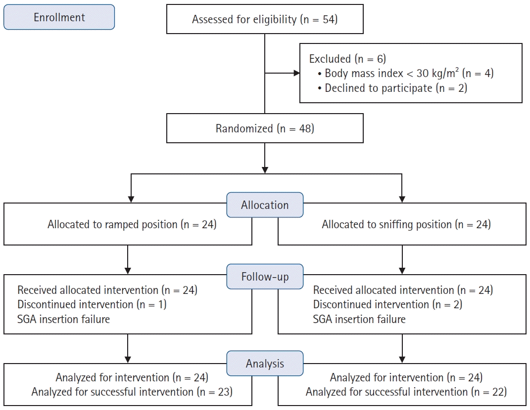
Of the 54 patients screened for eligibility, six were excluded from the study (Fig. 1). Forty-eight patients were randomized into two groups: ramped (n = 24) and sniffing (n = 24). Due to the failure of SGA insertion, one participant was excluded from the analysis in the ramped position group, and two participants were excluded from the analysis in the sniffing position group.
No significant differences exist in demographics and preoperative airway assessment factors between the groups (Table 1). Difficult mask ventilation (Warters scale ≥ 4) occurred in only one patient in the sniffing group, with a scale value of 8. The first-attempt success rate of AuraGain insertion was higher in the ramped group than in the sniffing group, although the difference was not statistically significant (22/24 vs. 18/24, P = 0.319) (Table 2). Insertion of the AuraGain was reported to be significantly easier in the ramped group, with 23/24 of insertions rated as ‘easy’ or ‘acceptable,’ versus 13/24 in the sniffing group (NNT = 2.4 [95% CI 1.6, 5.0], P = 0.003). Overall, insertion failure occurred in three patients: one in the ramped group and two in the sniffing group after three attempts each. These patients were intubated with an endotracheal tube and were excluded from subsequent analyses (Fig. 1).
The primary outcome of the study, the time required for AuraGain insertion, was significantly shorter in the ramped group (n = 23) compared to the sniffing group (n = 22) (13.0 s [11.0, 16.0] vs. 24.0 s [21.0, 28.0], P < 0.001) (Fig. 2). OLPs and Brimacombe scores were comparable between the groups (Table 3). No intraoperative desaturation, regurgitation, aspiration, laryngospasm, or bronchospasm events were observed in either group. The incidence of blood staining on the device and postoperative severity of sore throat, dysphonia, and dysphagia at 30 min and 24 h after surgery were not significantly different between the groups (Table 4).

Discussion

This study aimed to compare the ramped and sniffing positions for AuraGain insertion in patients with obesity. Our findings indicate that the ramped position provides significant advantages over the sniffing position in terms of both insertion time and ease of insertion while ensuring comparable safety and efficacy, as evidenced by the OLPs and complication rates. Although the insertion time was significantly shorter in the ramped group, the clinical implications may be relatively low because of the shorter insertion time in both groups. However, the ramped position facilitated SGA insertion more effectively, with an NNT of 2.4. This indicates that for every two to three patients positioned in the ramped position, one additional patient would experience easier SGA insertion than in the sniffing position.
The results of our study are consistent with and contribute to the growing body of literature emphasizing the benefits of the ramped position in airway management, especially in obese individuals. Previous research has shown that the ramped position improves mask ventilation and laryngeal view with direct laryngoscopy in patients [58]. This advantage extends to video laryngoscopy-guided tracheal intubation, where the ramped position has been shown to facilitate intubation and reduce intubation time in morbid patients with obesity [9]. While previous studies have extensively investigated the impact of ramped versus sniffing positions on ventilation and tracheal intubation [59], our study fills this gap by focusing specifically on SGA insertion. To our knowledge, this is the first study to compare the impact of these positions on SGA device insertion in patients with obesity.
The sniffing position is typically recommended for SGA insertion by manufacturers, regardless of the patient’s obesity [13,14]. However, studies have reported that the sniffing position that is regarded as the standard for tracheal intubation in non-patients with obesity [1820] may not be the ideal position for SGA insertion [1517]. Brimacombe and Berry [15] reported no significant differences in SGA insertion success rates between the sniffing and neutral positions. Jun et al. [17] reported no significant differences in the success rate or insertion time at the first attempt of SGA placement between the sniffing and simple head extension groups, even in patients with anticipated difficult airways. Another study indicated that the first-attempt success rate of SGA insertion was higher in patients with head elevation of 3 compared to those with head elevation of 6 cm head elevation in adult patients [16]. However, the authors admitted that the proper sniffing position that is composed of head extension and sufficient head elevation to achieve neck flexion on the thorax would be exceptionally advantageous in managing the airways of patients with obesity compared to simple head extension [17,23]. Our study validated that in patients with obesity, sufficiently elevating the head and shoulders in the ramped position is more advantageous for SGA insertion compared to the standard sniffing position.
The differences in the positioning required for tracheal intubation and SGA insertion may stem from the distinct anatomical challenges faced during these procedures [21]. For tracheal intubation, head extension combined with the head by 8–10 cm aligns the laryngeal, pharyngeal, and oral axes, providing an adequate laryngeal view and facilitating the passage of an endotracheal tube through vocal cords [20,2628]. However, SGA insertion relies more on smooth advancement through the oral, pharyngeal, and laryngeal areas without affecting the posterior pharyngeal wall [16]. This is particularly important in patients with obesity, who typically exhibit reduced posterior airway space behind the tongue [29]. Additionally, simple head extension on a flat surface was demonstrated to naturally cause a certain degree of neck flexion in non-patients with obesity with normal head extension [30]. Therefore, while simple head extension may be sufficient for SGA insertion in patients who are not obese, sufficient head elevation may be crucial for patients with obesity to compensate for greater body fat on the back and shoulders, thereby achieving adequate neck flexion for airway management [23]. The ramped position, achieved by adequate neck flexion and head extension in patients with obesity, could have facilitated the advancement of SGA through the oral, pharyngeal, and laryngeal areas with reduced resistance against the posterior pharyngeal wall [31]. This supports our findings that the ramped position reduces SGA insertion time and eases the difficulty of insertion in patients with obesity.
Previous studies on the impact of head positioning on SGA insertion reported no significant differences in airway sealing pressures, fiberoptic scores, or complications (including blood-tinged equipment, sore throat, and hoarseness) [1517]. In line with these studies, we confirmed that OLP, fiberoptic view, and complication rates were comparable between the groups.
This study has several limitations. First, our study included patients with a BMI ≥ 30 kg/m2 because of the relatively lower prevalence of morbid obesity in Asian populations. Targeting individuals with higher BMIs, such as those with morbid obesity, may yield more robust outcomes. This may have had a more clinically significant impact on the primary endpoint of SGA device insertion time. Second, because this was a single-center study, the generalizability of the results may have been limited. Third, owing to the nature of the interventions used in this study, data were collected by unblinded assessors. Fourth, the exclusive use of one type of SGA, the Ambu® AuraGainTM, warrants caution when applying these results to other SGAs. Therefore, future studies should include individuals with higher BMIs, conduct multicenter trials, and explore the use of various SGAs. This approach offers more comprehensive insight into the optimal positioning of SGA insertions in patients with obesity.
In conclusion, the ramped position was more advantageous than the sniffing position in terms of SGA insertion time and ease of insertion in patients with obesity while providing comparable airway sealing without an increase in adverse events. Therefore, the ramped position can be considered a safe and effective alternative for SGA device insertion in patients with obesity.

Notes

Funding

None.

Conflicts of Interest

No potential conflict of interest relevant to this article was reported.

Data Availability

The datasets generated during and/or analyzed during the current study are available from the corresponding author on reasonable request.

Author Contributions

Hye-won Jeong (Conceptualization; Data curation; Formal analysis; Investigation; Methodology; Project administration; Validation; Visualization; Writing – original draft)

Hong-Beom Bae (Conceptualization; Supervision)

Leyeoin Lee (Data curation; Formal analysis)

Woojeong Lee (Data curation; Formal analysis)

Joungmin Kim (Conceptualization; Data curation; Formal analysis; Investigation; Methodology; Project administration; Supervision; Validation; Visualization; Writing – original draft)

References

1. Chooi YC, Ding C, Magkos F. The epidemiology of obesity. Metabolism. 2019; 92:6–10.
2. Domi R, Laho H. Anesthetic challenges in the obese patient. J Anesth. 2012; 26:758–65.
3. Benumof JL. Comparison of intubating positions: the end point for position should be measured. Anesthesiology. 2002; 97:750.
4. Park SH, Park HP, Jeon YT, Hwang JW, Kim JH, Bahk JH. A comparison of direct laryngoscopic views depending on pillow height. J Anesth. 2010; 24:526–30.
5. Cattano D, Melnikov V, Khalil Y, Sridhar S, Hagberg CA. An evaluation of the rapid airway management positioner in obese patients undergoing gastric bypass or laparoscopic gastric banding surgery. Obes Surg. 2010; 20:1436–41.
6. Lee JH, Jung HC, Shim JH, Lee C. Comparison of the rate of successful endotracheal intubation between the "sniffing" and "ramped" positions in patients with an expected difficult intubation: a prospective randomized study. Korean J Anesthesiol. 2015; 68:116–21.
7. Lebowitz PW, Shay H, Straker T, Rubin D, Bodner S. Shoulder and head elevation improves laryngoscopic view for tracheal intubation in nonobese as well as obese individuals. J Clin Anesth. 2012; 24:104–8.
8. Collins JS, Lemmens HJ, Brodsky JB, Brock-Utne JG, Levitan RM. Laryngoscopy and morbid obesity: a comparison of the "sniff" and "ramped" positions. Obes Surg. 2004; 14:1171–5.
9. Lee S, Jang EA, Hong M, Bae HB, Kim J. Ramped versus sniffing position in the videolaryngoscopy-guided tracheal intubation of morbidly obese patients: a prospective randomized study. Korean J Anesthesiol. 2023; 76:47–55.
10. Apfelbaum JL, Hagberg CA, Connis RT, Abdelmalak BB, Agarkar M, Dutton RP, et al. 2022 American Society of Anesthesiologists Practice Guidelines for Management of the Difficult Airway. Anesthesiology. 2022; 136:31–81.
11. Frerk C, Mitchell VS, McNarry AF, Mendonca C, Bhagrath R, Patel A, et al. Difficult Airway Society 2015 guidelines for management of unanticipated difficult intubation in adults. Br J Anaesth. 2015; 115:827–48.
12. Ramachandran SK, Mathis MR, Tremper KK, Shanks AM, Kheterpal S. Predictors and clinical outcomes from failed Laryngeal Mask Airway Unique™: a study of 15,795 patients. Anesthesiology. 2012; 116:1217–26.
13. Instructions For Use – LMA® Supreme™ [Internet]. Westmeath, Ireland: Teleflex Medical; 2020 Nov [cited 2024 Apr 18]. Available from https://www.lmaco-ifu.com/sites/default/files/node/440/ifu/revision/12585/ifu-lma-supreme-us-paj2100003a.pdf.
15. Brimacombe J, Berry A. Laryngeal mask airway insertion. A comparison of the standard versus neutral position in normal patients with a view to its use in cervical spine instability. Anaesthesia. 1993; 48:670–1.
16. Yun MJ, Hwang JW, Kim SH, Hong HJ, Jeon YT, Park HP. Head elevation by 3 vs. 6 cm in ProSeal laryngeal mask airway insertion: a randomized controlled trial. BMC Anesthesiol. 2016; 16:57.
17. Jun JH, Baik HJ, Kim JH, Kim YJ, Chang RN. Comparison of the ease of laryngeal mask airway ProSeal insertion and the fiberoptic scoring according to the head position and the presence of a difficult airway. Korean J Anesthesiol. 2011; 60:244–9.
18. Horton WA, Fahy L, Charters P. Defining a standard intubating position using "angle finder". Br J Anaesth. 1989; 62:6–12.
19. Takenaka I, Aoyama K, Iwagaki T, Ishimura H, Kadoya T. The sniffing position provides greater occipito-atlanto-axial angulation than simple head extension: a radiological study. Can J Anaesth. 2007; 54:129–33.
20. El-Orbany M, Woehlck H, Salem MR. Head and neck position for direct laryngoscopy. Anesth Analg. 2011; 113:103–9.
21. Brain AI. Three cases of difficult intubation overcome by the laryngeal mask airway. Anaesthesia. 1985; 40:353–5.
22. Brimacombe J, Berry A. Mallampati classification and laryngeal mask airway insertion. Anaesthesia. 1993; 48:347.
23. Adnet F, Baillard C, Borron SW, Denantes C, Lefebvre L, Galinski M, et al. Randomized study comparing the "sniffing position" with simple head extension for laryngoscopic view in elective surgery patients. Anesthesiology. 2001; 95:836–41.
24. Warters RD, Szabo TA, Spinale FG, DeSantis SM, Reves JG. The effect of neuromuscular blockade on mask ventilation. Anaesthesia. 2011; 66:163–7.
25. Brimacombe J, Berry A. A proposed fiber-optic scoring system to standardize the assessment of laryngeal mask airway position. Anesth Analg. 1993; 76:457.
26. Isono S, Tanaka A, Ishikawa T, Tagaito Y, Nishino T. Sniffing position improves pharyngeal airway patency in anesthetized patients with obstructive sleep apnea. Anesthesiology. 2005; 103:489–94.
27. El-Orbany MI, Getachew YB, Joseph NJ, Salem MR, Friedman M. Head elevation improves laryngeal exposure with direct laryngoscopy. J Clin Anesth. 2015; 27:153–8.
28. Law JA, Duggan LV, Asselin M, Baker P, Crosby E, Downey A, et al. Canadian Airway Focus Group updated consensus-based recommendations for management of the difficult airway: part 2. Planning and implementing safe management of the patient with an anticipated difficult airway. Can J Anaesth. 2021; 68:1405–36.
29. Nashi N, Kang S, Barkdull GC, Lucas J, Davidson TM. Lingual fat at autopsy. Laryngoscope. 2007; 117:1467–73.
30. Adnet F, Borron SW, Dumas JL, Lapostolle F, Cupa M, Lapandry C. Study of the "sniffing position" by magnetic resonance imaging. Anesthesiology. 2001; 94:83–6.
31. Greenland KB, Edwards MJ, Hutton NJ. External auditory meatus-sternal notch relationship in adults in the sniffing position: a magnetic resonance imaging study. Br J Anaesth. 2010; 104:268–9.
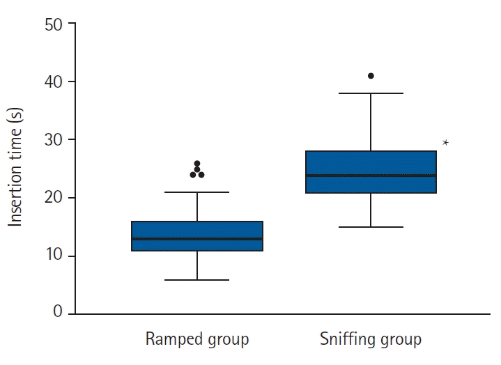
Fig. 1.
Flowchart of the study population. SGA: supraglottic airway.
kja-24255f1.tif
Fig. 2.
Time required for AuraGain insertion in the ramped (n = 23) and sniffing (n = 22) groups. Lines, boxes, and whiskers represent the median, interquartile range, and range, respectively. The circles represent outliers. *P < 0.05.
kja-24255f2.tif
Table 1.
Baseline Characteristics of the Study Participants
Variable Ramped group (n = 24) Sniffing group (n = 24) P value
Age (yr) 47.9 ± 14.4 44.6 ± 13.7 0.423
Sex (M) 6 11 0.227
Height (cm) 160.9 ± 8.4 163.5 ± 9.8 0.338
Weight (kg) 90.7 ± 14.0 90.4 ± 16.9 0.949
BMI (kg/m2) 34.8 (32.0, 36.6) 33.3 (31.6, 37.2) 0.650
Airway assessment factors
 Mallampati score (1/2/3/4) 17/7/0/0 15/7/2/0 0.661
 Neck circumference (cm) 39.5 (37.5, 43.0) 39.5 (38.0, 43.5) 0.764
 Sternomental distance (cm) 16.0 (14.2, 17.9) 16.0 (15.0, 18.0) 0.788
 Thyromental distance (cm) 8.0 (7.0, 10.0) 8.0 (7.5, 9.0) 0.640
 Interincisor distance (cm) 4.2 (4.0, 5.0) 4.5 (4.0, 5.0) 0.726
Warters grading scale (0/1/2/3/8) 15/8/1/0/0 10/6/5/2/1 0.107

Values are presented as mean ± SD, number or median (Q1, Q3). BMI: body mass index.

Table 2.
Characteristics of the AuraGain Insertion
Variable Ramped group (n = 24) Sniffing group (n = 24) NNT (95% CI) P value
Size of AuraGain used 0.174
 3 5 (21) 8 (33)
 4 18 (75) 12 (50)
 5 1 (4) 4 (17)
Insertion attempts (n) 0.319
 First attempt 22 (92) 18 (75)
 Second attempt 1 (4) 3 (13)
 Third attempt 1 (4) 3 (13)
Overall insertion success 1.000
 Success 23 (96) 22 (92)
 Failure 1 (4) 2 (8)
Reported ease of insertion 2.4 (1.6, 5.0)* 0.003
 Easy 16 (67) 5 (21)
 Acceptable 7 (29) 8 (33)
 Difficult 0 (0) 5 (21)
 Very difficult 0 (0) 4 (17)
 Impossible 1 (4) 2 (8)

Values are presented as numbers (%). NNT: number needed to treat. *Calculated as binary variables: ‘easy’ or ‘acceptable’ versus ‘difficult,’ ‘very difficult,’ or ‘impossible.’

Table 3.
Characteristics of Successful AuraGain Insertion
Variable Ramped group (n = 23) Sniffing group (n = 22) P value
OLP (cmH2O) 30.0 (30.0, 31.5) 30.0 (25.0, 35.0) 0.384
Brimacombe score 0.598
 1 0 2
 2 11 11
 3 4 4
 4 8 5

Values are presented as median (Q1, Q3) or number. OLP: oropharyngeal leak pressure.

Table 4.
Complications of Successful AuraGain Insertion
Variable Ramped group (n = 23) Sniffing group (n = 22) P value
Emergence from anesthesia
 Blood-tinged equipment 2 4 0.414
30 min after operation
 Sore throat (0/1/2/3)* 17/5/1/0 14/4/3/1 0.602
 Dysphonia (0/1/2/3) 18/4/1/0 20/0/2/0 0.130
 Dysphagia (0/1/2/3) 22/1/0/0 19/1/2/0 0.477
24 h after operation
 Sore throat (0/1/2/3) 19/4/0/0 18/3/1/0 1.000
 Dysphonia (0/1/2/3) 21/2/0/0 22/0/0/0 0.489
 Dysphagia (0/1/2/3) 21/1/1/0 22/0/0/0 1.000

Values are presented as numbers. *Severity of complications are shown as: 0, none; 1, mild; 2, moderate; 3, severe.

TOOLS
Similar articles