Journal List > Korean J Anesthesiol > v.77(5) > 1516088545

Lee, Park, Eum, Hwang, Ok, Sim, Perera, Ravald, Park, Wiedmer, and Sohn: Effect of lipid emulsion on vasoconstriction induced by epinephrine or norepinephrine in isolated rat aorta

Abstract

Background

Epinephrine (EPI) or norepinephrine (NOR) is widely used to treat cardiovascular collapse during lipid emulsion (LE) resuscitation for drug toxicity. However, the effect of LE on the vasoconstriction caused by EPI or NOR remains unknown. The purpose of this study was to examine the effect of an LE (Intralipid) on the vasoconstriction caused by EPI and NOR in isolated rat aorta.

Methods

The effect of LE on the vasoconstriction caused by EPI or NOR in isolated rat aorta was examined. Additionally, the effect of LE on the calcium increase caused by EPI or NOR was investigated. The distribution constant (KD: lipid to aqueous phase) of EPI or NOR between a LE (1%) and an aqueous phase was determined.

Results

LE (1 and 2%) did not significantly alter vasoconstriction caused by EPI or NOR in isolated endothelium-intact aorta. Moreover, the LE did not significantly alter the increased calcium level caused by EPI or NOR. The log KD of EPI in the LE (1%) was −0.71, −0.99, and −1.00 at 20, 50, and 100 mM ionic strength, respectively. The log KD of NOR in the LE (1%) was −1.22, −1.25, and −0.96 at 20, 50, and 100 mM ionic strength, respectively.

Conclusions

Taken together, the Intralipid emulsion did not alter vasoconstriction induced by EPI or NOR that seems to be due to the hydrophilicity of EPI or NOR, leading to sustained hemodynamic support produced by EPI or NOR used during LE resuscitation.

Introduction

Lipid emulsion (LE) is extensively utilized in the management of systemic toxicity resulting from local anesthetics [1]. Furthermore, it has been documented that LE, when used as an adjunctive medication, proves effective in addressing refractory cardiovascular collapse induced by toxic levels of non-local anesthetic drugs such as calcium channel blocker, anti-depressant, and anti-psychotic drug [24]. The widely accepted underlying mechanism of LE resuscitation is lipid shuttle that is basically associated with the lipophilicity of the compound [1]. Lipid shuttle states that the lipid phase of the LE absorbs highly lipophilic drugs (for example bupivacaine) from the brain and heart, and subsequently the LE with the lipophilic drugs, is transported to the liver, muscle, and adipose tissue for detoxification and storage [1]. In addition, LE directly produces the following effects: enhancement of cardiac contractility, provision of fatty acids, reduction of nitric oxide release, phosphorylation of glycogen synthase kinase-3β, and mitigation of mitochondrial dysfunction [1].
Vasopressors that include epinephrine (EPI) and norepinephrine (NOR) are widely used to treat cardiovascular collapse caused by a toxic dose of local anesthetics or non-local anesthetics [57]. Thus, these vasopressors (EPI or NOR) and LE are concurrently used to address severe cardiovascular collapse resulting from toxic levels of drugs that are difficult to manage. A recent study suggests that LE (Intralipid) may reduce plasma concentration and half-life of lipid soluble non-offending drug used for hemodynamic support during LE resuscitation for cardiovascular collapse caused by drug toxicity that may lead to a reduction in the therapeutic concentration of that drug and a subsequent attenuation of the hemodynamic support [8]. Moreover, some emergency physicians express concerns about whether LE may absorb beneficial vasopressors used to treat cardiovascular collapse during LE resuscitation for drug toxicity induced by local anesthetic or non-local anesthetic drugs. This absorption could potentially lead to a decrease in the vasopressor’s overall effectiveness. The extent to which LE reduces drug concentration is strongly positively correlated with the log P (or log Po/w) value (octanol/water partition coefficient) [9]. Log P is only one of several surrogate indicators that can be used to predict LE-induced reversal of cardiovascular collapse caused by a toxic dose of some drug, or the extent of LE-mediated absorption of offending drug to cause drug toxicity due to local anesthetic or non-local anesthetic drug [10]. However, it is important to note that the log P primarily reflects the distribution ratio of the non-ionized form of the compound in lipid (octanol) and aqueous phases [11]. Therefore, considering that the negative logarithm of the acid dissociation constants (pKa) of EPI and NOR is 8.59 and 8.58, respectively (Table 1), the calculated non-ionized fractions of total EPI and NOR using the Henderson-Hasselbalch equation are 6.06% and 6.20% at pH 7.4, respectively, and 3.13% and 3.21% at pH 7.1, respectively (Table 1). These percentages appear relatively small. Taking into consideration these calculated non-ionized fractions, it is worth noting that although EPI and NOR are water soluble (log P: EPI = −1.37, NOR = −1.24), these low log P values only represent the distribution of a small portion (non-ionized portion) of the total amount of EPI and NOR between the lipid and the aqueous phase (Table 1) [1113]. However, since all compounds, including local anesthetics, exist in both the ionized and non-ionized forms in vivo, the distribution coefficient that indicates the distribution ratio of both forms (ionized and non-ionized) in the lipid and aqueous phases holds more clinical relevance than log P [11]. An in vivo experiment using a rat model found that LE does not significantly change the peak increase in mean arterial pressure induced by EPI, but it does extend the time taken to reach this peak mean arterial pressure [14]. In addition, the effect of LE on the vasoconstriction caused by EPI and NOR, and the distribution coefficient of EPI and NOR is unknown. Moreover, due to ethical reasons, it is very difficult to examine the effect of LE on cardiovascular support induced by EPI and NOR during cardiopulmonary resuscitation using LE as an adjuvant drug. Thus, the goal of this study was to examine the effect of a LE (Intralipid) on the vasoconstriction caused by EPI and NOR in isolated rat aorta, and to get a better understanding of the underlying mechanism with particular focus on distribution constant. Based on the water-soluble properties of EPI and NOR, we tested the biological hypothesis that LE (1 and 2% Intralipid) does not alter vasoconstriction induced by EPI and NOR. If it is proven through this experiment that LE has a negligible effect on the vasoconstrictive support induced by EPI or NOR, this research may help alleviate some physicians’ concerns regarding LE-mediated sequestration of these drugs used for hemodynamic support.

Materials and Methods

The experimental procedure (GNU-211217-R0106, 22nd December, 2021) was approved by the Institutional Animal Care and Use Committee at Gyeongsang National University. All experimental methods followed the Guidelines for the Institutional Animal Care and Use Committee at Gyeongsang National University.

Materials

The commercially available compounds with highest purity were used. EPI, NOR, phenylephrine, acetylcholine, Nω-nitro-L-arginine methyl ester (L-NAME), and potassium chloride (KCl) were obtained from Sigma-Aldrich. Intralipid 200 mg/ml (20% weight/volume) was acquired from Fresenius Kabi AB. The buffer reagents disodium hydrogen phosphate dodecahydrate (Na2HPO4·12H2O) and sodium dihydrogen phosphate monohydrate (NaH2PO4·H2O) were from J.T. Baker and Merck, respectively. High performance liquid chromatography (HPLC) grade 99.5% dimethyl sulfoxide was from Labscan. LC-MS grade ≥ 99.9% methanol from Fisher Chemical was used as solvent for the analyte stock solutions. The standard pH solutions (4.0, 7.0, and 10.0) used for pH meter calibration were purchased from Merck.

Preparation of isolated rat aorta and isometric tension measurement

Male Sprague–Dawley rats weighing between 250 and 280 g obtained from Koatech underwent anesthesia with 100% carbon dioxide administered through a small opening in their cages. Subsequent to anesthesia, the isolation of rat aortas and isometric tension measurements were conducted following established procedures [15]. The thoracic cavity was surgically opened to retrieve the descending thoracic aorta that was subsequently immersed in Krebs solution. This solution contains the following concentrations: 118 mM of sodium chloride, 25 mM of sodium bicarbonate, 11 mM of glucose, 4.7 mM of KCl, 2.4 mM of calcium chloride, 1.2 mM of magnesium sulfate, and 1.2 mM of monopotassium phosphate. Removal of surrounding fat and connective tissues was performed under a microscope, and the isolated rat thoracic aortas were sliced into 2.5 mm-length segments. To eliminate the endothelium from some thoracic aortas, rolling movements were applied using two 25-gauge needles inserted into the aorta’s lumen. The isolated descending thoracic aortas were mounted on a Grass isometric transducer (model FT-03TM, Grass Instrument) within an organ bath maintained at 37°C. While a 90-min equilibrium period with a baseline resting tension of 24.5 mN was maintained, every 30 min, the Krebs solution was refreshed with freshly aerated Krebs solution (containing 95% oxygen and 5% carbon dioxide) to sustain a pH level of 7.4, as per previous protocols [15,16]. Endothelial integrity was confirmed by inducing sustained contractions with 10−7 M (0.1 µM) phenylephrine in the endothelium-intact thoracic aorta, followed by the addition of 10−5 M (10 µM) acetylcholine. An endothelium-intact thoracic aorta was defined as having more than 80% relaxation from phenylephrine-induced contraction. Endothelial denudation was verified by introducing acetylcholine (10−5 M; 10 µM) after stable contractions induced by 10−8 M (0.01 µM) phenylephrine, with less than 15% relaxation indicating an endothelium-denuded thoracic aorta. Aortas displaying acetylcholine-induced relaxation in both intact and denuded states were thoroughly washed with fresh Krebs solution to return to baseline resting tension. Subsequently, a contraction caused by isotonic 60 mM KCl solution was initiated to gauge the extent of vasoconstriction prompted by EPI or NOR. Once a sustained contraction was achieved through the addition of isotonic 60 mM KCl solution, the isolated aorta that displayed this contraction was thoroughly washed with fresh Krebs solution to return to its initial baseline tension. The subsequent experiment was then conducted.

Experimental protocols

The effect of LE (Intralipid) on the vasoconstriction caused by EPI or NOR was examined in isolated rat aorta. After pretreating rat aortas with intact or denuded endothelium with LE (1% and 2%) for 20 min, EPI (at concentrations ranging from 10−9 to 10−5 M) or NOR (at concentrations ranging from 10−8 to 10−5 M) was incrementally added to the organ bath to induce vasoconstriction, with or without the presence of LE. To mitigate the influence of residual endothelium on the contraction induced by EPI or NOR following endothelial denudation, all endothelium-denuded rat aortas were pre-treated with L-NAME (10−4 M) for 10 min prior to the addition of LE [17]

Determination of intracellular calcium concentration

To examine the effect of EPI and NOR treatment on intracellular calcium levels, we assessed the calcium concentration using a confocal laser microscope (IX70 FluoviewTM, Olympus) [18]. Vascular smooth muscle cells were seeded onto a confocal cell culture dish (SPL) and allowed to incubate overnight in media devoid of serum. To measure intracellular calcium concentrations, vascular smooth muscle cells were treated with 2.5 μM Fluo-4 AM (Invitrogen) in Hanks Balanced Salt Solution for 30 min. Subsequently, they were washed with phosphate-buffered saline solution and the Hanks Balanced Salt Solution was then replaced with fresh solution. Cells were treated with EPI (10−5 M; 10 µM) and NOR (10−5 M; 10 µM) alone, as well as with a mixture of EPI (10−5 M; 10 µM) with LE (Intralipid: 1% or 2%), and a mixture of LE (1% or 2%) with NOR (10−5 M; 10 µM). For 5 min, intracellular calcium concentrations were assessed every 2.5 s at wavelengths of 485 nm (excitation) and 520 nm (emission), respectively. Intracellular calcium levels were evaluated using images expressed by Fluo-4 AM, and calculated from the Flou-4 AM-induced fluorescence intensity (F) after EPI and NOR treatment divided by the fluorescence intensity without treatment (F0). The net alteration in calcium ions is expressed as (Fmax-F0)/F0, where Fmax denotes the fluorescence intensity at the peak calcium level after exposure to either EPI (10−5 M) or NOR (10−5 M) alone, or in combination with LE (1% or 2%) and EPI (10−5 M) or NOR (10−5 M).

Capillary electrophoresis (CE)

An Agilent Technologies 7100 CE system equipped with an UV/DAD absorbance detector was used. The capillary was a 68.5 cm fused coated silica capillary (Polymicro Technologies) with 60 cm to the detector and inner/outer diameters of 50/360 µm. The software was Agilent OpenLAB CDS ChemStation Edition c.01.05. The pH of the buffer solutions was checked with a WTW inoLab pH 7110 pH meter. A Sartorius BP301S analytical balance was used for weighing the analytes. Purified water was received from a water purification system Milli-Q Direct-Q 3 UV. The centrifuge used was a Micromax ThermoIEC with a Thermo IEC 851 rotor. In the CE runs, the capillary was flushed with 0.1 M sodium hydroxide for 3 min, with water for 2 min, and with phosphate buffer (PHB) for 5 min at high pressure (ca 940 mbar) before each sequence. Before each injection, the capillary was flushed with 0.1 M sodium hydroxide, water, and PHB solution for 1 min each. The applied separation voltage was 30 kV and the temperature was 37ºC. All the samples were injected at 100 mbar pressure at 10 s injection time. UV spectra of the runs was recorded at wavelengths 190–400 nm with 2 nm steps and the spectra of the analytes were saved in the UV spectrum library. Electropherograms were extracted at wavelengths 200 nm, 230 nm, 214 nm, and 254 nm. CE runs were conducted for both EPI and NOR samples at three different ionic strengths of PHB at pH 7.4 that were 20 mM, 50 mM, and 100 mM. EPI and NOR samples were prepared at concentrations of 100 µg/ml, dissolved in the respective PHB for each ionic strength. Dimethyl sulfoxide was used as the electroosmotic flow marker. The migration times and peak areas of all analyzed samples were recorded.

Liquid-liquid extraction

The commercial 20% Intralipid solution was diluted to 1% solution using the PHB with ionic strengths of 20 mM, 50 mM, and 100 mM. EPI and NOR were added to the Intralipid solutions separately, resulting in 1 ml solutions of 100 µg/ml concentration. This concentration corresponds to 5.46 × 10⁻⁴ M and 5.91 × 10⁻⁴ M of EPI and NOR, respectively. For both compounds, three separate samples were prepared for each ionic strength. The samples were shaken for 30 min, followed by 5 min centrifugation at 15,000 rpm (21,000 rcf). Part of the separated Intralipid layer was carefully removed so that the bottom aqueous layer could be obtained without contamination from the Intralipid layer.
The separated aqueous layers (bottom layers) of the samples were analyzed by CE with PHB at respective ionic strengths. The peak areas were recorded from three injections per sample, the migration time-corrected peak areas were calculated, and the compound concentrations were calculated using a calibration curve (six-point calibration curve at 5–200 µg/ml; each concentration was injected five times). The calibration curves were constructed by spiking the extracted aqueous phase (no drugs) with the studied compounds. The initial concentration and the EPI or NOR concentration in the separated aqueous layers were used to calculate the distribution. The distribution constant (KD), being the equilibrium constant for the distribution of a compound in two immiscible solvents, was calculated by equation (1),
KD = Cupper layer (lipid layer) Clower layer (aqueous layer)  = Cinitial - Caqueous layerCaqueous layer (1)
where cInitial is the initial concentration of the compound and caqueous Layer is the concentration of the compound in the separated (lower) aqueous layer. The percentual concentration (X%c(lipid layer)) of the compound in the lipid layer can be calculated from the KD using equation (2)
X%c lipid layer = 100KD1 +KD (2)

Statistical analysis

The primary endpoint of this study was to examine the effect of LE on the vasoconstriction caused by EPI or NOR. Sample size in each group was calculated using the resource equation method [19]. Normality test was performed using the Shapiro-Wilk test. The impact of LE on the vasoconstriction triggered by EPI and NOR was examined utilizing a linear mixed-effect model (Stata® version 14.2, StataCorp LLC) [20]. The impact of EPI, NOR, or a combination of LE and EPI, or LE and NOR on intracellular calcium levels was assessed using the Kruskal-Wallis test, followed by Dunn’s multiple comparison test, utilizing Prism® 5.0 software (GraphPad Inc.). A P value of less than 0.05 was considered statistically significant.

Results

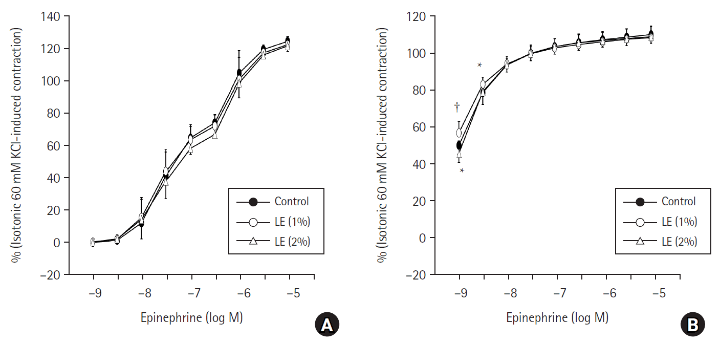
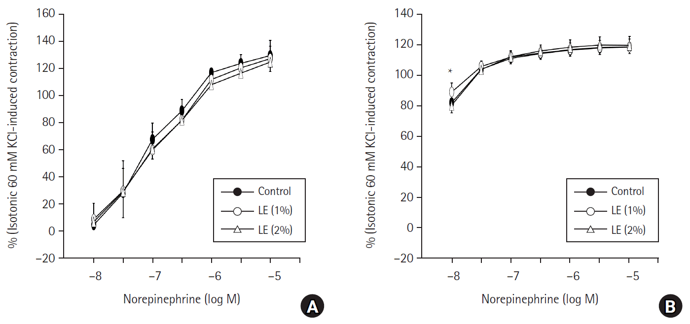
The presence of LE (1% and 2% Intralipid) did not produce a significant change in the contraction caused by EPI or NOR in endothelium-intact aorta (Figs. 1A and 2A). In addition, the LE (1% and 2%) did not significantly affect vasoconstriction induced by EPI (10−8 to 10−5 M) or NOR (3 x 10⁻⁸ to 10−5 M) (Figs. 1B and 2B) in endothelium-denuded aorta. However, in the endothelium-denuded aorta, the presence of 1% LE slightly increased the contraction induced by EPI (at concentrations of 10−9 and 3 x 10⁻⁹ M) (Fig. 1B). The P values were less than 0.001 and 0.021 versus the control at 10−9 and 3 x 10⁻⁹ M EPI, respectively, with the corresponding 95% CI (3.04, 10.76) and (0.67, 8.38). A similar trend was seen for NOR: 1% LE slightly increased 10−8 M NOR-induced contraction in endothelium-denuded aorta (Fig. 2B; P < 0.001 versus control: 95% CI [3.30, 10.57]). In contrast, 2% LE slightly decreased 10−9 M EPI-induced contraction in endothelium-denuded aorta (Fig. 1B; P = 0.030 versus control; 95% CI [−0.41, −8.13]).
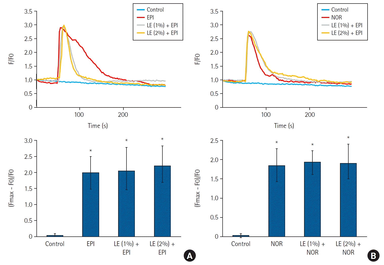
Both EPI (10−5 M; 10 µM) and NOR (10−5 M; 10 µM) elevated the intracellular calcium level in vascular smooth muscle cells (Figs. 3A and B; P < 0.001 versus control). Furthermore, the combination of LE (1% or 2%) with EPI (10−5 M), or LE (1% and 2%) with NOR (10−5 M), resulted in an elevation of intracellular calcium (Figs. 3A and B; P < 0.001 versus control). However, the mixture of LE and EPI or NOR did not significantly change the increase in calcium level caused by EPI or NOR alone (Figs. 3A and B).
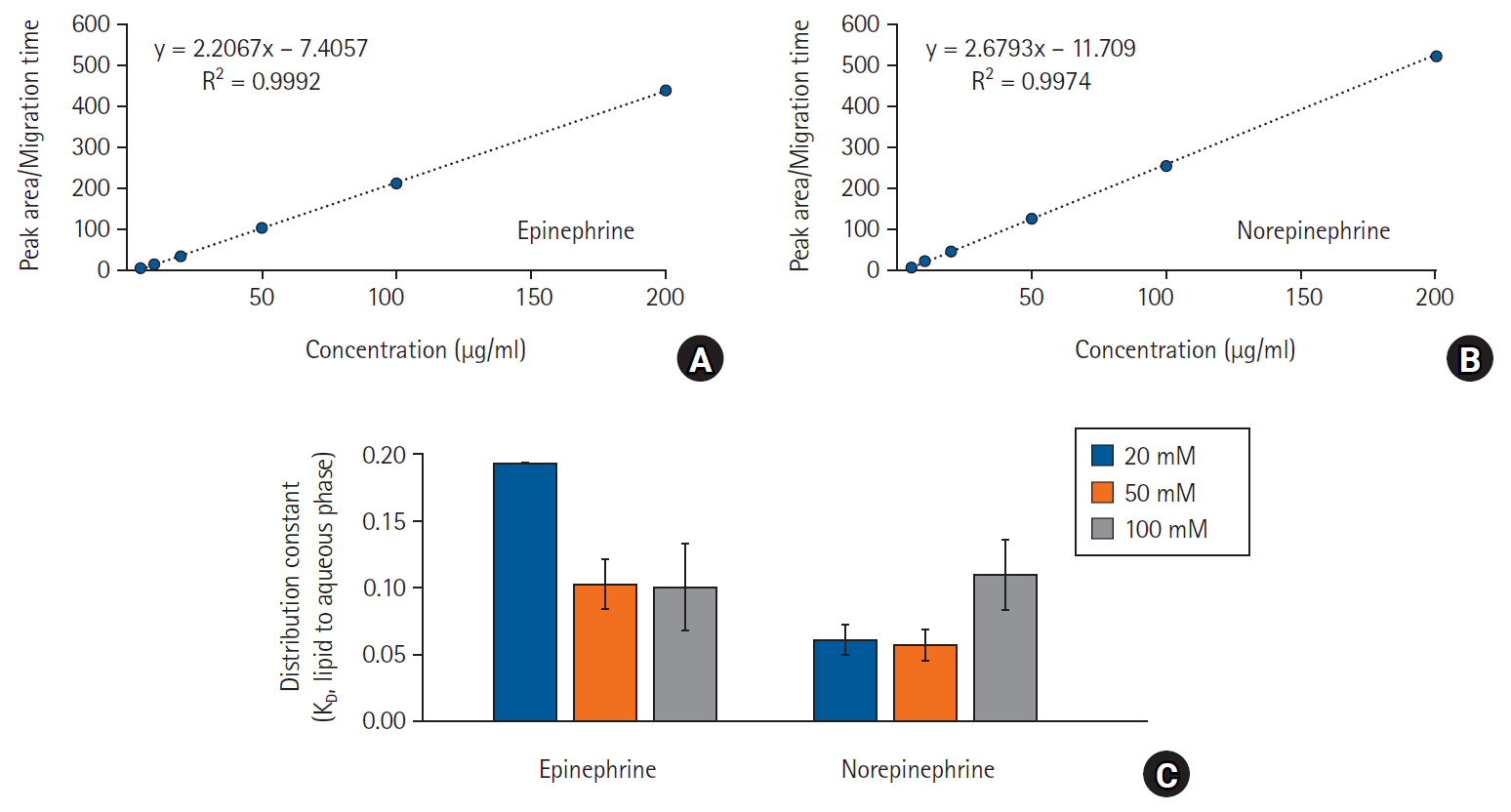
The KDs of EPI and NOR between a buffered aqueous phase and 1% LE were determined by liquid-liquid extraction. The concentrations in the aqueous phase after the extractions were quantitatively determined by CE with UV-absorption detection. The migration times and peak areas of EPI and NOR were recorded. The linearity curves for both compounds were determined in spiked aqueous buffers taken from liquid-liquid extractions without any drugs. The curves were highly linear (Figs. 4A and B) over the tested concentration range (5–200 µg/ml), giving a coefficient of correlation (R2) of more than 0.997 for both compounds. The corresponding linear equations were y = 2.21x – 7.41 and y = 2.68x – 11.71 for EPI and NOR, respectively (Figs. 4A and B), where y is the migration time-corrected analyte peak area and x is the analyte concentration (µg/ml). Using these calibration curves, the unknown concentrations were determined. From the obtained concentrations the KD values were determined using equation (1). The data in Fig. 4C shows that the KD values of EPI decreased slightly when changing the 20 mM PHB to higher ionic strength PHB solutions (50 and 100 mM). Even lower KD values were observed for NOR, with a slight increase in the value at the highest ionic strength (100 mM). The corresponding logarithmic values (log KD) of KD were negative for all investigated buffer solutions (20, 50, and 100 mM ionic strength), being in the range of −0.71 to −1.00 for EPI and −0.96 to −1.25 for NOR. The log KD values were in all cases clearly negative suggesting highly hydrophilic compounds.

Discussion

This study suggests that LE (Intralipid) does not significantly alter vasoconstriction induced by EPI or NOR. The major findings of this study are the following: 1) LE did not significantly affect vasoconstriction caused by EPI or NOR in endothelium-intact aorta; 2) LE did not significantly affect the calcium level increased by EPI or NOR in vascular smooth muscle cells; 3) EPI and NOR had only negligible interactions with the LE due to their hydrophilic properties.
EPI that is an alpha-1 adrenoceptor agonist and a beta-1 adrenoceptor agonist is used to treat hypotension and cardiac arrest [21]. The strong peripheral vasoconstrictor NOR that acts on alpha-1 and beta-1 adrenoceptor depending on the dose (less than 2 μg/min: beta-1 effect versus higher than 3 μg/min: alpha-1 effect) is used to treat hypotension unresponsive to fluid administration [22]. Thus, for managing refractory cardiovascular collapse caused by toxic doses of local or non-local anesthetics, LE and vasopressors like EPI or NOR can be concurrently administered. LE did not significantly alter EPI-induced contraction in endothelium-intact aorta. The results from liquid-liquid extraction experiments indicate that the portion of total EPI that is distributed into the LE at 20, 50, and 100 mM ionic strength is 16.16, 9.26, and 9.07%, respectively. These values are small, suggesting that EPI is strongly water soluble. Higher ionic strengths did not significantly affect the distribution that is of importance considering that the calculated ionic strength of the Krebs solution was 158 mM and that of plasma 161 mM. This LE mediated no significant change of EPI-induced vasoconstriction that can be explained by the relatively low KD of EPI. The KD of NOR at ionic strengths of 20 and 50 mM was even lower than that of EPI. The portion of total NOR distributed in the lipid phase was only 5.72 and 5.34% at 20 mM and 50 mM ionic strength, respectively. At 100 mM ionic strength the percentual distribution was slightly higher (9.83%). No significant alternation of NOR-induced contraction by LE was observed that can be explained by the very low KD at all studied ionic strengths (20 to 100 mM). Considering the discussion above, most parts of EPI and NOR will be distributed in the aqueous phase, indicating that the vasopressor effect induced by EPI or NOR, when used in conjunction with LE resuscitation for systemic toxicity due to local anesthetics or non-local anesthetics, may remain unchanged. LE suppresses the vasodilation mediated by nitric oxide that is stimulated by acetylcholine and the adenosine triphosphate-sensitive potassium channel agonist levcromakalim [23,24]. Moreover, LE impedes the phosphorylation of endothelial nitric oxide synthase induced by acetylcholine that plays a role in nitric oxide generation [23]. Furthermore, LE administration alone raises blood pressure and peripheral vascular resistance in humans, while it reduces flow-mediated vasodilation and vascular compliance in humans [25,26]. All these previous reports suggest that LEs inhibit endothelial nitric oxide release that is spontaneous or stimulated by some drugs [2326]. However, further in vivo studies regarding the effect of LE on the increased blood pressure caused by various doses of EPI or NOR are needed. Endothelial nitric oxide, generated from L-arginine by nitric oxide synthase, induces vasodilation through a pathway that involves guanylate cyclase, cyclic guanosine monophosphate (cGMP), and cGMP-dependent protein kinase [17]. Thus, endothelial nitric oxide attenuates vasoconstriction induced by an agonist. As residual endothelium after endothelial denudation affects contraction caused by EPI or NOR, the endothelium-denuded aortas were pretreated with L-NAME (10−4 M; 100 µM) to examine the effect of LE on the contraction caused by EPI or NOR in endothelium-denuded aorta. While this study employed L-NAME to reduce nitric oxide release triggered by the remaining endothelium following endothelial denudation, the extent of residual endothelium may produce a slight alteration in contraction induced by low concentrations of EPI or NOR solely in the endothelium-denuded aorta in response to LE. Furthermore, since long-chain fatty acids enhance voltage-dependent calcium currents, the slight increase in vasoconstriction induced by low doses of EPI or NOR when using LE might be attributed to the LE itself causing an increase in calcium levels [27]. Additional research is warranted to investigate the underlying mechanism behind the slight modification of vasoconstriction induced by low concentrations of EPI and NOR in endothelium-denuded aorta mediated by LE.
Contraction caused by agonists such as EPI or NOR produces increased intracellular calcium levels and subsequently vasoconstriction via activation of myosin light chain kinase that is mediated by calcium influx via receptor-operated calcium or voltage-operated calcium channels and calcium release from sarcoplasmic reticulum [28]. The Intralipid emulsion (1% and 2%) did not significantly alter the calcium increase caused by EPI or NOR. Thus, considering the results from the tension study and the low KD of EPI and NOR, these results suggest that the LE does not significantly absorb EPI or NOR, leading to sustained vasoconstriction produced by EPI or NOR used in LE resuscitation.
The study has the following limitations: First, blood pressure, predominantly influenced by peripheral vascular resistance and cardiac output, is mainly regulated by small resistance arterioles [29]. However, this study utilized the aorta that is a conduit vessel. Second, in vivo experiments would be more appropriate as the blood pressure is affected by the cardiac output and the sympathetic nervous system, and the LE increases the blood pressure in vivo. However, in this work in vitro experiments were done. Third, liquid-liquid extraction experiments used slightly higher concentrations of EPI (5.46 × 10⁻⁴ M) and NOR (5.91 × 10⁻⁴ M). One suggested dosing regimen of LE treatment as adjuvant drug for drug toxicity is 1% plasma triglyceride that provides positive inotropic and scavenging effect [3032]. Thus, we used 1% Intralipid emulsion in isometric tension measurements and liquid-liquid extractions. Even with these limitations, the results suggest that Intralipid emulsion has negligible effect on the vasoconstriction produced by EPI and NOR due to a clinically negligible amount of EPI or NOR sequestration into the LE. This leads to a beneficial sustained hemodynamic support produced by EPI and NOR used during LE resuscitation for intractable cardiovascular collapse caused by drug toxicity due to local anesthetic or non-local anesthetics. Taking these results into consideration, cardiovascular support induced by EPI or NOR remains unaltered in patients undergoing LE administration for parenteral nutrition in the intensive care unit.
In conclusion, based on our tension study and liquid-liquid extraction experiments, the studied LE (Intralipid) does not significantly alter the vasoconstriction induced by EPI or NOR due to their hydrophilic properties that may lead to a well sustained vasopressor effect during LE resuscitation for drug toxicity.

Notes

Funding

This research was supported by the Basic Science Research Program through the National Research Foundation of Korea (NRF), funded by the Ministry of Education (NRF-2021R1I1A3040332). This research was supported by the Basic Science Research Program through the NRF, funded by the Ministry of Education (IRIS RS-2023-00238572). This study was supported by a NRF grant funded by the Korean government (MSIT) (NRF-2021R1F1A1062363). This research was supported by funding from the Waldemar von Frenckell Foundation (grant no 4706562) and the Finnish Society of Sciences and Letters (grant no 4709946).

Conflicts of Interest

No potential conflict of interest relevant to this article was reported.

Data Availability

The data presented in this study are available on reasonable request from the corresponding author.

Author Contributions

Soo Hee Lee (Data curation; Funding acquisition; Investigation; Methodology; Software; Validation; Visualization; Writing – review & editing)

Kyeong-Eon Park (Data curation; Investigation; Methodology; Software; Validation; Visualization; Writing – review & editing)

Kibaek Eum (Data curation; Investigation; Methodology; Writing – review & editing)

Yeran Hwang (Data curation; Investigation; Methodology; Validation; Writing – review & editing)

Seong-Ho Ok (Conceptualization; Funding acquisition; Resources)

Gyujin Sim (Data curation; Formal analysis; Investigation; Methodology; Software; Validation; Visualization; Writing – review & editing)

Dumidu Perera (Data curation; Investigation; Methodology; Writing – original draft)

Henri K.M. Ravald (Data curation; Investigation; Methodology; Writing – original draft)

Youngho Park (Resources; Writing – review & editing)

Susanne K. Wiedmer (Conceptualization; Formal analysis; Funding acquisition; Project administration; Supervision; Writing – original draft; Writing – review & editing)

Ju-Tae Sohn (Conceptualization; Formal analysis; Funding acquisition; Methodology; Project administration; Supervision; Validation; Writing – original draft; Writing – review & editing)

References

1. Lee SH, Sohn JT. Mechanisms underlying lipid emulsion resuscitation for drug toxicity: a narrative review. Korean J Anesthesiol. 2023; 76:171–82.
2. Cao D, Heard K, Foran M, Koyfman A. Intravenous lipid emulsion in the emergency department: a systematic review of recent literature. J Emerg Med. 2015; 48:387–97.
3. Ok SH, Park M, Sohn JT. Lipid emulsion treatment of nonlocal anesthetic drug toxicity. Am J Ther. 2020; 28:e742–6.
4. Lee SH, Kim S, Sohn JT. Lipid emulsion treatment for drug toxicity caused by nonlocal anesthetic drugs in pediatric patients: a narrative review. Pediatr Emerg Care. 2023; 39:53–9.
5. Rosenblatt MA, Abel M, Fischer GW, Itzkovich CJ, Eisenkraft JB. Successful use of a 20% lipid emulsion to resuscitate a patient after a presumed bupivacaine-related cardiac arrest. Anesthesiology. 2006; 105:217–8.
6. Sirianni AJ, Osterhoudt KC, Calello DP, Muller AA, Waterhouse MR, Goodkin MB, et al. Use of lipid emulsion in the resuscitation of a patient with prolonged cardiovascular collapse after overdose of bupropion and lamotrigine. Ann Emerg Med. 2008; 51:412–5.
7. Rietjens SJ, de Lange DW, Donker DW, Meulenbelt J. Practical recommendations for calcium channel antagonist poisoning. Neth J Med. 2016; 74:60–7.
8. Li Z, Li M, Sun H, Yang Z, Huo Q, Bai Y, et al. Prediction of drug capturing by lipid emulsions in vivo for the treatment of a drug overdose. J Control Release. 2022; 346:148–57.
9. French D, Smollin C, Ruan W, Wong A, Drasner K, Wu AH. Partition constant and volume of distribution as predictors of clinical efficacy of lipid rescue for toxicological emergencies. Clin Toxicol (Phila). 2011; 49:801–9.
10. Wiedmer SK, Sohn JT. Lipophilicity of drugs, including local anesthetics, and its association with lipid emulsion resuscitation. Korean J Anesthesiol. 2024; 77:170–2.
11. Bhal SK, Kassam K, Peirson IG, Pearl GM. The rule of five revisited: applying log D in place of log P in drug-likeness filters. Mol Pharm. 2007; 4:556–60.
12. PubChem. Epinephrine [Internet]. Bethesda (MD): National Library of Medicine (US), National Center for Biotechnology Information [cited 2024 Jan 10]. Available from https://pubchem.ncbi.nlm.nih.gov/compound/Epinephrine#section=Solubility.
13. PubChem. Norepinephrine [Internet]. Bethesda (MD): National Library of Medicine (US), National Center for Biotechnology Information [cited 2024 Jan 10]. Available from https://pubchem.ncbi.nlm.nih.gov/compound/439260#section=Solubility.
14. Carreiro S, Blum J, Jay G, Hack JB. Intravenous lipid emulsion alters the hemodynamic response to epinephrine in a rat model. J Med Toxicol. 2013; 9:220–5.
15. Park KE, Lee S, Bae SI, Hwang Y, Ok SH, Ahn SH, et al. Theophylline-induced endothelium-dependent vasodilation is mediated by increased nitric oxide release and phosphodiesterase inhibition in rat aorta. Gen Physiol Biophys. 2023; 42:469–78.
16. Klöss S, Bouloumié A, Mülsch A. Aging and chronic hypertension decrease expression of rat aortic soluble guanylyl cyclase. Hypertension. 2000; 35:43–7.
17. Sohn JT, Kim HJ, Cho HC, Shin IW, Lee HK, Chung YK. Effect of etomidate on endothelium-dependent relaxation induced by acetylcholine in rat aorta. Anaesth Intensive Care. 2004; 32:476–81.
18. Ok SH, Kang D, Lee SH, Kim HJ, Ahn SH, Sohn JT. Lipid emulsions attenuate the inhibition of carnitine acylcarnitine translocase induced by toxic doses of local anesthetics in rat cardiomyoblasts. Hum Exp Toxicol. 2022; 41:9603271211065978.
19. Sohn JT. Resource equation method for sample size calculation in animal studies. Am J Emerg Med. 2023; 63:175–6.
20. Lavergne C, Martinez MJ, Trottier C. Empirical model selection in generalized linear mixed effects models. Comput Stat. 2008; 23:99–109.
21. Dalal R, Grujic D. Epinephrine [Internet]. Treasure Island (FL): StatPearls; 2023 May [cited 2024 Jan 10]. Available from https://www.ncbi.nlm.nih.gov/books/NBK482160/.
22. Smith MD, Maani CV. Norepinephrine [Internet]. Treasure Island (FL): StatPearls; 2023 May [cited 2024 Jan 10]. Available from https://www.ncbi.nlm.nih.gov/books/NBK537259/.
23. Ok SH, Lee SH, Yu J, Park J, Shin IW, Lee Y, et al. Lipid emulsion attenuates acetylcholine-induced relaxation in isolated rat aorta. Biomed Res Int. 2015; 2015:871545.
24. Lee SH, Kang D, Ok SH, Kim JY, Bae SI, Hwang Y, et al. Lipofundin MCT/LCT inhibits levcromakalim-induced vasodilation by inhibiting endothelial nitric oxide release. Int J Mol Sci. 2020; 21:1763.
25. Stojiljkovic MP, Zhang D, Lopes HF, Lee CG, Goodfriend TL, Egan BM. Hemodynamic effects of lipids in humans. Am J Physiol Regul Integr Comp Physiol. 2001; 280:R1674–9.
26. Gosmanov AR, Smiley DD, Peng L, Siquiera J, Robalino G, Newton C, et al. Vascular effects of intravenous intralipid and dextrose infusions in obese subjects. Metabolism. 2012; 61:1370–6.
27. Huang JM, Xian H, Bacaner M. Long-chain fatty acids activate calcium channels in ventricular myocytes. Proc Natl Acad Sci U S A. 1992; 89:6452–6.
28. Akata T. General anesthetics and vascular smooth muscle: direct actions of general anesthetics on cellular mechanisms regulating vascular tone. Anesthesiology. 2007; 106:365–91.
29. Clifford PS. Local control of blood flow. Adv Physiol Educ. 2011; 35:5–15.
30. Fettiplace MR, Ripper R, Lis K, Lin B, Lang J, Zider B, et al. Rapid cardiotonic effects of lipid emulsion infusion*. Crit Care Med. 2013; 41:e156–62.
31. Shin IW, Hah YS, Kim C, Park J, Shin H, Park KE, et al. Systemic blockage of nitric oxide synthase by L-NAME increases left ventricular systolic pressure, which is not augmented further by Intralipid®. Int J Biol Sci. 2014; 10:367–76.
32. Fettiplace MR, Akpa BS, Rubinstein I, Weinberg G. Confusion about infusion: rational volume limits for intravenous lipid emulsion during treatment of oral overdoses. Ann Emerg Med. 2015; 66:185–8.

Fig. 1.
Effect of lipid emulsion (LE) on the contraction caused by epinephrine in both endothelium-intact rat aorta (A) and endothelium-denuded rat aorta (B). Data (n = 5) are shown as mean ± SD and expressed as the percentage of isotonic 60 mM KCl-induced contraction. n indicates the number of rats from which isolated rat aortae were obtained. *P = 0.030 and 0.021 at 10−9 and 3 x 10⁻⁹ M epinephrine versus control, respectively. P < 0.001 versus control.
kja-24093f1.tif
Fig. 2.
Effect of lipid emulsion (LE) on the contraction caused by norepinephrine in both endothelium-intact rat aorta (A) and endothelium-denuded rat aorta (B). Data (n = 5) are shown as mean ± SD and expressed as the percentage of isotonic 60 mM KCl-induced contraction. n indicates the number of rats from which isolated rat aortae were obtained. *P < 0.001 versus control.
kja-24093f2.tif
Fig. 3.
Effect of lipid emulsion (LE) on the increased intracellular calcium level induced by 10−5 M epinephrine (EPI, A) or 10−5 M norepinephrine (NOR, B) in the vascular smooth muscle cells. Data (n = 4) are shown as median (Q1, Q3). n indicates the number of independent experiments. *P < 0.001 versus control. F: Flou-4 AM-induced fluorescence intensi­ty, F0: fluorescence intensity without treatment, Fmax: fluorescence intensity at the peak calcium level after exposure to either EPI or NOR alone, or in combination with either LE and EPI or LE and NOR.
kja-24093f3.tif
Fig. 4.
Calibration curves for epinephrine (A) and norepinephrine (B) by capillary electrophoresis. (C) Distribution constants (KD: lipid to aqueous phase) of epinephrine and norepinephrine at 25°C by liquid-liquid extraction at 20, 50, and 100 mM ionic strength of phosphate buffer. The data is presented as mean ± SD from three experiments.
kja-24093f4.tif
Table 1.
Physiochemical Properties of EPI and NOR
Drug log P* pKa NIF (%) at pH 7.4 NIF (%) at pH 7.3 NIF (%) at pH 7.2 NIF (%) at pH 7.1
EPI −1.37 8.59 6.06 4.88 3.91 3.13
NOR −1.24 8.58 6.20 4.99 4.00 3.21

*log P: log octanol/water partition coefficient from PubChem (https://pubchem.ncbi.nlm.nih.gov/). pKa: negative logarithm of the acid dissociation constant (Ka) from PubChem (https://pubchem.ncbi.nlm.nih.gov/). NIF (%): non-ionized fraction of total drug (EPI and NOR) is calculated using Henderson-Hasselbalch equation. EPI: epinephrine; NOR: norepinephrine.

TOOLS
Similar articles