Abstract
Background/Aims
During endoscopy, white spots (WS) are sometimes observed around benign or malignant colorectal tumors; however, few reports have investigated WS, and their significance remains unknown. Therefore, we investigated the significance of WS from clinical and pathological viewpoints and evaluated its usefulness in endoscopic diagnosis.
Methods
Clinical data of patients with lesions diagnosed as epithelial tumors from January 1, 2019, to December 31, 2020, were analyzed (n=3,869). We also performed a clinicopathological analysis of adenomas or carcinomas treated with endoscopic resection (n=759). Subsequently, detailed pathological observations of the WS were performed.
Results
The positivity rates for WS were 9.3% (3,869 lesions including advanced cancer and non-adenoma/carcinoma) and 25% (759 lesions limited to adenoma and early carcinoma). Analysis of 759 lesions showed that the WS-positive lesion group had a higher proportion of cancer cases and larger tumor diameters than the WS-negative group. Multiple logistic analysis revealed the following three statistically significant risk factors for carcinogenesis: positive WS, flat lesions, and tumor diameter ≥5 mm. Pathological analysis revealed that WS were macrophages that phagocytosed fat and mucus and were white primarily because of fat.
Colorectal cancer (CRC) is the third most common cause of cancer-related mortality worldwide, with over 1.85 million cases and 850,000 deaths annually.1 Therefore, improving the outcomes of CRC and preventing its development are crucial human health challenges.
The following three carcinogenic pathways have been proposed for CRC development: (1) adenoma-carcinoma sequence,2 (2) de novo pathway,3 and (3) serrated pathway.4 Of these, the adenoma-carcinoma sequence is considered the most relevant pathway.5 Therefore, the resection of adenomas, which are considered precancerous lesions of CRC, is reported to reduce CRC incidence by 76% to 90% and CRC-related mortality by 53%.6 Detecting and treating CRC at the adenoma stage is crucial to avoid death due to CRC.
Colonoscopy (CS) is important in the diagnosis and treatment of colorectal tumors. When a CS is performed, white-speckled mucosa around the colorectal tumors is occasionally observed. These observations are referred to as white spots (WS). WS, also known as chicken skin mucosa, have been reported to be an independent endoscopic predictor of advanced colorectal adenoma 7,8 and a potential indicator of submucosal invasion in carcinoma.9 However, WS can be found around benign lesions (such as adenomas or serrated polyps), indicating that they are not cancer-specific findings.
Muto et al.10 reported WS to be macrophages phagocytosing mucus and present not only on the mucosal surface but also in the deeper layers of the mucosa. Additionally, Hirota et al.11 compared the presence and absence of WS in patients with advanced carcinoma and reported significantly more lymph node metastasis in patients with WS, suggesting that WS reflects the degree of cancer progression. However, no large-scale studies have been conducted on WS, and its incidence, distribution trends, and clinical significance remain unknown.
Therefore, in this study, we aimed to investigate the significance of WS from a clinicopathological perspective and determine whether the presence or absence of WS around colorectal tumors could be a new indicator for endoscopic resection.
This was a single-center retrospective study that was based on an analysis of existing data, including the results of previous examinations and previously resected specimens.
The study period was two years (from January 1, 2019 to December 31, 2020). Overall, 3,990 lesions from 1,532 patients who underwent CS examination or treatment at Tohoku University Hospital during the study period were diagnosed as colorectal tumors.
If the quality of CS at a hospital is poor, the study will not be valid. Therefore, before this study, the quality of CS was evaluated based on the adenoma detection rate (ADR),12 cecum reach rate, and extraction time. ADR ≥30% for men, ≥20% for women, and ≥20% in total have been established as appropriate indicators for quality control.13 The cecum reach rate and extraction time should be ≥90% and ≥6 minutes, respectively.14
A flowchart of the sorting of the analyzed lesions is shown in Figure 1. Overall, 3,869 lesions diagnosed as adenomas or carcinomas through endoscopy were finally analyzed. Submucosal tumor (SMT), polyposis, and malignant lymphoma, whose pathological mode of origin differed from that of epithelial colorectal tumors, were excluded from this study. Patients who underwent chemoradiotherapy were excluded from the evaluation because they underwent therapeutic interventions. Cases in which the entire lesion could not be grasped owing to feces or bleeding were also excluded as they were difficult to evaluate.
Of the 3,869 lesions, the indications for resection of the 3,869 lesions were determined by consultation with several endoscopists according to the guidelines of the Japanese Society of Gastrointestinal Endoscopy.15,16
The indications for endoscopic resection were lesions that met one or more of the following criteria: (1) Tumor size greater than 5 mm; (2) Flat depressed tumor, regardless of size; (3) Tumors of type 2B or 3 based on The Japan NBI Expert Team classification, regardless of the size; (4) Tumors that are type V according to the pit pattern classification, regardless of the size; (5) CRC that is not advanced (CRC that has not progressed to the point where surgery is indicated). However, even if the lesion met the guideline criteria for endoscopic resection, we did not perform endoscopic resection if any of the factors listed in “(6) Patient factors” were present; (6) Patient factors: (i) The patient is undergoing treatment for prognostic factors other than colorectal tumor (e.g., on drug therapy for Stage IV cancer); (ii) The patient has an underlying disease that is intolerable to treatment, such as respiratory disease (e.g., severe chronic obstructive pulmonary disease) or hematologic disease (e.g., coagulation abnormalities); (iii) The patient cannot discontinue medications that interfere with endoscopic resection, such as antiplatelet or anticoagulant medications; (iv) The patient does not wish to undergo endoscopic resection.
During the study period, 850 lesions were endoscopically resected, of which 759 lesions that were histologically diagnosed as adenomas or carcinomas were subjected to clinicopathological analysis. Since the adenoma-carcinoma sequence is the most classic and common pathway and adenoma has been shown to be a precancerous lesion, the epithelial colorectal tumors analyzed in this study were limited to adenomas and carcinomas.
Patient clinical data were obtained from electronic medical records, and endoscopic image information was collected from an endoscopic database (Solemio ENDO; Olympus Co., Ltd.).
All patients were administered 2 to 3 L of polyethylene glycol-electrolyte solution for bowel preparation on the morning of the examination day. Butylscopolamine bromide or glucagon was administered in the absence of contraindications, and midazolam was used for conscious sedation only when the patient complained of discomfort or pain.
The endoscopies were performed using an electronic endoscopy system (LASEREO; Fujifilm Co., Ltd. or EVIS LUCERA ELITE; Olympus Co., Ltd.) with a magnifying CS (EC-L600ZP7; Fujifilm Co., Ltd. and PCF-H290ZI; Olympus Co., Ltd.).
In this study, WS were defined as mucosal abnormalities characterized by white-speckled patterns in the colon observed under conventional light endoscopy. The term “chicken skin mucosa” is not common in Japan. They are also recognized and referred to as WS in the Japanese glossary and are discussed as WS in this study; therefore, in this study, the definition of chicken skin mucosa is consistent with that in previous reports.7-9 WS-positive status was defined as the presence of WS, even partially, around tumors. We determined the presence or absence of WS by consensus of at least two endoscopists. A case where WS were present around the lesion was defined as positive (Fig. 2). The presence of a partially circumscribed area around the tumor rather than the entire circumference was considered positive. At least two endoscopists performed morphological evaluations of the tumors according to the Paris classification.17
Epithelial colorectal tumors resected using endoscopic submucosal dissection (ESD) or endoscopic mucosal resection were sliced at 2-mm intervals after overnight fixation in a 20% buffered formalin solution; subsequently, pathologists performed histological evaluation. In this study, Categories 3 and 4 were classified as adenomas and Category 5 as carcinomas according to the international Vienna classification.18,19 Advanced cancer was defined as T2 (Union for International Cancer Control 8th edition) or higher on pathological evaluation after surgical resection, and N- and M-factors were not considered. The pathologist used a measuring instrument to accurately determine the size of the excised lesions to one decimal place.
The WS samples were obtained from resected early-stage carcinomas. Mucosa with WS around the tumor was collected, cut into small sections, and immediately immersed in a fixing solution. The specimens were sent to Hanaichi Electron Microscopy Technical Laboratory Co., Ltd., for electron microscopic observation.
In a report by Muto et al.,10 WS were described as mucus-phagocytosed macrophages. Additionally, as electron microscopy revealed that WS has a high fatty component, we performed hematoxylin and eosin (H&E), periodic acid–Schiff (PAS), and adipophilin staining to observe WS in detail.
The aim of this study was to analyze whether WS are cancer-related findings and evaluate whether they can be applied to endoscopic diagnosis.
Statistical analyses were performed using JMP Pro 17 software (SAS Institute Inc.). Categorical variables were compared using the chi-squared test. Continuous variables are presented as medians (interquartile ranges) and were compared using the Mann-Whitney U-test. Multiple logistic regression analysis was used for multivariate analysis. Statistical significance was set at p-values <0.05.
The ADRs at our hospital for CS during the study period were 58.7% and 46.9% in men and women, respectively, with a combined ADR of 54.0%. The endoscopic cecum reach rate was 100%. All endoscopies required more than six minutes to observe the colon.
Table 1 presents the details of the 3,869 lesions analyzed. WS were present in approximately 9.3% of all lesions. Furthermore, 98.6% (353/358 lesions) of WS-positive lesions were distributed around the whole circumference. The sigmoid colon had the highest number of lesions in the WS-positive (43.6%) and WS-negative (26.0%) groups. When the positivity rate of WS was compared between the proximal (cecum, ascending colon, and transverse colon) and left (descending colon, sigmoid colon, and rectum) colons, the distal colon had a significantly higher rate of 15.0% (Fig. 3).
Details of the 759 resected lesions are presented in Table 2. Approximately 25% of the 759 lesions (189 lesions) had WS. The WS-positive group had a larger lesion size than the WS-negative group (12 [9.5–18] mm vs. 7 [5–10] mm).
Additionally, multiple analyses were performed on WS positivity rates for adenoma and carcinoma.
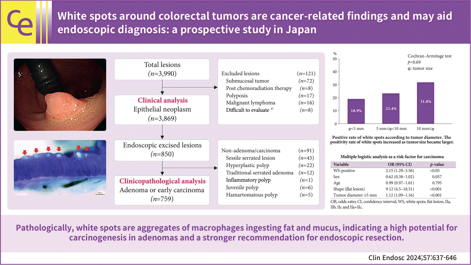
First, the positivity rate of WS was examined by classifying tumor diameter (φ) into the following three categories: (1) φ<5 mm, (2) 5 mm≤φ<10 mm, and (3) 10 mm≤φ. Although the number of WS-positive lesions increased with increasing tumor diameter, the increase was not statistically significant (Cochran-Armitage test, p=0.69; Fig. 4).
Second, a comparison of the percentages of cancers in the WS-positive and WS-negative groups showed that cancer was significantly more common in the WS-positive group than in the WS-negative group (30.2% and 12.3%, respectively).
For the third analysis, tumor diameter (φ) was divided into three groups ((1) φ<5 mm, (2) 5 mm≤φ<10 mm, and (3) 10 mm≤φ), and the proportions of cancers in the WS-positive and WS-negative groups were calculated and compared in each group (Table 3). No statistical difference was found between the proportion of cancers in the WS-positive and WS-negative groups in the groups of (1) φ<5 mm and (3) 10 mm≤φ; however, in the group of (2) 5 mm≤φ<10 mm, a statistically significant difference was observed between the two groups, with a higher proportion of carcinoma in the WS-positive group (WS-positive group: 16.3%, 13/80 lesions, WS-negative group: 8.0%, 21/262 lesions).
In the fourth study, a multiple logistic analysis was performed on 759 lesions (Table 4). In our analysis, the three statistically significant risk factors for cancer were WS-positive, flat lesions (IIa, IIb, IIc, and IIa+ IIc), and tumor diameter ≥5 mm. The odds ratios were 2.15, 9.12, and 1.12 for WS-positive, flat lesions, and tumor diameter ≥5 mm, respectively.
Electron microscopy revealed the presence of an almost homogeneous, electron-dense substance. This substance was considered an intracellular lipid in macrophages (Fig. 5).
We used ESD specimens to observe WS by pathological staining. WS were observed under an optical microscope after performing H&E, PAS, and immunohistochemical staining for adipophilin, which surrounds fat droplets and is involved in lipid storage (Fig. 6). Thus, we determined that the white deposits were foam fat cells within the cell structures. In addition to fat, mucus was found to be present inside the macrophages.
In this study, the positivity rate of WS was approximately 9.3%. This positivity rate included all adenomas, carcinomas, sessile serrated lesions (SSLs), and advanced cancers. As no previous reports exist on the positivity rate of WS, comparing the findings of this study with those of previous reports is impossible. However, we considered the rate of 9.3% to be credible because of the adequate quality indicators of CS at our institution (ADR, cecal reach rate, and extraction time).
The most common distribution of WS was in the sigmoid colon (43.6%), followed by the rectum (27.9%). This finding is consistent with the incidence of CRC (sigmoid colon was the most common [30.9%], followed by the rectum [20.7%]),11 and supports the possibility that WS are tumor-related findings.
The main factor influencing the carcinogenesis of adenoma is size.20-22 Liberman classified lesions >10 mm as advanced adenomas and concluded that advanced adenomas should be included in CS screening.23 In this study, the lesion size was larger in the WS-positive group than in the WS-negative group. The proportion of early-stage cancer cases was higher in the WS-positive group than in the WS-negative group. Therefore, we considered that WS are associated with malignancy.
Next, we discuss the results of our review of the 759 lesions that were endoscopically resected and pathologically diagnosed (adenoma or carcinoma). The positive rate of WS calculated only for adenoma and carcinoma (lesions that could be curatively resected through endoscopic resection) was approximately 25%; referring to three previous reports that calculated positive rates for adenoma and carcinoma, the rates were 30.7%,7 32.9%,8 and 31.3%.9 Our positive rate (25%) was also close to these percentages, and we believe that it is scientifically reasonable considering our quality indicators of CS. Globally, the carcinogenic rate of adenomas is mainly defined by tumor size >10 mm, resulting in a carcinogenic rate of 10% to 25%.20-22 Based on several analyses in this study, (1) the rate of WS positivity increases as tumor size increases; (2) tumor diameter (φ) in the WS-positive group is significantly larger than that in the WS-negative group; (3) in lesions with tumor diameter 5 mm≤φ<10 mm the rate of carcinoma in the WS-positive group is higher than that in the WS-negative group; (4) in treated lesions (n=759) as a whole, the rate of carcinoma in the WS-positive group is higher than that in the WS-negative group; and (5) in the multivariate analysis, WS positivity was extracted as a risk factor for cancer along with other factors (morphological type and tumor size).
These results raise the following question: Are WS indicators of tumor size (i.e., does WS increase with tumor size)? We established that the positivity rate of WS increases with increasing tumor size; however, this is not a statistically significant increasing trend. Other analyses conducted regarding indicators of malignancy showed statistically significant differences, and we conclude that WS can be scientifically considered an indicator of cancer rather than simply an indicator of tumor size.
Notably, in the analysis of 759 lesions, we found that in lesions with a tumor diameter of 5 mm or more but less than 10 mm, the proportion of carcinoma was significantly higher in the WS-positive group than in the WS-negative group (Table 3). As previously mentioned, the carcinogenic rate of adenomas depends mainly on the tumor size. Based on the assumption that WS is an indicator of cancer, the results of this study suggest that lesions <5 mm in diameter have not yet undergone cancerous transformation, while lesions >10 mm are presumed to have already completed cancerous transformation in many lesions; therefore, there may be no significant difference between the WS-positive and WS-negative groups.
However, lesions with a tumor diameter of ≥5 mm and <10 mm are all similar in size and exist in the same histological type of adenoma; conversely, various types of lesions exist, including those that are not pathologically cancerous but have already undergone cellular changes at the molecular level toward cancer formation and those that remain as adenomas. We considered that WS may be one of these phenotypes. As shown in Figure 7, when only the tumors were excised without WS, subsequent endoscopic observation revealed that the WS at the excision site disappeared (Fig. 7). This phenomenon supports our hypothesis that WS may be one of these phenotypes. We assumed that the disappearance of the tumor affected the macrophages surrounding the tumor owing to the disappearance of factors such as cytokines emitted by the tumor.
Next, we discuss the pathological observations of WS. WS were proposed to be mainly a collection of macrophages in 1981.10 Subsequently, no detailed morphological or pathological observations of WS have been made.
In this study, we demonstrated that WS are lipid-phagocytosing macrophages, as observed using electron microscopy. Based on these results, we performed PAS and adipophilin staining in addition to H&E staining to observe this pathology. Overall, the electron microscopy and histopathological staining findings led us to the conclusion that WS are clusters of macrophages that phagocytose mucus and lipids. As macrophages are components of the tumor microenvironment,24 pathological observations support our hypothesis that WS are tumor-related findings. We also speculate that the white color of WS is due to the bubbling of lipids taken up by macrophages and that the seemingly varying degrees of white color on endoscopic observation reflect differences in the amount of mucus.
The fact that WS is more frequently distributed in tumors of the distal colon may be largely related to macrophages. The colonic transit time is longer in the distal colon,25 and abnormal macrophage activation and increased recruitment occur because of the secretion of various cytokines in the distal colon under prolonged stretch.8 Therefore, the distal colon and rectum might have a higher chance of bowel inflammation by retaining stool, bacteria, and macrophages.9 The inflammatory microenvironment is important for cancer development, and tumor-associated macrophages exert pro-tumor functions in this microenvironment.7 We speculate that this minor chronic inflammation of the distal colon may be the reason for the high incidence of tumors with WS.
This study has some limitations. First, the pathology under consideration was limited to adenomas and carcinomas. This study only analyzed the clinical and pathological characteristics of WS in adenomas and carcinomas and did not examine the relationship between WS and lesions such as SSLs and SMTs. In our study, WS were not present around the SMT; however, there were lesions in SSL that were positive for WS. Therefore, future studies should focus on the relationship between SSL and WS. Second, endoscopic resection of epithelial colorectal tumors is indicated according to the Guidelines for Colorectal Polyp Practice 2014/2020 in Japan,15,16 and resection is basically considered for lesions with a diameter of 5 mm or greater. For lesions with a diameter of <5 mm, follow-up is acceptable if they do not exhibit endoscopic features that suggest malignancy, such as concavity formation, irregularity, and microvascular or surface structures. Although the results of our study showed that WS are cancer-related findings, there is a lack of evidence as to whether follow-up is acceptable or whether resection should be performed for WS-positive adenomas <5 mm in diameter. Third, the reasons for the high distribution in the distal colon and the possibility that the 5- to 10-mm lesions may be cancerous at the molecular biological level would be more convincing if they were verified by basic experiments using co-cultures, such as organoids; however, this was not performed in this study. Despite these limitations, we believe that our study may contribute to endoscopic diagnostics, particularly in guiding treatment decisions for lesions 5 to 10 mm in size, which are frequently difficult to determine whether to resect.
In conclusion, WS are cancer-related findings, and we posit that the potential for carcinogenesis is high in adenomas with WS. This result suggests that adenomas with WS have a more positive indication for endoscopic resection. Remarkable progress has been made in endoscopic diagnostics with advances in endoscopic equipment and image-processing technology. Therefore, endoscopists should receive adequate training to master advanced techniques. However, WS can be confirmed without the use of special techniques and can be easily observed even by inexperienced endoscopists. Therefore, if the presence or absence of WS can be applied to the treatment strategy for colorectal epithelial tumors, particularly adenomas, CRC development and CRC-related mortality may be prevented.
Notes
Author Contributions
Conceptualization: KK; Data curation: KK, YS, HN; Formal analysis: KK; Investigation: KK; Methodology: KK, TN; Project administration: KK; Resources: KK, HS; Software: IT, FF; Supervision: KK, YKa, YKi, AM; Validation: IT, HS; Visualization: RM; Writing–original draft: KK; Writing–review & editing: all authors.
REFERENCES
1. Biller LH, Schrag D. Diagnosis and treatment of metastatic colorectal cancer: a review. JAMA. 2021; 325:669–685.
2. Hill MJ, Morson BC, Bussey HJ. Aetiology of adenoma: carcinoma sequence in large bowel. Lancet. 1978; 1:245–247.
3. Goto H, Oda Y, Murakami Y, et al. Proportion of de novo cancers among colorectal cancers in Japan. Gastroenterology. 2006; 131:40–46.
4. Leggett B, Whitehall V. Role of the serrated pathway in colorectal cancer pathogenesis. Gastroenterology. 2010; 138:2088–2100.
5. Jass JR, Whitehall VL, Young J, et al. Emerging concepts in colorectal neoplasia. Gastroenterology. 2002; 123:862–876.
6. Zauber AG, Winawer SJ, O’Brien MJ, et al. Colonoscopic polypectomy and long-term prevention of colorectal-cancer deaths. N Engl J Med. 2012; 366:687–696.
7. Chung EJ, Lee JY, Choe J, et al. Colonic chicken skin mucosa is an independent endoscopic predictor of advanced colorectal adenoma. Intest Res. 2015; 13:318–325.
8. Lee YM, Song KH, Koo HS, et al. Colonic chicken skin mucosa surrounding colon polyps is an endoscopic predictive marker for colonic neoplastic polyps. Gut Liver. 2022; 16:754–763.
9. Zhang YJ, Wen W, Li F, et al. Chicken skin mucosa surrounding small colorectal cancer could be an endoscopic predictive marker of submucosal invasion. World J Gastrointest Oncol. 2023; 15:1062–1072.
10. Muto T, Kamiya J, Sawada T, et al. Clinical and histological studies on white spots of colonic mucosa around colonic polyps with special reference to diagnosis of early carcinoma. Gastroenterol Endosc. 1981; 23:241–247.
11. Hirota S, Hosobe S, Ikeda M, et al. Possibility of defense against colorectal tumor by foamy cells. J Clin Gastroenterol. 1997; 24:82–86.
12. Corley DA, Jensen CD, Marks AR, et al. Adenoma detection rate and risk of colorectal cancer and death. N Engl J Med. 2014; 370:1298–1306.
13. van Doorn SC, Klanderman RB, Hazewinkel Y, et al. Adenoma detection rate varies greatly during colonoscopy training. Gastrointest Endosc. 2015; 82:122–129.
14. Rex DK, Schoenfeld PS, Cohen J, et al. Quality indicators for colonoscopy. Am J Gastroenterol. 2015; 110:72–90.
15. Tanaka S, Saitoh Y, Matsuda T, et al. Evidence-based clinical practice guidelines for management of colorectal polyps. J Gastroenterol. 2015; 50:252–260.
16. Tanaka S, Saitoh Y, Matsuda T, et al. Evidence-based clinical practice guidelines for management of colorectal polyps. J Gastroenterol. 2021; 56:323–335.
17. Lambert R, Lightdale CJ. The Paris endoscopic classification of superficial neoplastic lesions: esophagus, stomach, and colon. Gastrointest Endosc. 2003; 58:S3–S43.
18. Schlemper RJ, Riddell RH, Kato Y, et al. The Vienna classification of gastrointestinal epithelial neoplasia. Gut. 2000; 47:251–255.
19. Schlemper RJ, Kato Y, Stolte M. Review of histological classifications of gastrointestinal epithelial neoplasia: differences in diagnosis of early carcinomas between Japanese and Western pathologists. J Gastroenterol. 2001; 36:445–456.
20. Sakamoto T, Matsuda T, Nakajima T, et al. Clinicopathological features of colorectal polyps: evaluation of the ‘predict, resect and discard’ strategies. Colorectal Dis. 2013; 15:e295–e300.
21. Gschwantler M, Kriwanek S, Langner E, et al. High-grade dysplasia and invasive carcinoma in colorectal adenomas: a multivariate analysis of the impact of adenoma and patient characteristics. Eur J Gastroenterol Hepatol. 2002; 14:183–188.
22. O’Brien MJ, Winawer SJ, Zauber AG, et al. The National Polyp Study. Patient and polyp characteristics associated with high-grade dysplasia in colorectal adenomas. Gastroenterology. 1990; 98:371–379.
23. Lieberman DA, Weiss DG, Harford WV, et al. Five-year colon surveillance after screening colonoscopy. Gastroenterology. 2007; 133:1077–1085.
24. Lewis CE, Pollard JW. Distinct role of macrophages in different tumor microenvironments. Cancer Res. 2006; 66:605–612.
25. Wagener S, Shankar KR, Turnock RR, et al. Colonic transit time: what is normal? J Pediatr Surg. 2004; 39:166–169.
Fig. 1.
Flowchart of the review and analysis. Duplicate lesions from the same patient during the study period were excluded. “n” indicates the number of lesions, not the number of patients. WS, white spots. a)Difficult to evaluate because obtaining a whole image of the lesion was impossible owing to fecal matter or bleeding.
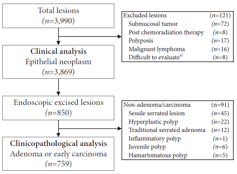
Fig. 2.
Endoscopic findings of the white spot-positive colorectal epithelial tumor. (A) White spots are distributed around the whole circumference of early-stage colorectal cancer (sigmoid colon, T1b). (B) White spots are distributed around the adenoma (sigmoid colon, low-grade tubular adenoma). The patient provided oral informed consent for the publication and use of her/his images.
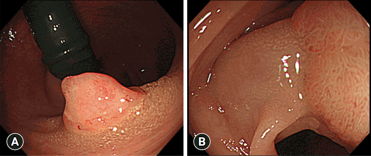
Fig. 3.
Comparison of the positivity rate of white spots (WS) between proximal colon and distal colon. WS were more frequently present in the distal colon than in the proximal colon (Fisher exact test, *p<0.001). Proximal colon: cecum, ascending colon, and transverse colon. Distal colon: descending colon, sigmoid colon, and rectum.
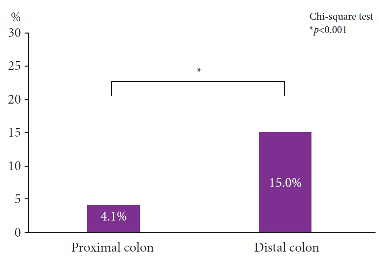
Fig. 4.
Positive rate of white spots according to tumor diameter. The positivity rate of white spots increased as tumor size became larger.
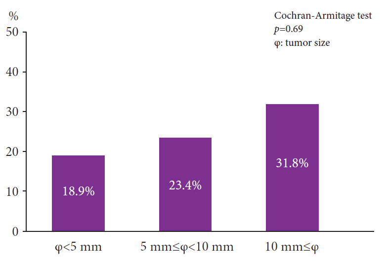
Fig. 5.
Electron microscopic observations of white spots (WS). Electron microscopic examination of the WS revealed a cluster of macrophages in an area that appeared to coincide with the WS (the object surrounded by the dotted line represents a macrophage). Inside the cells, vacuolation, which is a result of fat denaturation, is present. a, nuclear; b, fat; c, lysosome; d, myelin body (possibly).
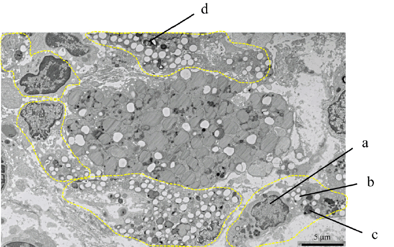
Fig. 6.
Pathological staining image of white spots (WS). (A) The specimen was excised through endoscopic submucosal dissection, and the WS was observed on the lateral side. Magnified observation revealed that the area corresponding to the WS aligned with the substance was deposited spherically just below the epithelium (arrows). (B) Hematoxylin and eosin (H&E) staining (×100): Histiocytes are clustered immediately below the epithelium in the area consistent with the gross magnified image of WS (corresponding to the area surrounded by the dotted line). (C) Periodic acid–Schiff (PAS) staining (×100): The same area where a cluster of histiocytes was observed in H&E staining was positive (corresponding to the area surrounded by the dotted line). (D) Immunohistochemistry (IHC) with anti-adipophilin antibody (×100): IHC was performed by diluting the antibody to 1/50, and the area matching the H&E and PAS stain-positive areas was considered positive (corresponding to the area surrounded by the dotted line).
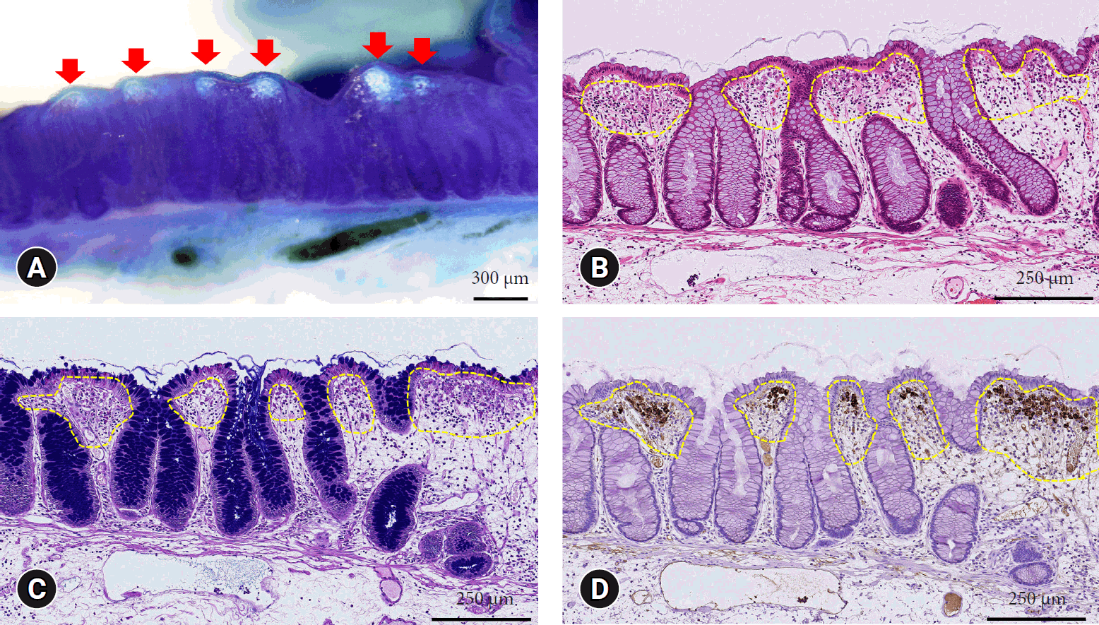
Fig. 7.
Changes in endoscopically resected tumors with white spots (WS) over time. (A) Colonoscopy (CS) of endoscopic mucosal resection: The lesion was resected with sufficient endoscopic margins; however, the WS remained in the mucosa (corresponding to the area surrounded by the dotted line). The pathological diagnosis in this case was pTis, cN0, cM0, and G1; pStage 0 (Union for International Cancer Control 8th edition), and both the horizontal and vertical margins were negative. No lymphovascular invasion was observed, and the patient was cured using endoscopic resection. (B) CS at 1.5 years after treatment: The resection area was scarred (corresponding to the area surrounded by the dotted line), and the WS had disappeared.
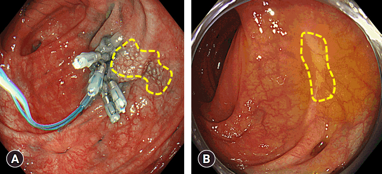
Table 1.
Clinical characteristics of patients with white spots-positive and -negative lesion (n=3,869)
| Characteristic | WS-positive | WS-negative | p-value |
|---|---|---|---|
| Lesions | 358 | 3,511 | |
| Sex | <0.001a) | ||
| Male | 230 (64.2) | 2,566 (73.1) | |
| Female | 128 (35.8) | 945 (26.9) | |
| Age (yr) | 68 (60–74) | 71 (64–76) | <0.001b) |
| Macroscopic type | <0.001a) | ||
| 0-Is, Isp, and Ip | 210 (58.7) | 3094 (88.1) | |
| 0-IIa,IIb, IIc, and IIa+IIc | 43 (12.0) | 401 (11.4) | |
| Type 1–5: advanced cancer | 105 (29.3) | 16 (0.5) | |
| Tumor location | <0.001a) | ||
| Cecum | 3 (0.8) | 269 (7.7) | |
| Ascending colon | 34 (9.5) | 786 (22.4) | |
| Transverse colon | 45 (12.6) | 886 (25.2) | |
| Descending colon | 20 (5.6) | 365 (10.4) | |
| Sigmoid colon | 156 (43.6) | 912 (26.0) | |
| Rectum | 100 (27.9) | 293 (8.3) |
Table 2.
Summary of lesions that were endoscopically resected and evaluated (n=759)
| WS-positive | WS-negative | p-value | |
|---|---|---|---|
| Lesions | 189 | 570 | |
| Sex | 0.15a) | ||
| Male | 69 (36.5) | 176 (30.9) | |
| Female | 120 (63.5) | 394 (69.1) | |
| Age (yr) | 69 (61.5–75) | 70 (62–76) | 0.08b) |
| Macroscopic type | 0.08a) | ||
| 0-Is, Isp, and Ip | 147 (77.8) | 407 (71.4) | |
| 0-IIa, IIb, IIc, and IIa+IIc | 42 (22.2) | 163 (28.6) | |
| Histological type | <0.001a) | ||
| Low-grade adenoma | 121 (64.0) | 475 (83.3) | |
| High-grade adenoma | 11 (5.8) | 25 (4.4) | |
| Well differentiated tubular adenocarcinoma | 43 (22.8) | 57 (10.0) | |
| Moderately differentiated tubular adenocarcinoma | 14 (7.4) | 13 (2.3) | |
| Tumor diameter (mm) | 12 (9.5–18) | 7 (5–10) | <0.001b) |
| Carcinoma depth | <0.05a) | ||
| Intramucosal | 39 (68.4) | 60 (85.7) | |
| Submucosal | 18 (31.6) | 10 (14.3) |
Table 3.
Percentage of carcinoma calculated based on tumor size (n=759)
| WS-positive group | WS-negative group | Total (n) | p-valuea) | |
|---|---|---|---|---|
| φ<5 mm | 3.1% (1/32 lesions)b) | 2.9% (4/136 lesions)b) | 168 | 0.66 |
| 5 mm≤φ<10 mm | 16.3% (13/80 lesions)b) | 8.0% (21/262 lesions)b) | 342 | <0.05 |
| 10 mm≤φ | 44.2% (34/77 lesions)b) | 33.7% (58/172 lesions)b) | 249 | 0.15 |


 PDF
PDF Citation
 Print
Print


 XML Download