Endoscopic ultrasound (EUS) through-the-needle biopsy (EUS-TTNB) has been reported as a tissue sampling method for pancreatic cystic lesions (PCLs).1,2 The diagnostic yield of this method is higher than that of EUS-guided fine needle aspiration (EUS-FNA).3 Particularly, its utility has been reported for PCLs such as intraductal papillary mucinous neoplasm (IPMN) and mucinous cystic neoplasm. However, data on solid tumors with cystic degeneration are limited. Herein, we report a case in which EUS-TTNB was useful for the preoperative histological diagnosis of a pancreatic neuroendocrine neoplasm (PNEN) with a cystic component.
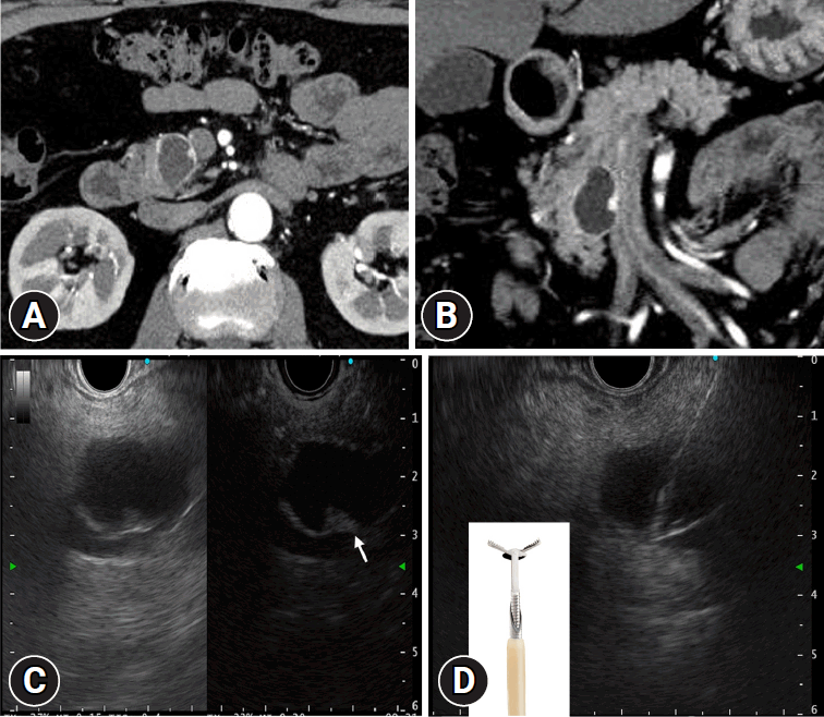
A 61-year-old man was referred to our hospital for further management of a cystic lesion in the pancreatic head. Contrast-enhanced computed tomography showed a 2-cm cystic lesion with enhancement effects on the cyst wall and a 4-mm hypervascular nodule (Fig. 1A, B). EUS revealed an anechoic cyst with a 4-mm nodule on the cyst wall, which showed early enhancement with Sonazoid contrast (GE Healthcare) (Fig. 1C). Considering its hypervascular nature, we suspected a cystic PNEN. However, the differential diagnoses included IPMN with mural nodules, pancreatic pseudocysts, and cystic degeneration of other solid tumors. Therefore, EUS-TTNB was performed to identify the lesion. The pancreatic cyst was punctured using a 19-gauge EUS-FNA needle (EZ Shot3; Olympus). Following the puncture, the stylet was withdrawn and 0.75-mm micro biopsy forceps (Moray Micro forceps; STERIS) were introduced through the FNA needle. Targeted biopsy of the solid nodule was performed thrice (Fig. 1D). The biopsy led to a diagnosis of PNEN (Fig. 2A, B), and pancreaticoduodenectomy was performed. The resected specimens showed densely proliferating small tumor cells in the cystic wall, confirming the diagnosis of PNEN (Fig. 2C, D). To the best of our knowledge, reports on EUS-TTNB for PNEN with cystic degeneration are few, and this case highlights its utility in preoperative diagnosis. We obtained approval of an informed consent waiver from the Institutional Ethics Committee of the Shizuoka Cancer Center (approval number: J2023-253-2023-1-3).
REFERENCES
1. Nakai Y, Isayama H, Chang KJ, et al. A pilot study of EUS-guided through-the-needle forceps biopsy (with video). Gastrointest Endosc. 2016; 84:158–162.
2. Barresi L, Crinò SF, Fabbri C, et al. Endoscopic ultrasound-through-the-needle biopsy in pancreatic cystic lesions: a multicenter study. Dig Endosc. 2018; 30:760–770.
3. Yang D, Trindade AJ, Yachimski P, et al. Histologic analysis of endoscopic ultrasound-guided through the needle microforceps biopsies accurately identifies mucinous pancreas cysts. Clin Gastroenterol Hepatol. 2019; 17:1587–1596.
Fig. 1.
(A, B) Axial and coronal contrast-enhanced computed tomography scans reveal a 20-mm cystic lesion in the head of the pancreas during the arterial phase, with contrast enhancement observed in the cyst wall. A 4-mm nodule with strong contrast enhancement is also detected. (C) Contrast-enhanced endoscopic ultrasound using Sonazoid reveals contrast (GE Healthcare) enhancement in the cystic wall and nodule (arrow). (D) After puncturing with a 19-gauge fine needle aspiration needle, a biopsy is performed using 0.75-mm micro biopsy forceps (Moray Micro forceps; STERIS).

Fig. 2.
(A) Biopsy specimen shows that tumor cells with round nuclei proliferated in a trabecular or alveolar pattern (hematoxylin & eosin [H&E] stain, ×10). Immunohistochemical staining is positive for chromogranin A, leading to the diagnosis of pancreatic neuroendocrine neoplasm. (B) In the loupe image of the resected specimen, a cyst with a thin capsule is observed. (C) In the magnified image of the red-framed area of the image (B) where the biopsy specimen is taken, densely proliferating tumor cells are observed in the cyst wall (H&E stain, ×100). (D) In the loupe image of immunostaining with chromogranin A, positivity is observed in the cyst wall, leading to the diagnosis of cystic degeneration of pancreatic neuroendocrine neoplasm.


 PDF
PDF Citation
 Print
Print


 XML Download