Journal List > Clin Endosc > v.57(5) > 1516088532

AbiMansour, Jaruvongvanich, Velaga, Law, Storm, Topazian, Levy, Alexander, Vargas, Bofill-Garica, Martin, Petersen, Abu Dayyeh, and Chandrasekhara: Coaxial plastic stent placement within lumen-apposing metal stents for the management of pancreatic fluid collections: a systemic review and meta-analysis

Abstract

Background/Aims:

Coaxial placement of double pigtail plastic stents (DPPS) through lumen-apposing metal stents (LAMSs) is commonly performed to reduce the risk of LAMS obstruction, bleeding, and stent migration when used for the drainage of pancreatic fluid collections (PFCs). A systematic review and meta-analysis were performed to compare the outcomes of LAMS alone and LAMS with coaxial DPPS placement in the management of PFCs.

Methods:

A systematic review was conducted to identify studies comparing LAMS and LAMS/DPPS for PFC drainage. Primary outcomes included the rate of clinical success, overall adverse events (AEs), bleeding, infection, occlusion, and stent migration. The pooled effect size was summarized using a random-effects model and compared between LAMS and LAMS/DPPS by calculating odds ratios (ORs).

Results:

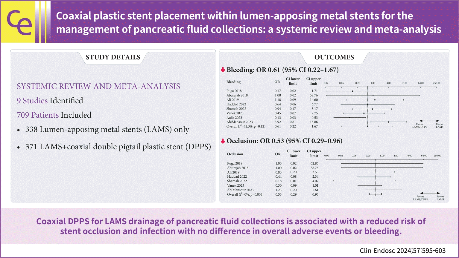
Nine studies involving 709 patients were identified (338 on LAMS and 371 on LAMS/DPPS). LAMS/DPPS was associated with a reduced risk of stent obstruction (OR, 0.59; p=0.004) and infection (OR, 0.55; p=0.001). No significant differences were observed in clinical success (OR, 0.96; p=0.440), overall AEs (OR, 0.57; p=0.060), bleeding (OR, 0.61; p=0.120), or stent migration (OR, 1.03; p=0.480).

Conclusions:

Coaxial DPPS for LAMS drainage of PFCs is associated with a reduced risk of stent occlusion and infection; however, no difference was observed in the overall AE rates or bleeding.

Graphical abstract

INTRODUCTION

Inflammatory pancreatic fluid collections (PFCs) result from disruption of the pancreatic duct and can occur as a sequela of acute or chronic pancreatitis. The majority of PFCs resolve spontaneously; however, intervention may be required in case they become symptomatic, infected, or develop local complications.1 Minimally-invasive approaches, including endoscopic and percutaneous drainage, have replaced surgery as the preferred first-line interventions.2-5 An endoscopic step-up paradigm has been associated with high rates of clinical success, lower rates of re-intervention, and shorter duration of hospital stay, establishing it as the primary drainage modality in facilities with access to endoscopic expertise.6-9
Advancement in endoscopic ultrasound (EUS) technology has enabled real-time assessment and ultrasound-guided formation of a cystogastrostomy or enterostomy.10 Lumen-apposing metal stents (LAMSs) with or without an integrated electrocautery-enhanced delivery system represent the most recent development designed for PFC drainage with interventional EUS.11 The stents available in the United States (AXIOS Stent and Electrocautery Enhanced Delivery System; Boston Scientific Corporation) are United States Food and Drug Administration-cleared for drainage of pseudocysts and walled-off necrosis adherent to the bowel wall.12
In the initial encounters with LAMS, effective drainage of PFCs was observed13,14; however, the placement was associated with an increased risk of bleeding within the cavity and pseudoaneurysm formation.15 Recent data suggests that bleeding risk is independent of stent type; however, it remains a major clinical concern, alongside adverse events (AEs) related to stent migration, stent obstruction, and infection of the collection. This underscores the importance of close patient monitoring.16 Placement of a coaxial double pigtail plastic stent (DPPS) through the LAMS is believed to minimize these risks by limiting damage to the contralateral cavity wall after the collection has decompressed. Furthermore, coaxial DPPS may decrease the risk of LAMS migration and PFC infection by providing an anchor and deeper drainage into the collection, while maintaining patency through the LAMS. However, this practice has not been studied in large patient cohorts that are sufficiently powerful for detecting noticeable differences in event rates. Therefore, this study aimed to perform a systematic review and meta-analysis to compare the clinical efficacy and safety of LAMS with or without coaxial DPPS for the treatment of PFCs.

METHODS

Data sources and search strategies

A comprehensive search of several databases from inception to September 2023 for relevant English-language publications was conducted. These databases included the Cochrane Central Register of Controlled Trials, Cochrane Database of Systematic Reviews, Embase, Ovid, Medline, and Scopus. Manual searches of relevant journals and abstracts on conference proceedings were also conducted. The search strategy was designed and conducted by an experienced librarian (L.P.) with input from the principal investigator (V.C.). Supplementary Material 1 outlines the search strategy.

Eligible studies for meta-analysis

Eligible studies meeting all the following criteria were included in this meta-analysis: (1) only studies comparing LAMS and DPPS in EUS-guided drainage for PFC, including the pancreatic pseudocyst and walled-off necrosis; and (2) studies including data and details for at least technical success and clinical success. The exclusion criteria were as follows: (1) articles published in languages other than English; (2) case reports, reviews, guidelines, or letters; (3) studies without adequate outcomes; and (4) non-human studies. Two authors (JA and SV) independently reviewed and evaluated the eligibility of the retrieved articles. Differences in the study eligibility were resolved by discussion with a third individual (VJ) until a consensus was reached. The quality of the included studies and risk of bias were evaluated using an assessment tool established by the National Institute of Health for observational studies17 and the Revised Cochrane risk-of-bias tool for randomized studies (RoB2).18

Statistical analysis

Success rates and AEs were calculated as proportions in each study. The pooled effect size and 95% confidence intervals (CIs) were estimated using a random-effects model with the DerSimonian and Laird approach. The between-study heterogeneity of effect size estimates across studies was quantified using the Q statistic and I2. The heterogeneity of the effect size estimates across these studies was quantified using the Q statistic, its p-value, and I2 (p<0.10 was considered significant). A value of I2 of 0% to 50% indicated insignificant heterogeneity and 51% to 75% indicated significant heterogeneity. The use of funnel plots was deemed inappropriate given that the power of the test was too low to distinguish chance from actual funnel plot asymmetry. All analyses were performed using Open Meta Analyst software (CEBM; Brown University). Statistical significance was set at p<0.05.

RESULTS

In total, 1221 potentially relevant articles were identified from the outlined search strategy, with 300 duplicate records removed prior to screening and 886 excluded after abstract and title review (Supplementary Fig. 1). The nine identified studies reported the outcomes of 709 patients in total, including 338 patients who underwent LAMS only and 371 who underwent LAMS/DPPS (Table 1).19-27 Details of the stent types used are outlined in Supplementary Table 1.19-27 The studies were evaluated for the risk of bias, with concerns in observational studies largely driven by small sample sizes and a lack of blinding (Supplementary Table 2).19-27 The single randomized controlled trial displayed some concerns about bias related to a lack of blinding; however, the overall risk of bias was low (Supplementary Fig. 2).
Clinical success (Fig. 1A) and technical success (Fig. 1B) were not significantly different between the two cohorts with a pooled odds ratio (OR) of 0.96 (95% CI, 0.48–1.89; I2=17.7%; p=0.440) and 1.08 (95% CI, 0.59–1.96; I2=0%; p=0.390), respectively (Table 2). No statistically significant difference was observed in the overall AE rate (OR, 0.57; 95% CI, 0.25–1.29; I2=56.6%; p=0.060) (Fig. 1C). The overall incidence of stent occlusion (Fig. 1D) and infection of the collection (Fig. 1E) was lower in patients who underwent coaxial DPPS placement with ORs of 0.53 (95% CI, 0.29–0.96; I2=0%; p=0.004) for stent occlusion and 0.55 (95% CI, 0.35–0.84; I2=0%; p=0.001) for infection compared to the patients who underwent LAMS only. There was no significant difference in rates of bleeding (OR, 0.61; 95% CI, 0.22–1.67; I2=42.3%; p=0.120) (Fig. 1F) or stent migration (OR, 1.03; 95% CI, 0.36–2.90; I2=0%; p=0.480) (Fig. 1G) between the two groups. No significant heterogeneity was noted in the outcomes of interest (I2<50%) except for overall AEs which showed significant heterogeneity among the included studies (I2=56.6%).

DISCUSSION

Several technical modifications to PFC drainage have been described to improve outcomes and limit AEs, including multimodal therapy28 and multi-gated drainage.29 However, the rationale for coaxial DPPS placement to mitigate AEs remains predominantly theoretical owing to the limited rigorous data available, and the technique has not been described in initial prospective studies.30,31 Placement of a coaxial DPPS likely evolved from initial experiences with self-expanding metal stents for the drainage of PFCs.32 LAMS, on the other hand, have large bilateral flanges to aid with tissue apposition and prevent migration, which can obviate the requirement for coaxial stent placement.5 However, the potential benefit of a coaxial DPPS extends beyond mere anchoring, as the soft and rounded ends provide a level of protection after the collection collapses. Furthermore, coaxial DPPS can preserve patency, as the inner channel of the LAMS becomes occluded by solid necrosis or food debris, and maintain a potential space within a cavity, especially in case of rapid decompression of the collection. The latter is particularly helpful in situations where a patient is suspected of having a disconnected pancreatic duct, for which management entails long-term transmural DPPS drainage.33-35
Data on the use of coaxial plastic stenting are largely limited to single-center retrospective studies, as highlighted in the systemic database review. A previously performed meta-analysis included 460 patients and suggested no difference in clinical outcomes of interest.36 However, our study improves on prior reports with an updated and more robust cohort of 709 patients and demonstrates lower rates of occlusion and infection with the use of coaxially-placed DPPS for the management of PFC. This finding is clinically plausible considering that impaired drainage resulting from stent obstruction is a mechanism for the onset of collection infection. It aligns with the findings of the only randomized trial on the subject, which observed lower rates of LAMS occlusion in patients who underwent coaxial DPPS placement, albeit no difference in infection was seen.25 Despite a lower incidence of obstruction and infection, no difference was noted in the cumulative rate of overall AEs. This may be due to the varying definitions of AE used in the included studies, and the overall AE rate was the only outcome of interest that exhibited significant heterogeneity, which limited the interpretability.
Notably, no difference was observed in the incidence of bleeding between groups. Initial single-center studies investigating the use of LAMS suggested an increased risk of bleeding-related AEs and the need for unplanned intervention, with an incidence of 2.5% to 25%.15,16,37 A theorized benefit of coaxial DPPS placement is that the soft and rounded edges of the plastic stent limit erosion into the nearby vasculature compared to the metal flanges of LAMS. The risk of bleeding may have been overestimated in earlier cohorts in which LAMS was routinely maintained until clinical resolution. Because experience with LAMS and related AE, such as bleeding, increased over time, prompt LAMS removal after PFC resolution was prioritized. Therefore, modern practices focusing on timely LAMS removal may blunt any protective benefit from bleeding with coaxial DPPS placement.
Downsides of DPPS placement include the risk of migration of the DPPS itself, as well as added cost, time, and the necessity to remove/replace the stent to access the fluid cavity. Smaller caliber plastic stents may be more prone to migration than larger ones (e.g., 5 Fr versus 10 Fr). No difference in stent migration was observed in this study, although the definitions for stent migration varied from incidental migration of plastic stents to symptomatic LAMS migration, limiting the external validity of this finding. While the risk of plastic stent migration must be considered, plastic stent migration did not seem to negatively impact patient outcomes in one study that tracked incidental migration.19
The main limitation of this meta-analysis is the quality of the included studies. Most studies have not protocolized follow-ups, with practice variations between individual centers and endoscopists. This included the timing of repeat clinical evaluation, imaging, and necrosectomy. The definitions of each outcome of interest varied among studies that introduced heterogeneity, particularly for reporting overall AEs. However, comparing the included cohorts is still beneficial, because heterogeneity is likely to exist in both arms of each study. Notably, the incidence of AEs may be related to other fluid collection variables, including size, presence of infection, and paracolic extension, which were not accounted for in this analysis, particularly in the absence of patient-level data.38 Lastly, publication bias was not formally evaluated, considering that a funnel plot would be inadequately powered to provide useful conclusions. Several of the included studies did not report any differences in the outcomes, which is notable because publication bias generally introduces bias against the null hypothesis.

CONCLUSIONS

In summary, this meta-analysis demonstrated that coaxial DPPS placement with LAMS for PFC drainage is associated with a reduced risk of LAMS occlusion and PFC infection. However, no differences in the overall AEs, or more specifically, bleeding, were observed. This study provides key information for endoscopists in the absence of robust randomized data or technical guidelines. Endoscopists should consider the placement of a coaxial DPPS to reduce the risk of LAMS occlusion and infection while also considering patient anatomy, imaging, and other risk factors.

Supplementary Material

Supplementary Material 1. Search strategy.
ce-2023-297-Supplementary-Material-1.pdf
Supplementary Fig. 1. PRISMA 2020 flow diagram for new systematic reviews which included searches of databases, registers and other sources.
ce-2023-297-Supplementary-Fig-1.pdf
Supplementary Fig. 2. Bias assessment of randomized trial.
ce-2023-297-Supplementary-Fig-2.pdf
Supplementary Table 1. Details on stent type in included studies.
ce-2023-297-Supplementary-Table-1.pdf
Supplementary Table 2. Bias assessment of observational cohorts.
ce-2023-297-Supplementary-Table-2.pdf
Supplementary materials related to this article can be found online at https://doi.org/10.5946/ce.2023.297.

Notes

Ethical Statements

Not applicable.

Conflicts of Interest

Andrew C. Storm is a consultant for Apollo Endosurgery and received research support from Apollo Endosurgery and Boston Scientific. Ryan Law is a consultant for ConMed and Medtronic, and receives royalties from UpToDate. Bret T. Petersen is a consultant for Olympus America and an investigator for Boston Scientific and Ambu. Barham K. Abu Dayyeh is a consultant for Endogenex, Endo-TAGSS, Metamodix, and BFKW; a consultant and received grant/research support from USGI, Apollo Endosurgery, Medtronic, Spatz Medical, EndoGastric Solutions, Aspire Bariatrics, and Boston Scientific; speaker roles with Olympus and Johnson and Johnson; and received research support from Cairn Diagnostics. Vinay Chandrasekhara is a consultant for Covidien LP and Boston Scientific and is a shareholder in Nevakar Corporation. The other authors have no potential conflicts of interest.

Funding

None.

Author Contributions

Conceptualization: VC; Data curation: JA, SV; Formal analysis: VJ; Investigation: VJ, SV, RL, ACS, MT, MJL, RA, EJV, ABG, JAM, BTP, BKAD; Methodology: VJ; Supervision: VC; Writing–original draft: JA; Writing–review & editing: JA, RL, ACS, MT, MJL, RA, EJV, ABG, JAM, BTP, BKAD, VC.

REFERENCES

1. Baron TH, DiMaio CJ, Wang AY, et al. American Gastroenterological Association clinical practice update: management of pancreatic necrosis. Gastroenterology. 2020; 158:67–75.
2. van Santvoort HC, Besselink MG, Bakker OJ, et al. A step-up approach or open necrosectomy for necrotizing pancreatitis. N Engl J Med. 2010; 362:1491–1502.
3. van Brunschot S, van Grinsven J, van Santvoort HC, et al. Endoscopic or surgical step-up approach for infected necrotising pancreatitis: a multicentre randomised trial. Lancet. 2018; 391:51–58.
4. Hollemans RA, Bakker OJ, Boermeester MA, et al. Superiority of step-up approach vs open necrosectomy in long-term follow-up of patients with necrotizing pancreatitis. Gastroenterology. 2019; 156:1016–1026.
5. ASGE Standards of Practice Committee, Muthusamy VR, Chandrasekhara V, et al. The role of endoscopy in the diagnosis and treatment of inflammatory pancreatic fluid collections. Gastrointest Endosc. 2016; 83:481–488.
6. Keane MG, Sze SF, Cieplik N, et al. Endoscopic versus percutaneous drainage of symptomatic pancreatic fluid collections: a 14-year experience from a tertiary hepatobiliary centre. Surg Endosc. 2016; 30:3730–3740.
7. Khan MA, Hammad T, Khan Z, et al. Endoscopic versus percutaneous management for symptomatic pancreatic fluid collections: a systematic review and meta-analysis. Endosc Int Open. 2018; 6:E474–E483.
8. Hookey LC, Debroux S, Delhaye M, et al. Endoscopic drainage of pancreatic-fluid collections in 116 patients: a comparison of etiologies, drainage techniques, and outcomes. Gastrointest Endosc. 2006; 63:635–643.
9. Chandrasekhara V, Elhanafi S, Storm AC, et al. Predicting the need for step-up therapy after EUS-guided drainage of pancreatic fluid collections with lumen-apposing metal stents. Clin Gastroenterol Hepatol. 2021; 19:2192–2198.
10. Park DH, Lee SS, Moon SH, et al. Endoscopic ultrasound-guided versus conventional transmural drainage for pancreatic pseudocysts: a prospective randomized trial. Endoscopy. 2009; 41:842–848.
11. Siddiqui AA, Adler DG, Nieto J, et al. EUS-guided drainage of peripancreatic fluid collections and necrosis by using a novel lumen-apposing stent: a large retrospective, multicenter U.S. experience (with videos). Gastrointest Endosc. 2016; 83:699–707.
12. U.S. Food and Drug Administration, Center for Drug Evaluation and Research. AXIOS Stent with Delivery System 510(k) K153088. 20998/S007 approval letter; 2015. [cited 2023 Oct 1]. Available from: https://www.accessdata.fda.gov/cdrh_docs/pdf15/K153088.pdf.
13. Shah RJ, Shah JN, Waxman I, et al. Safety and efficacy of endoscopic ultrasound-guided drainage of pancreatic fluid collections with lumen-apposing covered self-expanding metal stents. Clin Gastroenterol Hepatol. 2015; 13:747–752.
14. Rinninella E, Kunda R, Dollhopf M, et al. EUS-guided drainage of pancreatic fluid collections using a novel lumen-apposing metal stent on an electrocautery-enhanced delivery system: a large retrospective study (with video). Gastrointest Endosc. 2015; 82:1039–1046.
15. Bang JY, Hasan M, Navaneethan U, et al. Lumen-apposing metal stents (LAMS) for pancreatic fluid collection (PFC) drainage: may not be business as usual. Gut. 2017; 66:2054–2056.
16. Chandrasekhara V, Barthet M, Devière J, et al. Safety and efficacy of lumen-apposing metal stents versus plastic stents to treat walled-off pancreatic necrosis: systematic review and meta-analysis. Endosc Int Open. 2020; 8:E1639–E1653.
17. Goff D, Lloyd-Jones D; Risk Assessment Working Group. Assessing cardiovascular risk: systematic evidence review from the Risk Assessment Work Group. United States Department of Health and Human Services/National Institutes of Health/National Heart, Lung, and Blood Institute;2013. [cited 2023 Oct 1]. Available from: https://www.nhlbi.nih.gov/health-topics/assessing-cardiovascular-risk.
18. Higgins JP, Altman DG, Gøtzsche PC, et al. The Cochrane Collaboration's tool for assessing risk of bias in randomised trials. BMJ. 2011; 343:d5928.
19. AbiMansour JP, Jaruvongvanich V, Velaga S, et al. Lumen-apposing metal stents with or without coaxial plastic stent placement for the management of pancreatic fluid collections. Gastrointest Endosc. 2024; 99:104–107.
20. Puga M, Consiglieri CF, Busquets J, et al. Safety of lumen-apposing stent with or without coaxial plastic stent for endoscopic ultrasound-guided drainage of pancreatic fluid collections: a retrospective study. Endoscopy. 2018; 50:1022–1026.
21. Aburajab M, Smith Z, Khan A, et al. Safety and efficacy of lumen-apposing metal stents with and without simultaneous double-pigtail plastic stents for draining pancreatic pseudocyst. Gastrointest Endosc. 2018; 87:1248–1255.
22. Ali SE, Benrajab K, Mardini H, et al. Anchoring lumen-apposing metal stent with coaxial plastic stent for endoscopic ultrasound-guided drainage of pancreatic fluid collections: any benefit? Ann Gastroenterol. 2019; 32:620–625.
23. Haddad JD, Tielleman T, Fuller A, et al. Safety and efficacy of lumen-apposing metal stents with and without coaxial plastic stents for pancreatic fluid collections. Tech Innov Gastrointest Endosc. 2023; 25:113–118.
24. Shamah SP, Sahakian AB, Chapman CG, et al. Double pigtail stent placement as an adjunct to lumen-apposing metal stentsfor drainage of pancreatic fluid collections may not affect outcomes: a multicenter experience. Endosc Ultrasound. 2022; 11:53–58.
25. Vanek P, Falt P, Vitek P, et al. EUS-guided transluminal drainage using lumen-apposing metal stents with or without coaxial plastic stents for treatment of walled-off necrotizing pancreatitis: a prospective bicentric randomized controlled trial. Gastrointest Endosc. 2023; 97:1070–1080.
26. Aujla P, Aleem A, Subramanian SK, et al. Stent within a stent: when lumen apposing metal stent meets its match in coaxial double pigtail stent. Gastrointest Endosc. 2023; 97:AB920.
27. Perez Estrada C, Adalberto R. Efficacy and safety of lumen-apposing metal stents in endoscopic ultrasound-guided drainage of abdominal fluid collections and the influence of inserting a coaxial double pigtail plastic stent. United Eur Gastroenterol J. 2022; 10(Suppl 8):984–985.
28. Gluck M, Ross A, Irani S, et al. Dual modality drainage for symptomatic walled-off pancreatic necrosis reduces length of hospitalization, radiological procedures, and number of endoscopies compared to standard percutaneous drainage. J Gastrointest Surg. 2012; 16:248–257.
29. Varadarajulu S, Phadnis MA, Christein JD, et al. Multiple transluminal gateway technique for EUS-guided drainage of symptomatic walled-off pancreatic necrosis. Gastrointest Endosc. 2011; 74:74–80.
30. Bakker OJ, van Santvoort HC, van Brunschot S, et al. Endoscopic transgastric vs surgical necrosectomy for infected necrotizing pancreatitis: a randomized trial. JAMA. 2012; 307:1053–1061.
31. Varadarajulu S, Bang JY, Sutton BS, et al. Equal efficacy of endoscopic and surgical cystogastrostomy for pancreatic pseudocyst drainage in a randomized trial. Gastroenterology. 2013; 145:583–590.
32. Penn DE, Draganov PV, Wagh MS, et al. Prospective evaluation of the use of fully covered self-expanding metal stents for EUS-guided transmural drainage of pancreatic pseudocysts. Gastrointest Endosc. 2012; 76:679–684.
33. Bang JY, Mel Wilcox C, Arnoletti JP, et al. Importance of disconnected pancreatic duct syndrome in recurrence of pancreatic fluid collections initially drained using lumen-apposing metal stents. Clin Gastroenterol Hepatol. 2021; 19:1275–1281.
34. van Dijk SM, Timmerhuis HC, Verdonk RC, et al. Treatment of disrupted and disconnected pancreatic duct in necrotizing pancreatitis: a systematic review and meta-analysis. Pancreatology. 2019; 19:905–915.
35. Wang L, Elhanafi S, Storm AC, et al. Impact of disconnected pancreatic duct syndrome on endoscopic ultrasound-guided drainage of pancreatic fluid collections. Endoscopy. 2021; 53:603–610.
36. Giri S, Harindranath S, Afzalpurkar S, et al. Does a coaxial double pigtail stent reduce adverse events after lumen apposing metal stent placement for pancreatic fluid collections? A systematic review and meta-analysis. Ther Adv Gastrointest Endosc. 2023; 16:26317745231199364.
37. Lang GD, Fritz C, Bhat T, et al. EUS-guided drainage of peripancreatic fluid collections with lumen-apposing metal stents and plastic double-pigtail stents: comparison of efficacy and adverse event rates. Gastrointest Endosc. 2018; 87:150–157.
38. Dayyeh BK, Chandrasekhara V, Shah RJ, et al. Combined drainage and protocolized necrosectomy through a coaxial lumen-apposing metal stent for pancreatic walled-off necrosis: a prospective multicenter trial. Ann Surg. 2023; 277:e1072–e1080.
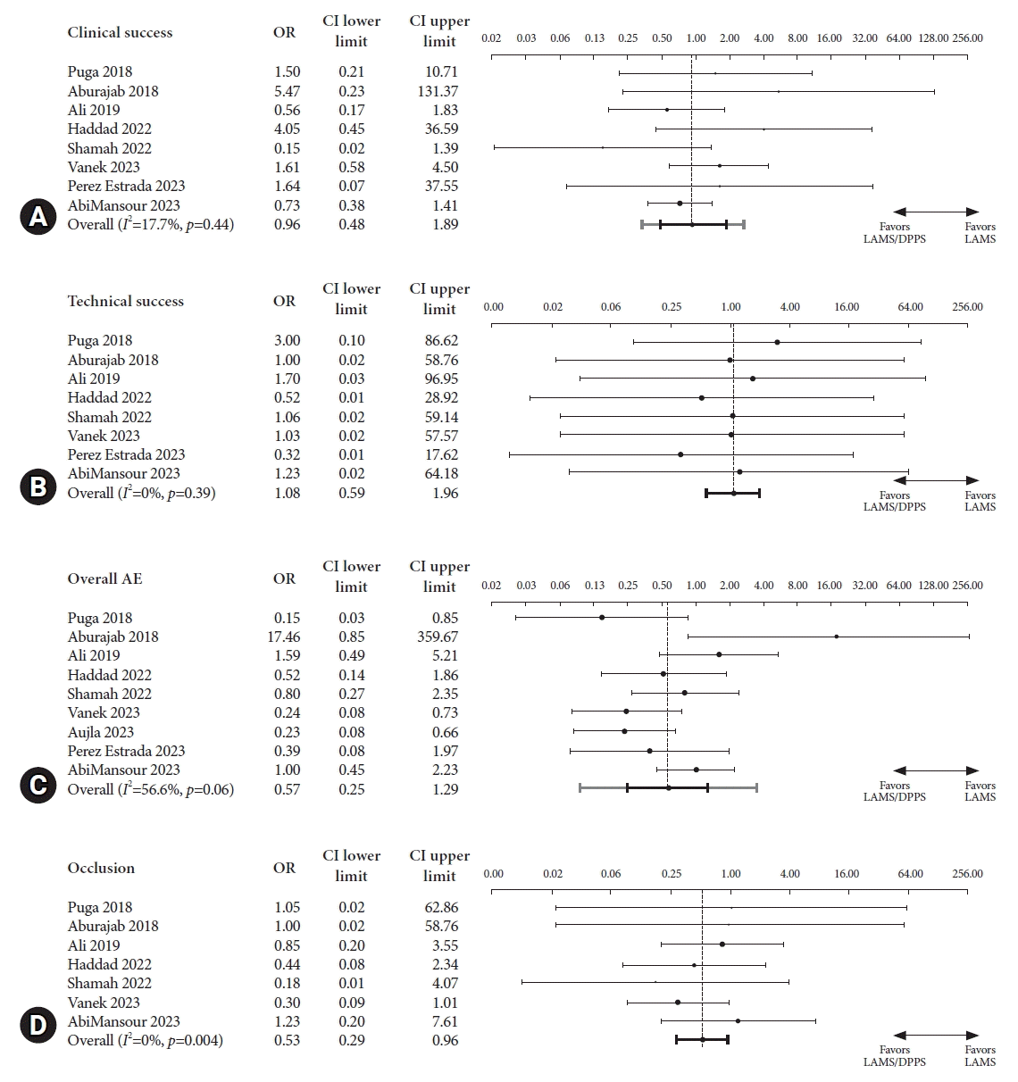
Fig. 1.
Pooled odds ratio (OR) for (A) clinical success, (B) technical success, (C) overall adverse events, (D) occlusion, (E) infection, (F) bleeding, and (G) migration. CI, confidence interval; LAMS, lumen-apposing metal stent; DPPS, double pigtail plastic stent; AE, adverse event.
ce-2023-297f1.tif
ce-2023-297f2.tif
Table 1.
Details of studies identified on systematic review and included in the meta-analysis
Study Year Counrty Total patient (n)
Walled-off necrosis (n)
Clinical success (n)
Overall AE (n)
LAMS LAMS/DPPS LAMS LAMS/DPPS LAMS LAMS/DPPS LAMS LAMS/DPPS
Puga et al.20 2018 Spain 21 20 11 12 18 18 9 2
Aburajab et al.21 2018 USA 23 23 0 0 21 23 0 6
Ali et al.22 2019 USA 21 36 14 29 15 21 6 14
Haddad et al.23 2023 USA 45 23 29 13 38 22 13 4
Shamah et al.24 2022 USA 33 35 6 11 32 29 10 9
Vanek et al.25 2023 USA 33 34 33 34 10 14 17 7
Aujla et al.26 2023 USA 21 80 NR NR NR NR 10 14
Perez Estrada et al.27 2022 Spain 58 18 NR NR 56 18 14 2
AbiMansour et al.19 2024 USA 83 102 62 80 63 71 13 16

AE, adverse event; LAMS, lumen-apposing metal stent; DPPS, double pigtail plastic stent; NR, not reported.

Table 2.
Meta-analysis results with pooled OR for lumen-apposing metal stent with coaxial stent placement compared to lumen-apposing metal stent placement alone
Meta analysis results Pooled OR (95% CI) I2(%)
Clinical success 0.96 (0.48–1.89) 17.7
Overall AE 0.57 (0.25–1.29) 56.6
Bleeding 0.61 (0.22–1.67) 42.3
Migration 1.03 (0.36–2.90) 0
Infection 0.55 (0.35–0.84) 0
Occlusion 0.53 (0.29–0.96) 0

OR, odds ratio; CI, confidence interval; AE, adverse event.

TOOLS
Similar articles