Journal List > Clin Endosc > v.57(5) > 1516088529

Ohno, Fujimori, Kaku, Matsumoto, Murakami, Teramatsu, Ueda, Hijioka, Aso, and Ogawa: Puncture angle on an endoscopic ultrasound image is independently associated with unsuccessful guidewire manipulation of endoscopic ultrasound-guided hepaticogastrostomy: a retrospective study in Japan

Abstract

Background/Aims

Although endoscopic ultrasound-guided hepaticogastrostomy (EUS-HGS) is performed globally, the procedure remains challenging. Guidewire manipulation is the most difficult step, and there are few reports on the factors associated with unsuccessful guidewire manipulation. This study aimed to assess the significance of the puncture angle on EUS images and identify the most effective guidewire rescue method for patients with unsuccessful guidewire manipulation.

Methods

We retrospectively enrolled 115 patients who underwent EUS-HGS between May 2016 and April 2022 at two centers. The puncture angle between the needle and the intrahepatic bile duct was measured through EUS movie records.

Results

Guidewire manipulation was unsuccessful in 28 patients. Receiver operating characteristic (ROC) curves identified an optimal puncture angle cutoff value of 85° (cutoff value, 85°; area under the ROC curve, 0.826; sensitivity, 85.7%; specificity, 81.6%). Multivariate analysis demonstrated that a puncture angle <85° was a significant risk factor for unsuccessful guidewire manipulation (odds ratio, 19.8; 95% confidence interval, 6.42–61.5; p<0.001). Among the 28 unsuccessful cases, 24 patients (85.7%) achieved successful guidewire manipulation using various rescue methods.

Conclusions

The puncture angle observed on EUS is crucial for guidewire manipulation. A puncture angle of <85° was associated with unsuccessful guidewire manipulation.

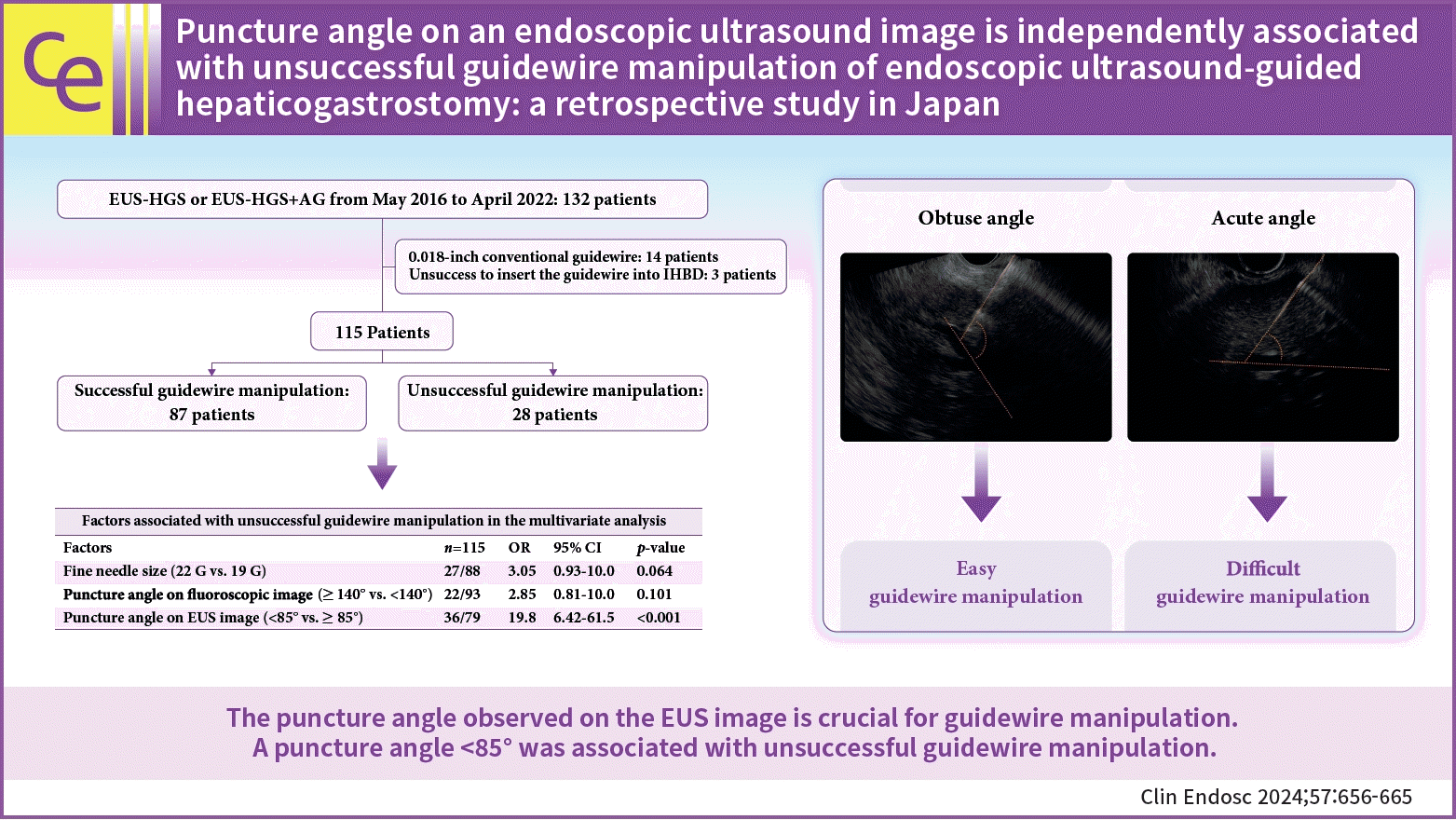
Graphical abstract

INTRODUCTION

Endoscopic ultrasound-guided hepaticogastrostomy (EUS-HGS) is an alternative biliary drainage method to endoscopic retrograde cholangiopancreatography (ERCP).1,2 Recent reports have revealed high technical and clinical success.3-5 However, unlike ERCP, many EUS-HGSs are conducted by endoscopic experts in high-volume centers. Therefore, EUS-HGS procedures are yet to be standardized, and some reports indicate their difficulty and adverse events (AEs).5-7 Generally, the steps for EUS-HGS are as follows: (1) intrahepatic bile duct (IHBD) puncture, (2) cholangiography, (3) guidewire manipulation, (4) tract dilation, and (5) stent placement. Among these procedures, guidewire manipulation is the most challenging.8,9 Although several rescue methods for patients with unsuccessful guidewire manipulation during EUS-HGS have been reported, the optimal rescue method remains uncertain.10-12 Ogura et al.9 previously established that the angle between the fine needle and the scope of the fluoroscopic image is a factor in successful guidewire manipulation. Furthermore, we recently indicated the influence of the angle between the needle and IHBD on fluoroscopic images when assessing the need for tract dilatation.13 However, the puncture angle on the fluoroscopic image was clarified after the puncture of the IHBD and contrast injection. Predicting procedural success prior to puncture is crucial in clinical practice. The puncture line was determined based on the EUS images obtained before the puncture. Therefore, this novel study aimed to evaluate whether the angle between the needle and the IHBD on EUS images is associated with successful guidewire manipulation. Furthermore, reports on the rescue methods used for difficult guidewire manipulation are scarce. Therefore, we investigated the management of patients in whom it is difficult to manipulate the guidewire into the hilar bile duct during EUS-HGS.

METHODS

Patients and study protocol

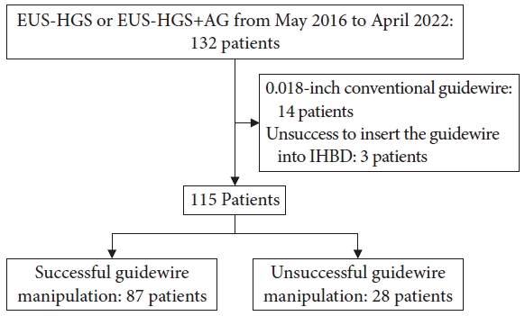
This retrospective study was conducted at Kyushu University Hospital and Kyushu Medical Center using data obtained from 132 patients who underwent EUS-HGS between May 2016 and April 2022. A total of 115 patients were enrolled; 14 patients were excluded owing to poor maneuverability attributed to a 0.018-inch conventional guidewire (NovaGold; Boston Scientific).14,15 Additionally, three patients were excluded because of difficulty in inserting the guidewire into the IHBD (Fig. 1). Comprehensive data from all the patients were retrieved using the medical reporting system. All patients were followed up until study completion or death, with a final follow-up date of September 30, 2022.

Procedures

All patients received antibiotics before the procedure. Sedation was achieved with midazolam and pentazocine. Two experienced endoscopists at each center, skilled in interventional EUS, performed the EUS-HGS procedures. The assistant operators during EUS-HGS were experienced in ERCP procedures (>500) and served as assistant operators in ERCP procedures (>250).
An echoendoscope using a GF-UCT260 (Olympus), EG-580UT (Fujifilm Medical System), or EG-740UT (Fujifilm Medical System), was inserted into the stomach, and the puncture line was determined using EUS and fluoroscopic images. The IHBD was punctured using a fine needle (19 or 22 G) while avoiding the blood vessels. After aspiration of the bile juice, a sufficient amount of contrast medium was administered to confirm the bile duct orientation. A guidewire (19 G fine needle: 0.025-inch VisiGlide2, Olympus; 22 G fine needle, 0.018-inch Fielder; Olympus) was carefully inserted into the bile duct. As previously reported, dilation procedures were performed, if required, using a balloon and/or mechanical dilator (REN; Kaneka or ES dilator; Zeon Medical).13 Finally, plastic stents (Type IT; Gadelius Medical) or metallic stents (partially covered: Niti-S; Taewoong Medical, EGIS; S&G Biotech Inc. or fully covered: HANARO Benefit, Boston Scientific) were placed over the guidewire via the EUS-HGS route. When the EUS-HGS+antegrade (AG) stenting was performed, an uncovered metallic stent (ZILVER; Cook Medical, ZEOSTENT V; ZEON Medical, or BileRush; Piolax Medical Devices) was deployed to the AG route.
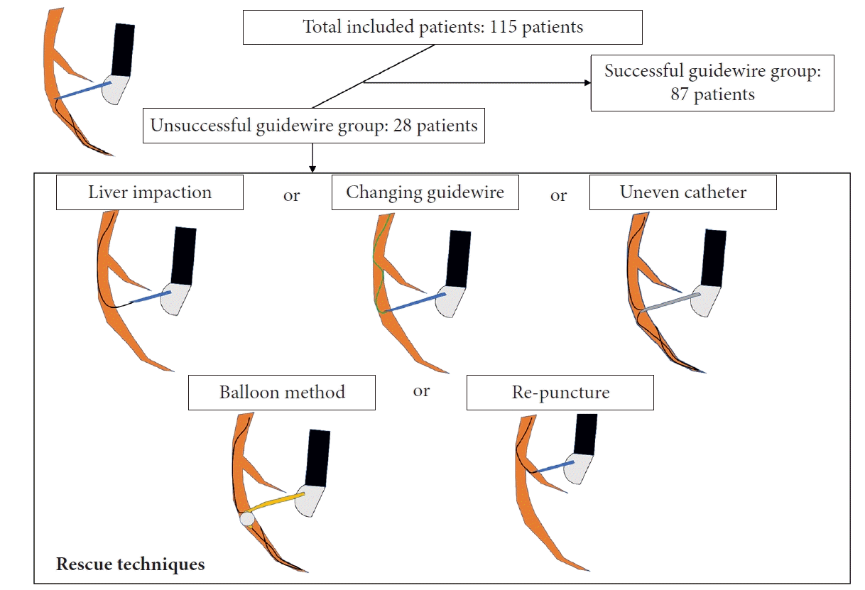
In case of difficulty during guidewire insertion into the hilar bile duct, the following guidewire rescue method was performed: (1) changing to another guidewire (from a 0.025-inch VisiGlide2 or a 0.018-inch Fielder to a 0.025-inch MICHISUJI, Kaneka or Endoselecter, Boston Scientific); (2) liver impaction by pulling the puncture needle to the liver parenchyma and manipulating the guidewire10; (3) uneven catheter method by inserting an uneven double-lumen cannula (Piolax Medical Devices) into the IHBD and manipulating the guidewire from the proximal port11; (4) balloon method by inserting a multi-lumen balloon catheter (Bouncer; Cook Medical) into the IHBD, inflating the balloon in the bile duct, and manipulating the guidewire from the second lumen12; and (5) re-puncturing another IHBD. If any of the five steps failed, the appropriate method was used. The guidewire rescue method was selected at the discretion of the physician (Fig. 2).

Outcomes and definitions

The primary outcome of this study was to assess whether the puncture angle on EUS images was associated with unsuccessful guidewire manipulation. The puncture angle between the fine needle and the IHBD on the EUS image was measured using the EUS video and images (Fig. 3A, C, Supplementary Videos 1, 2). Secondary outcomes included procedure time, AEs, clinical success, technical success, and the most effective guidewire rescue method. We confirmed the type of guidewire rescue method by analyzing fluoroscopic videos, EUS videos, and outcome reports. Procedure time was defined as the insertion and removal of the endoscope. AEs were defined based on the American Society for Gastrointestinal Endoscopy lexicon.16 Clinical success was based on the TOKYO criteria as follows: 50% decrease or normalization of bilirubin level within 2 weeks post-procedure.17 The technical success of EUS-HGS was defined as the success of the stent application, which was different from that for guidewire manipulation, defined as guidewire guidance to the hilum bile duct. However, unsuccessful guidewire manipulation was described as insertion of the first guidewire into the peripheral bile duct alone. Patients in whom any rescue method was employed were included in the unsuccessful group. Factors associated with unsuccessful guidewire manipulation included age, sex, underlying disease, the reason for EUS-HGS, the presence of cholangitis, needle size, puncture site, the diameter of IHBD, the distance of hepatic parenchyma from the liver surface to the puncture point of the IHBD, puncture angle as observed on the fluoroscopic image, and puncture angle as observed in the EUS image.

Statistical analyses

The optimal cutoff values for the diameter of the IHBD, distance of the hepatic parenchyma, puncture angle on the fluoroscopic image, and puncture angle on the EUS image were determined using the area under the receiver operating characteristic (ROC) curve (AUC). Successful and unsuccessful guidewire manipulation groups were compared using paired t-test. Univariate and multivariate logistic analyses were performed to determine factors associated with guidewire manipulation. The significance level was set at p<0.05. JMP 17 (SAS Institute Inc.) and R ver. 4.1.3 (R Core Team, 2022) were used for statistical analyses.

Ethics approval statement

The Institutional Review Boards (IRB) of Kyushu University Hospital (approval number: 22126-00) and Kyushu Medical Center (approval number: 22OR-23) approved this study. The study was conducted in accordance with the principles of the Declaration of Helsinki. The need for informed consent was waived due to the retrospective nature of the study.

RESULTS

Patients

A flowchart of the study is shown in Figure 1. A total of 115 consecutive patients who underwent EUS-HGS were enrolled in this study. The clinical features of the patients are summarized in Table 1. The median age was 71.0 years (range, 36–93 years), and 48 patients (41.7%) were female. Pancreatic cancer was the most common disease (41.7%), followed by bile duct cancer (27.0%). The indications for EUS-HGS were unsuccessful ERCP in 34 (29.6%) patients, surgically altered anatomy in 44 (38.3%), and duodenal obstruction in 37 (32.2%). In 88 patients (76.5 %), a 19 G fine needle was used, and the most common puncture site was the B3 (73.0%). The median diameter of the IHBD was 5.0 mm, the median hepatic parenchyma distance was 2.4 cm, and the median puncture angles on the fluoroscopic and EUS images were 100° and 95°, respectively. AEs occurred in 16 patients and included bile peritonitis (n=7), hemorrhage (n=3), acute cholecystitis (n=2), acute pancreatitis (n=3), and liver abscess (n=1). Guidewire manipulation was unsuccessful in 28 patients (24.3%) who required a rescue method.

Clinical characteristics and outcomes of guidewire manipulation

Table 2 presents the clinical characteristics and outcomes of EUS-HGS in the successful guidewire manipulation group (SGMG) and unsuccessful guidewire manipulation group (UGMG). A 19 G fine needle was used more frequently in the SGMG than in the UGMG (p=0.028). However, the puncture angle on the EUS image was lower in the UGMG than in the SGMG (p<0.001). The technical and clinical success rates of EUS-HGS were significantly higher in SGMG than in UGMG (p=0.001 and p=0.022, respectively). The procedure time for the SGMG was significantly shorter than that for the UGMG (p<0.001). There was no significant difference between the SGMG and UGMG in terms of the rate of AEs (11.5% vs. 21.4%, p=0.204).

Factors associated with unsuccessful guidewire manipulation

The optimal cutoff value of the puncture angle on the EUS image was determined using ROC curves (Fig. 4), resulting in an AUC of 0.826 (95% confidence interval [CI], 0.733–0.920). The sensitivity and specificity of the 85° angle for predicting unsuccessful guidewire manipulation were 85.7% and 81.6%, respectively. The optimal cutoff value of the puncture angle on the fluoroscopic image was 140°. Table 3 presents the results of both univariate and multivariate analyses of the factors associated with unsuccessful guidewire manipulation during EUS-HGS. In the univariate analysis, 22 G fine needle (odds ratio [OR], 2.87; 95% CI, 1.13–7.29; p=0.026), puncture angle ≥140° on the fluoroscopic image (OR, 2.69; 95% CI, 1.00–7.24; p=0.049), and puncture angle <85° on the EUS image (OR, 19.1; 95% CI, 6.56–55.6; p<0.001) were identified as significant factors associated with unsuccessful guidewire manipulation in EUS-HGS. In the multivariate analysis, a puncture angle <85° on the EUS image (OR, 19.8; 95% CI, 6.42–61.5; p<0.001) was the only significant factor associated with unsuccessful guidewire manipulation in EUS-HGS.

Details of the unsuccessful guidewire group

In the UGMG (28 patients), a guidewire was inserted into the hilar bile duct in 24 patients using one of the guidewire rescue methods (Table 4). Notably, the success rates of the changing guidewire method and liver impaction methods were lower than those of the other rescue methods (11.8% and 27.3%, respectively). Of the five cases in which the re-puncture method failed, four resulted in unsuccessful guidewire manipulation, while one case achieved success using the balloon method.

DISCUSSION

Previous studies have indicated that guidewire manipulation is the most difficult step during the EUS-HGS procedure.9,18 A report suggested that successful guidewire manipulation is influenced by the echoendoscope angle evaluated using fluoroscopic imaging.9 However, to our knowledge, no reports have assessed the factors contributing to unsuccessful guidewire manipulation using EUS images. In this novel study, we retrospectively evaluated the influence of the puncture angle between the fine needle and the IHBD on EUS images. Our findings reveal that the puncture angle observed on EUS is an independent factor associated with successful guidewire manipulation during EUS-HGS.
While there was no difference in the AE rate between the SGMG and UGMG groups, SGMG was superior to UGMG in terms of procedure time, technical success, and clinical success. These results imply that successful guidewire manipulation during the first IHBD puncture is pivotal for improving EUS-HGS outcomes. Consequently, identifying factors associated with unsuccessful guidewire manipulation may contribute to the establishment of EUS-HGS procedures.
Our novel finding reveals that a puncture angle <85° between the fine needle and IHBD on the EUS image was independently associated with unsuccessful guidewire manipulation (OR, 19.8; 95% CI, 6.42–61.5; p<0.001). Hence, a better IHBD was selected for successful guidewire manipulation before puncture. In this study, the IHBD diameter and hepatic parenchyma distance were not associated with unsuccessful guidewire manipulation. Oh et al.18 previously showed that IHBD diameter ≤5 mm and hepatic portion length >3 cm were the factors associated with a low technical success rate. However, it is essential to recognize that the outcome of this previous report was not focused on the technical success of guidewire manipulation, but on the overall technical success of EUS-HGS. In our study, the rate of technical success of EUS-HGS was 94.8%, consistent with previous reports (93.7%–96.0%).3-5 In line with our present study, Ogura et al.9 focused on the technical success of guidewire manipulation and reported the importance of echoendoscope angle in EUS-HGS. They found that an angle exceeding 135° between the fine needle and scope was a determining factor for successful guidewire manipulation. However, the angle observed on a fluoroscopic image is typically determined after puncturing the IHBD, whereas the puncture point is determined using EUS imaging. Notably, in our study, the puncture site was not significantly associated with unsuccessful guidewire manipulation. In addition, there was no correlation between the fluoroscopic puncture and EUS puncture angles according to Pearson correlation coefficient analysis (p=0.854). Thus, we considered this to be owing to differences in bile duct runnability. In the cases shown in Figure 3, the angles are both obtuse on the fluoroscopic image (Fig. 3B, D), but the guidewire was easily inserted into the hilar side only in the case shown in Figure 3B. However, in these cases, the angles of the EUS images were significantly different (Fig. 3A, C) from those of the fluoroscopic images. The difference in the influence of the angle between the fluoroscopic and EUS images was possibly due to bile duct runnability. Another reason for the lack of a correlation between the puncture site and unsuccessful guidewire manipulation may be the patient’s background and our EUS-HGS strategy. We selected a B3 puncture because of the risk of esophageal puncture associated with a B2 puncture.19,20 Therefore, the conditions appeared to be more challenging with a B2 puncture in our study. Among the eight patients with B2 puncture and unsuccessful guidewire manipulation in our study, the echoendoscope was directed toward the peripheral bile duct in EUS-HGS due to left lobe swelling in four patients (Fig. 5). Therefore, we concluded that the B2 puncture was not associated with successful guidewire manipulation. However, the number of patients who underwent B2 puncture was limited (n=31), and additional cases should be included for further validation.
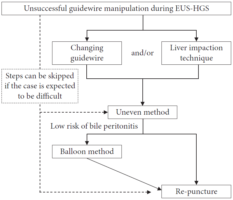
We examined the details of the UGMG and found that the success rates of the individual guidewire rescue methods were not equivalent (Table 4, Supplementary Table 1). Conversely, the low success rate of the guidewire changing method and liver impaction can be attributed to their inability to alter the angle between the puncture line and IHBD (11.8% and 27.3%, respectively). The uneven method exhibited a higher success rate than the guidewire changing or liver impaction methods. This could be attributed to the ability of the double lumen of the uneven catheter to alter the guidewire insertion angle (44.4%). A similar rationale applies to the balloon method. Although the balloon method demonstrated a 100% success rate, only two cases underwent that as the balloon method was selected at the third or fourth step if changing guidewire or liver impaction was difficult (Supplementary Table 1). This was due to the risk of bile peritonitis caused by the balloon catheter diameter (6.6 Fr). The moving-scope technique has also been reported as another method of changing the angle by pushing the scope downwards.21 Matsubara et al.20 reported a moving-scope technique when the guidewire was inserted into the peripheral bile duct. Based on the aforementioned results, we propose a rescue strategy for unsuccessful guidewire manipulation (Fig. 6). However, the optimal guidewire rescue method remains unknown. Guidewire rescue methods should be properly performed in this case. Unsuccessful guidewire manipulation led to technical and clinical success. In fact, in the UGMG group (28 patients), we were also unable to insert the guidewire into the hilar bile duct in four patients despite attempting various rescue methods. Therefore, to improve the technical success rate, selecting the optimal puncture site before performing EUS-HGS is crucial. The optimal puncture site was selected based on the puncture angle on the EUS image before the puncture. Careful manipulation of the endoscope prior to puncture is crucial because the puncture angle on the EUS image can be easily changed by moving the scope and/or elevator lever.
This study has some limitations. First, it was a retrospective study. Second, although the total number of patients in this study was comparatively large (n=115), only a limited number underwent B2 puncture using a 22 G fine needle. Therefore, a multicenter prospective study is necessary to validate these results. Third, accurately measuring the puncture angle during EUS-HGS in real-time can be challenging. However, understanding that the puncture angle on EUS images strongly influences guidewire manipulation can aid in the selection of the most suitable puncture site.
In conclusion, the angle between the fine needle and the IHBD observed on EUS imaging may be an effective tool for preventing unsuccessful guidewire manipulation. Hence, it is crucial to carefully assess the angle on EUS images before puncturing during EUS-HGS.

Supplementary Material

Supplementary Table 1. The details of the rescue methods.
ce-2023-244-Supplementary-Table-1.pdf
Supplementary Video 1. Successful guidewire manipulation into the hilar bile duct with an obtuse angle between the fine needle and intrahepatic bile duct on endoscopic ultrasonography.
Supplementary Video 2. Unsuccessful guidewire manipulation into the hilar bile duct with an acute angle between the fine needle and intrahepatic bile duct on endoscopic ultrasound.
Supplementary materials related to this article can be found online at https://doi.org/10.5946/ce.2023.244.

Notes

Conflicts of Interest

The authors have no potential conflicts of interest.

Funding

This work was supported in part by JSPS KAKENHI (grant number: JP18K09945) and the Grant of The Clinical Research Promotion Foundation (2022).

Author Contributions

Conceptualization: AO, NF, TK; Data curation: AO, NF, TK, KM, MM, KT, KU, MH, AA; Formal analysis: AO, NF, TK; Investigation: AO, NF, TK; Methodology: AO, NF, TK; Project administration: AO, NF, TK; Resources: AO, NF, TK; Software: AO, NF, TK; Supervision: NF, TK, YO; Validation: NF, TK; Visualization: AO; Writing–original draft: AO; Writing–review & editing: all authors.

REFERENCES

1. Lee TH, Choi JH, Park do H, et al. Similar efficacies of endoscopic ultrasound-guided transmural and percutaneous drainage for malignant distal biliary obstruction. Clin Gastroenterol Hepatol. 2016; 14:1011–1019.
2. Sharaiha RZ, Khan MA, Kamal F, et al. Efficacy and safety of EUS-guided biliary drainage in comparison with percutaneous biliary drainage when ERCP fails: a systematic review and meta-analysis. Gastrointest Endosc. 2017; 85:904–914.
3. Artifon EL, Marson FP, Gaidhane M, et al. Hepaticogastrostomy or choledochoduodenostomy for distal malignant biliary obstruction after failed ERCP: is there any difference? Gastrointest Endosc. 2015; 81:950–959.
4. Uemura RS, Khan MA, Otoch JP, et al. EUS-guided choledochoduodenostomy versus hepaticogastrostomy: a systematic review and meta-analysis. J Clin Gastroenterol. 2018; 52:123–130.
5. Wang K, Zhu J, Xing L, et al. Assessment of efficacy and safety of EUS-guided biliary drainage: a systematic review. Gastrointest Endosc. 2016; 83:1218–1227.
6. Khan MA, Akbar A, Baron TH, et al. Endoscopic ultrasound-guided biliary drainage: a systematic review and meta-analysis. Dig Dis Sci. 2016; 61:684–703.
7. Moole H, Bechtold ML, Forcione D, et al. A meta-analysis and systematic review: success of endoscopic ultrasound guided biliary stenting in patients with inoperable malignant biliary strictures and a failed ERCP. Medicine (Baltimore). 2017; 96:e5154.
8. Vila JJ, Pérez-Miranda M, Vazquez-Sequeiros E, et al. Initial experience with EUS-guided cholangiopancreatography for biliary and pancreatic duct drainage: a Spanish national survey. Gastrointest Endosc. 2012; 76:1133–1141.
9. Ogura T, Nishioka N, Ueno S, et al. Effect of echoendoscope angle on success of guidewire manipulation during endoscopic ultrasound-guided hepaticogastrostomy. Endoscopy. 2021; 53:369–375.
10. Ogura T, Masuda D, Takeuchi T, et al. Liver impaction technique to prevent shearing of the guidewire during endoscopic ultrasound-guided hepaticogastrostomy. Endoscopy. 2015; 47(S 01):E583–E584.
11. Kawakami H, Kubota Y, Makiyama H, et al. Uneven double-lumen cannula for rescue guidewire technique in endoscopic ultrasonography-guided hepaticogastrostomy. Endoscopy. 2017; 49:E264–E265.
12. Ohno A, Kaku T, Fujimori N. Balloon guidewire technique during EUS-guided hepaticogastrostomy. Endosc Ultrasound. 2022; 11:330–331.
13. Ohno A, Fujimori N, Kaku T, et al. Feasibility and efficacy of endoscopic ultrasound-guided hepaticogastrostomy without dilation: a propensity score matching analysis. Dig Dis Sci. 2022; 67:5676–5684.
14. Ogura T, Okuda A, Ueno S, et al. Prospective comparison study between 19-gauge needle with .025-inch guidewire and 22-gauge needle with novel .018-inch guidewire during EUS-guided transhepatic biliary drainage (with video). Gastrointest Endosc. 2022; 96:262–268.
15. Hara K, Okuno N, Haba S, et al. How to perform EUS-guided hepaticogastrostomy easier and safer. J Hepatobiliary Pancreat Sci. 2020; 27:563–564.
16. Cotton PB, Eisen GM, Aabakken L, et al. A lexicon for endoscopic adverse events: report of an ASGE workshop. Gastrointest Endosc. 2010; 71:446–454.
17. Isayama H, Hamada T, Yasuda I, et al. TOKYO criteria 2014 for transpapillary biliary stenting. Dig Endosc. 2015; 27:259–264.
18. Oh D, Park DH, Song TJ, et al. Optimal biliary access point and learning curve for endoscopic ultrasound-guided hepaticogastrostomy with transmural stenting. Therap Adv Gastroenterol. 2017; 10:42–53.
19. Fugazza A, Colombo M, Spadaccini M, et al. Relief of jaundice in malignant biliary obstruction: when should we consider endoscopic ultrasonography-guided hepaticogastrostomy as an option? Hepatobiliary Pancreat Dis Int. 2022; 21:234–240.
20. Matsubara S, Nakagawa K, Suda K, et al. Practical tips for safe and successful endoscopic ultrasound-guided hepaticogastrostomy: a state-of-the-art technical review. J Clin Med. 2022; 11:1591.
21. Ueno S, Ogura T, Higuchi K. Moving scope technique for guidewire insertion during endoscopic ultrasound-guided hepaticogastrostomy. Dig Endosc. 2021; 33:e109–e110.

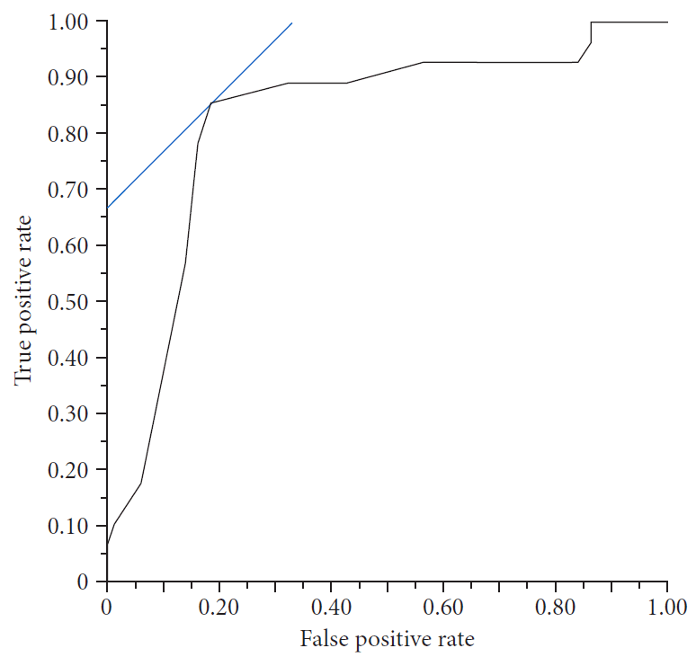
Fig. 1.
Flow chart of the study participants. EUS-HGS, endoscopic ultrasound-guided hepaticogastrostomy; AG, antegrade; IHBD, intrahepatic bile duct.
ce-2023-244f1.tif
Fig. 2.
Flow chart of the rescue guidewire technique.
ce-2023-244f2.tif
Fig. 3.
Measurements of the puncture angle between the fine needle and intrahepatic bile duct on the endoscopic ultrasound (EUS) images (A, C), and the puncture angle between the fine needle and echoendoscope (B, D) on the fluoroscopic images. Successful guidewire manipulation cases: The puncture angle was obtuse (110°) on the EUS image (A) and was also obtuse (135°) on the fluoroscopic image (B). Unuccessful guidewire manipulation cases: The puncture angle was acute (55°) on the EUS image (C) and was also obtuse (135°) on the fluoroscopic image (D).
ce-2023-244f3.tif
Fig. 4.
Receiver operating characteristic curves for the evaluation of the impact of the puncture angle between the fine needle and the intrahepatic bile duct and the determination of the optimal cutoff value.
ce-2023-244f4.tif
Fig. 5.
Endoscopic ultrasound and fluoroscopic images of a patient with unsuccessful guidewire manipulation in the B2 puncture group. (A) Abdominal computed tomography reveals swelling of the left lobe. (B) The first puncture angle between the needle and the intrahepatic biliary tract was acute. (C) The guidewire was only inserted into the peripheral bile duct. The scope was not directed toward the hilar side. (D) We re-punctured another bile duct, and the puncture angle between the needle and the intrahepatic biliary tract was obtuse. (E) The guidewire was easily inserted into the hilar bile duct. The scope was toward the peripheral side.
ce-2023-244f5.tif
Fig. 6.
Rescue methods for unsuccessful guidewire manipulation. EUS-HGS, endoscopic ultrasound-guided hepaticogastrostomy.
ce-2023-244f6.tif
ce-2023-244f7.tif
Table 1.
Characteristics of the enrolled patients
Characteristic Value
No. of patients 115 (100.0)
Age (yr) 71.0 (36–93)
Sex
 Male 67 (58.2)
 Female 48 (41.7)
Diseases
 Pancreatic cancer 48 (41.7)
 Bile duct cancer 31 (27.0)
 Others 36 (31.3)
Reason for EUS-HGS
 Unsuccessful ERCP 34 (29.6)
 Surgically altered anatomy 44 (38.3)
 Duodenal obstruction 37 (32.2)
Procedure type
 HGS 78 (67.8)
 HGS+AG 37 (32.2)
Presence of cholangitis 46 (40.0)
Fine needle size
 19 G 88 (76.5)
 22 G 27 (23.5)
Puncture site
 B3 84 (73.0)
 B2 31 (27.0)
Dilation procedure
 Yes 58 (50.4)
 No 57 (49.6)
Diameter of intrahepatic bile duct (mm) 5.0 (1–11)
Distance of hepatic parenchyma (cm) 2.4 (1–4.2)
Puncture angle on fluoroscopic image (°) 100 (20–195)
Puncture angle on EUS image (°) 95 (25–175)
Median procedure time (min) 53.0 (20–153)
Technical success 109 (94.8)
Clinical success 104 (90.4)
Adverse events 16 (13.9)
 Bile peritonitis 7
 Hemorrhage 3
 Acute cholecystitis 2
 Acute pancreatitis 3
 Liver abscess 1
Guidewire manipulation
 Success 87 (75.7)
 Unsuccess 28 (24.3)

Values are presented as number (%) or median (range).

EUS-HGS, endoscopic ultrasound-guided hepaticogastrostomy; ERCP, endoscopic retrograde cholangiopancreatography; AG, antegrade.

Table 2.
Clinical characteristics and outcomes between the successful and unsuccessful guidewire manipulation groups in hepaticogastrostomy
Characteristic SGMG (n=87) UGMG (n=28) p-value
Age (yr) 71 (36–93) 70 (50–87) 0.769
Female, sex 35 (40.2) 13 (46.4) 0.564
Diseases (pancreatic cancer) 38 (43.7) 10 (35.7) 0.454
Reason for EUS-HGS (unsuccessful ERCP) 25 (28.7) 9 (32.1) 0.732
Procedure type (HGS) 57 (65.5) 20 (71.4) 0.636
Presence of cholangitis 35 (40.2) 11 (39.3) 0.929
Fine needle size (19 G) 71 (81.6) 17 (60.7) 0.028
Puncture site (B3) 64 (73.6) 20 (71.4) 0.825
Intrahepatic bile duct diameter (mm) 5.0 (2–11) 4.0 (1–9) 0.117
Distance of hepatic parenchyma (cm) 2.5 (1.0–4.2) 2.25 (1.2–3.6) 0.735
Puncture angle on fluoroscopic image (°) 90 (20–155) 110 (30–195) 0.102
Puncture angle on EUS image (°) 100 (60–170) 75 (25–135) <0.001
 Puncture angle on EUS image (°) in the B2 group 23/140 (70–170) 8/85 (25–135)
 Puncture angle on EUS image (°) in the B3 group 64/92.5 (25–160) 20/75 (60–80)
Technical success of EUS-HGS 86 (98.9) 23 (82.1) 0.001
Clinical success 82 (94.3) 22 (78.6) 0.022
Procedure time (min) 47.0 (20–120) 70.0 (27–153) <0.001
Adverse events 10 (11.5) 6 (21.4) 0.204

Values are presented as median (range), number (%), or number/median (range).

SGMG, successful guidewire manipulation group; UGMG, unsuccessful guidewire manipulation group; EUS-HGS, endoscopic ultrasound-guided hepaticogastrostomy; ERCP, endoscopic retrograde cholangiopancreatography.

Table 3.
Factors associated with unsuccessful guidewire manipulation in the univariable and multivariable analyses
Factors Total (n=115) Univariable analysis
Multivariable analysis
OR (95% CI) p-value OR (95%CI) p-value
Age (<75 yr vs. ≥75 yr) 74/41 1.22 (0.49–3.03) 0.656
Sex (female vs. male) 48/67 1.28 (0.54–3.03) 0.563
Disease (others vs. pancreatic cancer) 67/48 1.39 (0.57–3.37) 0.458
Reason for EUS-HGS (unsuccessful ERCP vs. others) 34/81 1.17 (0.46–2.94) 0.731
Presence of cholangitis (no vs. yes) 69/46 1.04 (0.43–2.48) 0.929
Fine needle size (22 G vs. 19 G) 27/88 2.87 (1.13–7.29) 0.026 3.05 (0.93–10.0) 0.064
Puncture site (B2 vs. B3) 31/84 1.11 (0.43–2.87) 0.824
Diameter of intrahepatic bile duct (≤3 mm vs. >3 mm) 26/89 2.46 (0.95–6.33) 0.061
Distance of hepatic parenchyma (≤3.2 cm vs. >3.2 cm) 98/17 6.08 (0.76–48.1) 0.087
Puncture angle on fluoroscopic image (≥140° vs. <140°) 22/93 2.69 (1.00–7.24) 0.049 2.85 (0.81–10.0) 0.101
Puncture angle on EUS image (<85° vs. ≥85°) 36/79 19.1 (6.56–55.6) <0.001 19.8 (6.42–61.5) <0.001

OR, odds ratio; CI, confidence interval; EUS-HGS, endoscopic ultrasound-guided hepaticogastrostomy; ERCP, endoscopic retrograde cholangiopancreatography.

Table 4.
Details of unsuccessful guidewire manipulation group
Outcome % (n/ total n)
Technical success (guidewire manipulation) 85.7 (24/28)
Clinical success 78.6 (22/28)
Adverse events 21.4 (6/28)
Guidewire rescue methodsa)
 1) Changing guidewire type 11.8 (2/17)
 2) Liver impaction 27.3 (3/11)
 3) Uneven catheter 44.4 (8/18)
 4) Balloon method 100.0 (2/2)
 5) Re-puncture 64.3 (9/14)

a) Success rate.

TOOLS
Similar articles