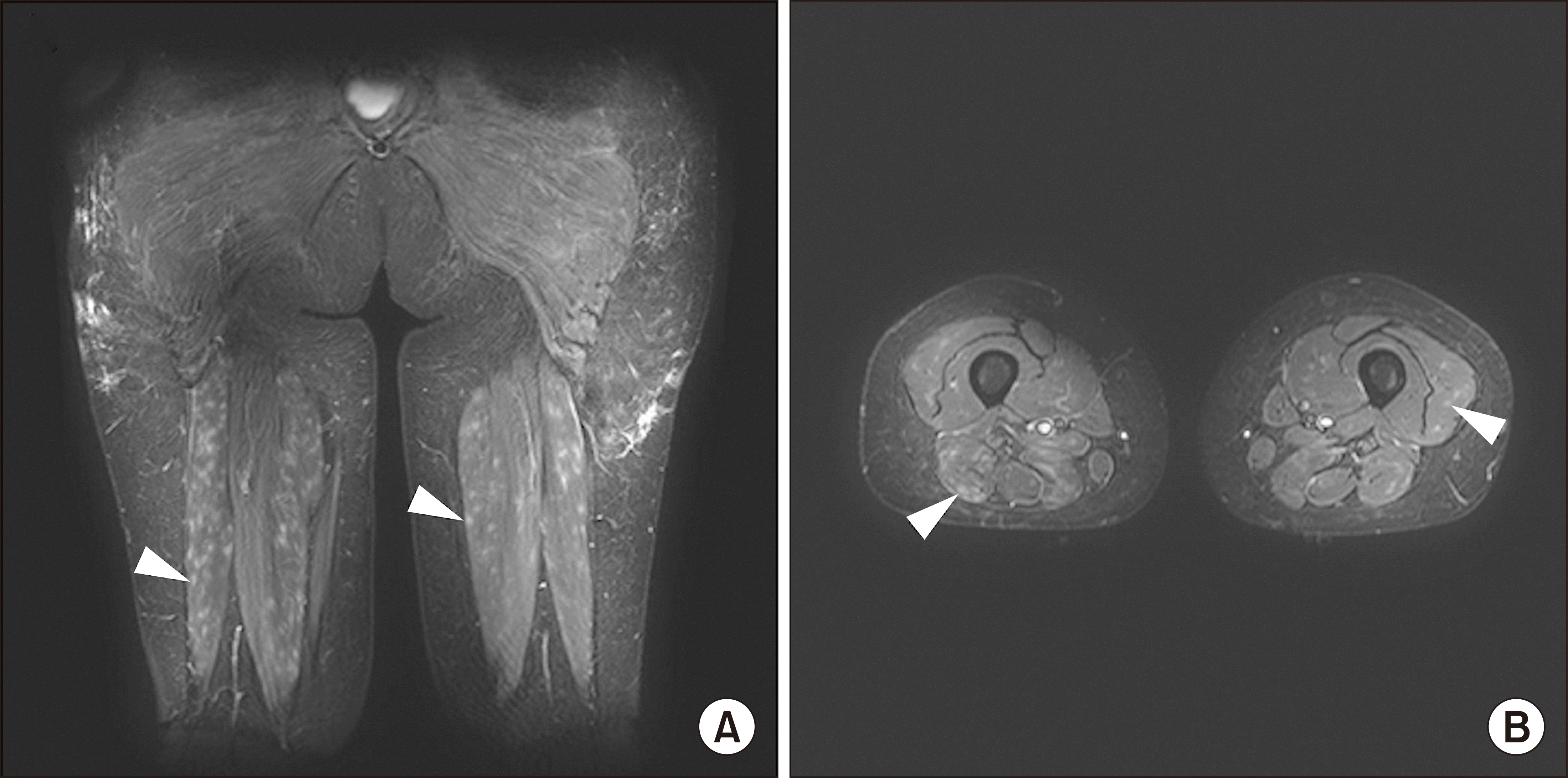
A Japanese female in her thirties with Sjögren’s syndrome (SS) showed myalgia in her lower legs and extremities. One year and 5 months after classification of SS, systemic lupus erythematosus (SLE) [1] was classified based on anti-nuclear antibody with homogeneous and speckled pattern (1:320), bicytopenia, hypocomplementemia, elevated anti-double-stranded (ds) DNA, and anti-Sm antibodies. Elevated lactate dehydrogenase (LDH) (579 U/L, normal 124~322 U/L), creatine phosphokinase (CK) (513 U/L, normal 41~153 U/L), aldolase (16.9 U/L, normal 2.1~6.1 U/L), and C-reactive protein with myalgia that lasted for 3 months were observed. A manual muscle test of the biceps brachii and quadriceps femoris decreased to 4+/5, indicating muscle weakness. A positive electromyogram and muscle biopsy with high intensity on magnetic resonance imaging (MRI) [2] in her thighs and lower legs indicated myositis (Figure 1A and 1B) in SLE or SS, although myositis-related autoantibodies including anti-aminoacyl tRNA synthetase antibody; anti-melanoma differentiation-associated gene 5 antibody; anti-transcriptional intermediary factor 1-γ antibody; anti-Mi-2 antibody; anti-mitochondrial M2 antibody that were measured by enzyme immunoassay methods were negative. Pathologically, inflammatory cell infiltration around muscle fibers was observed in the biceps brachii biopsy tissue, consistent with myositis (Figure 1C). The myositis was improved by treatment with hydroxychloroquine and glucocorticoid (0.5 mg/kg). When prednisolone was reduced to 7 mg/day, MRI was performed again due to a recurrence of thigh pain and severe fatigue with butterfly rash 13 months after first myalgia, progressing hypocomplementemia, and anti-dsDNA antibody elevation. High intensity on MRI consistent with lupus vasculitis was observed in the thigh-muscle group (Figure 2A and 2B) with elevated LDH (314 U/L) without elevations of CK (102 U/L) or aldolase (4.4 U/L) with exacerbated lupus, although myeloperoxidase-anti-neutrophil cytoplasmic antibody (ANCA) and proteinase 3 ANCA were negative even at the two episodes of myalgia. The introduction of 0.8 mg/kg prednisolone and 480 mg (10 mg/kg) of intravenous belimumab improved the myalgia and LDH and MRI findings. Currently, myalgia, muscle weakness, and fatigue have disappeared, and complement levels, anti-dsDNA antibody, and anti-Sm antibody have returned to normal.
Although MRI images of four cases of SLE-associated myositis were published [3], we have found no report describing typical myositis and vasculitis over time. This patient’s different myalgia episodes were considered to be distinct pathologies based on the MRI image pattern and the presence/absence of myogenic enzyme elevation.
The approval of the Itabashi Hospital Ethics Committee was waived because this manuscript does not contain personally identifiable photos. The patient provided written informed consent for the publication of her data and images.
REFERENCES
1. Aringer M, Costenbader K, Daikh D, Brinks R, Mosca M, Ramsey-Goldman R, et al. 2019; 2019 European League Against Rheumatism/American College of Rheumatology classification criteria for systemic lupus erythematosus. Ann Rheum Dis. 78:1151–9. DOI: 10.1136/annrheumdis-2018-214819. PMID: 31383717.
2. Tomasová Studynková J, Charvát F, Jarosová K, Vencovsky J. 2007; The role of MRI in the assessment of polymyositis and dermatomyositis. Rheumatology (Oxford). 46:1174–9. DOI: 10.1093/rheumatology/kem088. PMID: 17500079.
3. Elessawy SS, Abdelsalam EM, Abdel Razek E, Tharwat S. 2016; Whole-body MRI for full assessment and characterization of diffuse inflammatory myopathy. Acta Radiol Open. 5:2058460116668216. DOI: 10.1177/2058460116668216. PMID: 27708860. PMCID: PMC5034335.
Figure 1
MRI and pathological findings in the stages of myositis. (A) STIR (short-tau inversion recovery) findings on a coronal section of the patient's lower leg including the soleus muscle, gastrocnemius muscle, or flexor digitorum longus at the first episode of myopathy. (B) Axial section of the same imaging period. (C) Biceps brachii muscle biopsy image: Hematoxylin and eosin staining (x100 magnification). MRI, magnetic resonance imaging.

Figure 2
MRI findings of the vasculitis phase. (A) STIR (short-tau inversion recovery) findings on a coronal section of muscle groups including the rectus femoris, adductor magnus, sartorius muscle, and biceps femoris that make up the thigh at the patient's second episode of limb pain. Arrowheads: the high-intensity signal. (B) Axial section obtained during the patient's second limb-pain episode. MRI, magnetic resonance imaging.


 PDF
PDF Citation
 Print
Print


 XML Download