This article has been
cited by other articles in ScienceCentral.
Abstract
Osteopoikilosis (OPK) is a rare benign congenital genetic-mediated sclerosing skeletal disease, characterized by the formation of osteosclerosis foci. OPK is usually clinically asymptomatic, but some patients (15%~20%) may have arthralgia and synovitis. OPK may be associated with rheumatic diseases and might lead to unreasonable over-examination in real clinical practice. Single cases of the OPK together with ankylosing spondylitis (AS) have been described. Here we present a 33-year-old patient diagnosed with AS coexisting with OPK. In the case considered, the combination of AS and OPK accompanied with a high activity of inflammation, peripheral arthritis, a rapid rate of structural progression in axial skeleton, inefficiency of disease-modifying antirheumatic drugs and nonsteroidal anti-inflammatory drugs, a lack of response to anti interleukin-17 and a good response to a tumor necrosis factor inhibitor golimumab. We describe the important points of differential diagnosis associated with the identification of focal changes in bone tissue, especially neoplastic lesion. Foci revealed had typical localization, so, acquaintance of practicing doctors with such rare cases would minimize unnecessary examinations.
Go to :

Keywords: Osteopoikilosis, Ankylosing spondylitis, Biologic
INTRODUCTION
Osteopoikilosis (OPK) is a rare benign congenital genetic-mediated sclerosing skeletal disease, characterized by the formation of osteosclerosis foci [
1]. OPK is found incidentally during an X-ray study [
2] and is usually clinically asymptomatic, but some patients (15%~20%) may have arthralgia and synovitis. OPK may be associated with rheumatic diseases, such as rheumatoid arthritis, systemic lupus erythematosus, spondyloarthritis, fibromyalgia, scleroderma or Familial Mediterranean Fever. Single cases of the OPK together with ankylosing spondylitis (AS) have been described [
3-
5]. Significant mutual influence of these diseases has not been proved. In the case considered, the combination of AS and OPK accompanied with a high activity of inflammation, peripheral arthritis, a rapid rate of structural progression in axial skeleton, a lack of response to disease-modifying antirheumatic drugs, anti interleukin-17 (IL-17) and a good response to a tumor necrosis factor (TNF) inhibitor golimumab. The coexisting of AS and OPK might lead to unreasonable over-examination in real clinical practice.
Go to :

CASE REPORT
A male 33-year-old patient was admitted to our rheumatology department. He suffered from AS, which had been confirmed according to the modified 1984 New York Criteria. The disease occured at the age of 30 and was manifested in the inflammatory low back pain, prolonged morning stiffness and peripheral symptoms (pain and swelling in the knee and hip joints). The presence of
HLA-B27 (human leukocyte antigen-B27), a high inflammation activity (erythrocyte sedimentation rate [ESR] 66 mm/h, C-reactive protein [CRP] 77.5 mg/L, the Bath AS Disease Activity Index [BASDAI] 7.5), a limited range of motion in the cervical, thoracic and lumbar spine were revealed. The stage IV of sacroiliitis and pronounced syndesmophytes were diagnosed in all spine regions via X-ray. At the same time, many osteoscrlerotic foci were found in the hip and pelvic bones (
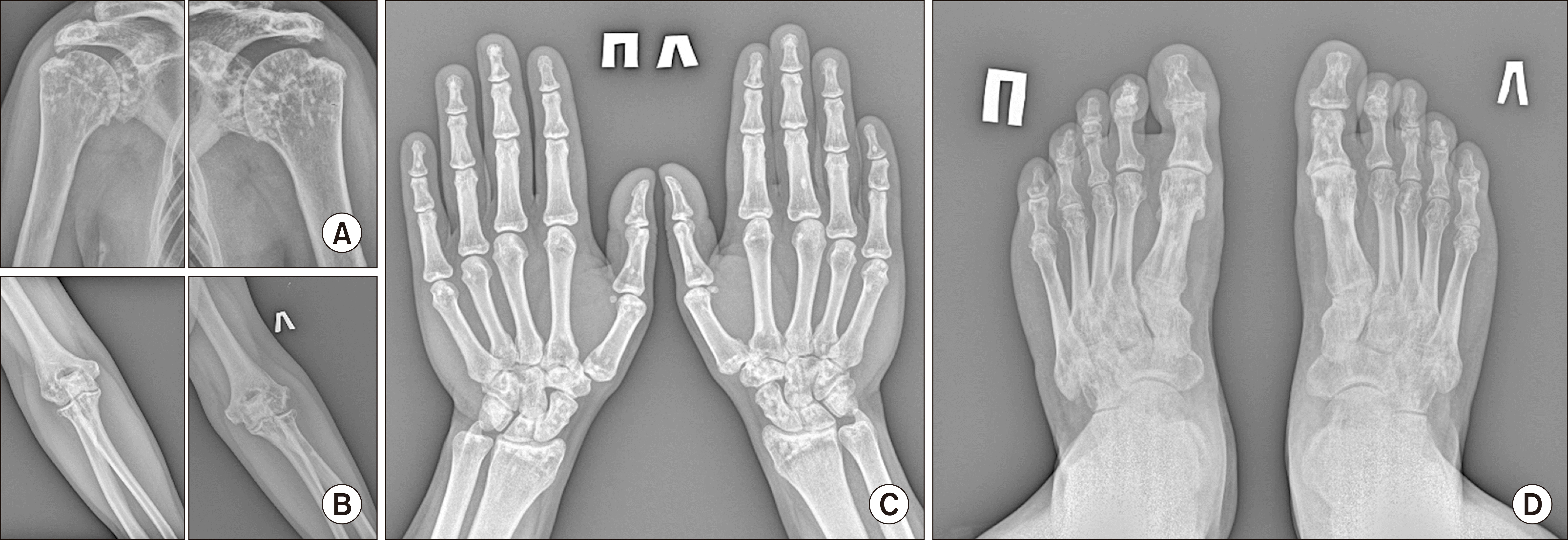
Figure 1). OPK was diagnosed on the basis of characteristic changes in various parts of the skeleton, especially in phalanges and wrists (
Figure 2). Pain or inflammation didn’t appear more severe in joints where OPK was present.
 | Figure 1X-rays of pelvis and spine. (A) X-ray of pelvis bone reveals a stage IV of sacroiliitis and osteosclerotic foci in femurs and pelvis bones. (B~E) X-ray of cervical, thoracic and lumbar spine demonstrates pronounced syndesmophytes and ankylosing without any osteopoikilosis foci in vertebrae. 
|
 | Figure 2Many osteoclerotic foci are found in X-ray of shoulder (A) and elbow (B) joints. (C) X-ray of hands reveals symmetric osteoclerotic foci. No bone erosions are found. (D) X-ray of feet demonstrates foci in phalanges and metatarsal bones. Many erosions and extra-articular ossification are seen that in accordance with arthritis due to ankylosing spondylitis. 
|
Nonsteroidal anti-inflammatory drugs (NSAIDs) were prescribed for continuous use. The patient received NSAIDs during the entire observation period. Sulfasalazine was prescribed as a disease-modifying therapy in view of the extra-axial manifestations (synovitis). Subsequently, it was replaced for methotrexate (up to 20 mg per week) because of the lack of efficacy and the persisting of arthritides. Due to the persistence of the high activity of the disease (a high level of pain in the spine, ESR 60 mm/h, CRP 107 mg/L, BASDAI 7.2) secukinumab 150 mg s/c per month was administered. As the structural changes in axial skeleton were progressing rapidly, IL-17A inhibitors was chosen. It demonstrated a good effect (normal CRP, low back pain and absence of synovitis) and a low disease activity had been being observed for 11 months. No changes in osteosclerotic foci and axial lesion were found. The CRP elevation and an increase in pain levels allowed to identify secukinumab’s inefficacy. Consequently, the switching on golimumab 50 mg s/c per month was performed. The occurrence of arthritis and an increase in ESR and CRP led to the addition of sulfasalazine 2 g per day. A constantly low disease activity was achieved and persisted for 1 year of observation. The size of the lesions did not change.
Informed written consent was obtained from the patient for publication of this report and any accompanying images. The Local Ethics Committee of the RICEL (Research Institute of Clinical and Experimental Lymрhology) - Branch of the IC&G SB RAS (Institute of Cytology and Genetics, Siberian Branch of Russian Academy of Sciences) approved the study (No. 178).
Go to :

DISCUSSION
OPK is a rare disease with a formation of multiple small non-aggressive sclerotic foci in extremities bones. The association with mutation in
LEMD3 (LEM domain-containing protein 3) gene encoding the inner nuclear membrane protein MAN1 was demonstrated. OPK may be found both in men and women of any age with the same frequency. The OPK incidence is 1 in 50,000 people. The lesions are from 2 to 10 mm in size, circular/oval in shape and form clusters of lamellar-like bone within the cancellous substance. They are located in the ischial, pubic bones, metaepiphyseal regions of the tubular bones. Most often they are located in the phalanges of the fingers (100%), bones of the wrist (97.4%), metacarpal bones (92.5%), phalanges of the toes (87.2%), metatarsal bones (84.4%), bones forearm (84.6%), pelvic bones (74.4%), radius (66.7%), ulna (66.7%), sacrum (58.9%), humerus (28.2% ), tibia (20.5%), fibula (2.8%) and do not disappear during human life [
6]. The axial skeleton is usually not affected. The changes revealed during the X-ray study were somehow similar to those that are usually found in metastatic bone lesions, mastocytosis and tuberous sclerosis, so differential diagnosis is required [
7]. Metastatic lesion should be excluded by the magnetic resonance imaging or positron emission tomography–computed tomography. They are usually asymmetric, localized in the axial skeleton and associated with bone destruction. In our case foci of osteosclerosis without a perifocal reaction were found mainly in the femurs, lateral sacral masses and ilia on the both sides, and were not found in the vertebrae. There were no signs of metastatic lesions of the lymph nodes. No additional formations were found in soft tissues. There wasn’t weight loss or other tumor-associated ‘red flags.’ The patient had an adequate prostate-specific antigen level. Changes in mastocytosis are usually more diffuse and would include damage to the axial skeleton, as well as a characteristic clinical feature [
8]. In tuberous sclerosis the lesion may be periarticular close to the sacroiliac joints, but have a patchy appearance. In addition, there were no clinical signs of tuberous sclerosis such as epileptic seizures, mental retardation or skin lesions [
9]. The patient did not have any endocrine pathology or low-energy fractures, the alkaline phosphatase level was adequate. Tuberculosis and other infections had been excluded by the specific tests.
It is worth mentioning that OPK might be combined with skin lesions such as connective tissue nevi (Buschke-Ollendorff syndrome) and discoid lupus erythematosus [
10]. In the case above, no significant abnormalities were revealed in the patient as a result of dermatological examination.
OPK usually has predominantly asymptomatic course and does not require special treatment. Nevertheless, arthralgia and synovitis without any changes in laboratory parameters have been demonstrated in a small percentage of cases. In our case, a considerable increase in the inflammation markers with a distinctive clinical feature (inflammatory low back pain, prolonged stiffness and arthritis) were manifested at the onset of the disease. Taking in consideration the stage IV of sacroiliitis, formed syndesmophytes in all regions of spine, limitation of the range of motion in the spine, there was no doubt that the patient suffered from AS. Despite a short duration of the disease (3 years), an advanced stage of AS had been developed. So, based on characteristic changes in various parts of the skeleton, especially in phalanges and wrists and with excluding other reasons (especially metastatic lesions), only AS with OPK was present. Significant effect of TNF-α inhibitors was exemplified by another patient suffering from a high AS activity combined with OPK [
5]. Nevertheless, the ineffectiveness of one and the effectiveness of another drug may only be a feature of the patient’s response to treatment, because OPK does not involve the axial spine.
Go to :

SUMMARY
This case of AS and OPK combination is curious because of the patient having a high inflammation activity, a rapid structural changes progression, IL-17A inhibitors medication inefficacy and significant effect of TNF-α inhibitors. The growth/reduce and change in the number of intraosseous foci have been excluded during the follow-up. No specific treatment of OPK is needed. Acquaintance of practicing doctors with such rare cases would minimize unnecessary examinations.
Go to :


 PDF
PDF Citation
 Print
Print


 XML Download