Journal List > J Rheum Dis > v.31(4) > 1516088517

Bang and Shim: Early human migration determines the risk of being attacked by wolves: ethnic gene diversity on the development of systemic lupus erythematosus

Abstract

The prevalence of systemic lupus erythematosus (SLE) varies significantly based on ethnicity rather than geographic distribution; thus, the prevalence is higher in Asian, Hispanic, and Black African populations than in European populations. The risk of developing lupus nephritis (LN) is the highest among Asian populations. Therefore, we hypothesize that human genetic diversity between races has occurred through the early human migration and human genetic adaptation to various environments, with a particular focus on pathogens. Additionally, we compile the currently available evidence on the ethnic gene diversity of SLE and how it relates to disease severity. The human leukocyte antigen (HLA) locus is well established as associated with susceptibility to SLE; specific allele distributions have been observed across diverse populations. Notably, specific amino acid residues within these HLA loci demonstrate significant associations with SLE risk. The non-HLA genetic loci associated with SLE risk also varies across diverse ancestries, implicating distinct immunological pathways, such as the type-I interferon and janus kinase–signal transducers and activators of transcription (JAK–STAT) pathways in Asians, the type-II interferon signaling pathway in Europeans, and B cell activation pathway in Africans. Furthermore, assessing individual genetic susceptibility using genetic risk scores (GRS) for SLE helps to reveal the diverse prevalence, age of onset, and clinical phenotypes across different ethnicities. A higher GRS increases the risk of LN and the severity of SLE. Therefore, understanding ethnic gene diversity is crucial for elucidating disease mechanisms and SLE severity, which could enable the development of novel drugs specific to each race.

INTRODUCTION

Systemic lupus erythematosus (SLE) is a chronic autoimmune disease with a broad spectrum of clinical manifestations, ranging from mild joint pain to life-threatening damage to various organs such as kidney, central nervous, and cardiovascular system. SLE is characterized by the production of various autoantibodies to numerous cellular constituents, especially nucleic acids and proteins [1]. SLE develops in genetically susceptible individuals exposed to environmental, sex-related, or endogenous triggers.
Geographic variations contribute to the diversity in the risk and severity of SLE. The SLE incidence rates vary across regions, ranging from 1.5 to 7.4 per 100,000 person-years in Europe, 1.4 to 6.3 in South America, 3.7 to 49.0 in North America, and 2.5 to 8.6 in Asia [2]. Moreover, the prevalence of SLE varies significantly based on ethnicity rather than geographic distribution. Indeed, the prevalence of Asian, Hispanic, and Black African populations is 2-fold higher than that of European population [2,3]. The risk of developing lupus nephritis (LN), one of the leading causes of mortality, was the highest in Asian population [4].
There are still many unmet needs in treating SLE [5,6]. Current treatments for SLE, including corticosteroids, cytotoxic drugs, and immunosuppressants, present various side effects. Thus, refractory disease manifestations, such as nephritis, alongside neurological involvement, require new drugs with greater efficacy and safety. However, most clinical trials over the past 60 years that tested the effectiveness of novel drugs for SLE have yet to be successful. One reason may be the heterogeneity of the disease associated with genetic variants in different races. Several clinical trials on lupus patients showed different treatment responses across races. Sub-analyses in the study of mycophenolate mofetil (MMF) vs. intravenous cyclophosphamide (IVC) as induction therapies of LN revealed that statistically significantly fewer patients responded to IVC compared to MMF in mixed-race African and Latin Americans. Similarly, fewer Hispanic patients responded to IVC than MMF [7]. Further, more patients in the African ancestry group were withdrawn from the study than those in the Caucasian and Asian groups because of adverse events from IVC, while more patients in the Asian subgroup were withdrawn due to side effects caused by MMF [8]. In analyzing B cell targeted therapies clinical trials, patients of African descent responded better to rituximab [9] but were less responsive to belimumab compared to those of other ethnicities, probably due to their higher serum levels of B cell-activating factor of the tumor necrosis factor family [10].
In this review, we hypothesize that the early human migration and human genetic adaptation to various environments, particularly pathogens, contribute to human genetic diversity between races. Moreover, we compiled the current evidence on the ethnic gene diversity related to SLE and its severity.

MAIN SUBJECTS

The race of Homo sapiens: the plausible theory of human genetic diversity between races

1) The early human migration

Humans migrated from East Africa into Northern Europe and Asia between 200,000 and 60,000 years ago (Figure 1). Two conflicting hypotheses have been raised on human evolution: the interbreeding and the replacement theory [11]. According to the replacement theory, Sapiens and other humans, such as Neanderthal and Erectus, would not have had sexual interest in one another. Therefore, their genes should have disappeared following extinction since they had not merged. The replacement theory had been common knowledge until recent decades.
However, the increasing availability of data on DNA in recent years has greatly facilitated the study of human history, showing that Neanderthal-derived DNA accounts for 1% to 4% of modern genomes in people outside sub-Saharan Africa [12]; it is highest in East Asians, intermediate in Europeans, and the lowest in Africans [13]. In conclusion, Sapiens, from African immigrants, bred with Neanderthals so that the two populations merged. Subsequently, today’s Eurasians are a mixture of Sapiens and Neanderthals. As Sapiens reached East Asia, they further interbred with the local Erectus, culminating in the Chinese and Koreans being a mixture of Sapiens, Neanderthals, and Erectus.

2) Human genetic adaptation to various environments, including pathogens

(1) The evolution of immune genes
Human evolution has been affected by different climates, food sources, and pathogens. Subsequently, human skin became lighter, the metabolism adapted to new food sources, and the immune system had to manage different pathogens. Moreover, Homo sapiens have evolved into various subspecies and races [14].
When it comes to pathogens, the first bacteria and virus evolved 3 billion and 1.5 billion years ago, respectively [15]. Pathogens were first observed in Africa 200,000 to 300,000 years ago, spreading to other continents over the last 60,000 to 100,000 years and more rapidly through the emergence of agriculture 10,000 years ago [16].
Infectious diseases are the leading cause of mortality in human history [17]. Since a formal link was established between infectious diseases and natural selection in previous studies showing that thalassemia and sickle cell disease can protect against malaria [18], various evidence has accumulated concerning the genetic determinants of infectious, inflammatory, and autoimmune disorders [19,20].
Furthermore, genome-wide association studies (GWAS) have supported the hypothesis that microbes have significantly affected human evolution [16,20,21].
Recently, statistics of the COVID-19 infection showed racial differences in morbidity and mortality, which are significantly lower in developing countries compared to the developed countries in Europe and North America. A total of 37% of all mortalities during the pandemic occurred in the European Union (EU) [22], even though the population of the EU forms only about 9.78% of the global population. A striking difference in mean per million mortality was reported between Asian and European countries (2.7 vs. 197 deaths per million population, p<0.001). For example, mortality rates were less than 1 death/million in Taiwan, Vietnam, and Thailand compared to 1,112 in Belgium [23].
The antagonistic pleiotropy hypothesis was considered in the pathogenesis of chronic inflammatory diseases, which is supported by the overlap between loci underlying infectious and inflammatory traits [24,25]. Additionally, several pleiotropic variants were reported to confer protection against infectious diseases alongside susceptibility to some chronic inflammatory diseases [26-28]. Indeed, the human leukocyte antigen (HLA) locus is a representative example, whereby variants of the HLA are considered to be under pathogen-driven positive selection [29] and can increase the risk of autoinflammatory diseases, such as ankylosing spondylitis and type 1 diabetes [30-34]. Another example is the common TYK2 P1104A variant, which protects against autoimmune phenotypes (odds ratio [OR]=0.1~0.3) but, in the homozygous state, confers a predisposition to mycobacterium-related infectious diseases (OR >10) [28,35-41]. Thus, selection has likely led to a higher genetic risk of inflammatory gastrointestinal disorders since the Bronze Age. The main risk variants for inflammatory bowel disease are located close to key immunity genes (IRF1, IKZF1, FUT2, and SH2B3), for which monogenic lesions confer susceptibility or resistance to infectious diseases [42-44].
These results suggest that recent temporal changes in pathogen exposure probably caused positive selection to target the regulatory machinery underlying immune cell variation [45,46].
(2) The evolution of major histocompatibility complex polymorphisms
The major histocompatibility complex (MHC) is the most polymorphic gene locus in vertebrates [47]. A co-evolutionary arms race with pathogens is responsible for the high levels of genetic diversity [48]. The HLA system includes MHC class I genes (HLA-A, -B, and -C as classical genes; HLA-E, -F, and -G as nonclassical genes), MHC class II genes (HLA-DP, -DQ, and -DR as classical genes; HLA-DM and -DO as non-classical genes), and MHC class III genes (complement and cytokine genes). HLA-DRB1 is the most variable region in HLA class II genes, with over 2,000 registered alleles [49,50] since HLA-DRA is basically invariant. HLA-DRB1 shows the strongest general signature of selection among HLA class II loci [51] and has evolved and diversified very rapidly [52] after ancestral human migration owing to encounters with many novel pathogens [53]. HLA-DRB1 is more highly expressed on the cell surface than other HLA class II molecules [54].
Another significant variation in MHC molecules is the epitope repertoire size, with an up to 16-fold variation in the number of bound peptides [55]. A group of promiscuous MHC molecules that enhance the immune response against a broader range of pathogens are called generalists, in contrast to specialists, which exhibit a response against fewer pathogens [56]. A tiny difference in the amino acid sequence shows massive promiscuity range differences. Even though the HLA-DRB1*13 group alleles present an amino acid sequence identity to each other of over 98%, the promiscuity levels vary 57-fold [57]. High pathogen diversity increases the MHC diversity in relation to their average genomic diversity [29], and the diversity of MHC class II molecules is found in the molecular positions that define promiscuity level, which is under positive selection [57]. Interestingly, HLA-DRB1*12:02 has the highest promiscuity level among HLA-DRB1 alleles in Southeast Asia, which protects against pulmonary tuberculosis and typhoid fever—endemic diseases in this area [58]. Therefore, the high allele frequency of DRB1*12:02 reflects the pathogen-driven selection that occurred during the migration of Mongolians to South China [59].

3) Space invaders: endogenous retroviral sequences in systemic lupus erythematosus

Human endogenous retroviruses (HERVs) are viral elements in the human genome that could be derived from retroviruses. Most HERVs transitioned into the human genome around 25 million years ago [60], and about 8% of the human genome consists of retroelements containing long terminal repeats, a group that includes endogenous retroviruses [61]. Some HERVs are still active, and various factors, including ultraviolet radiation, inflammatory cytokines, steroid hormones, and exogenous virus products, could reactivate their expression [62]. HERVs have been considered to induce multiple autoimmune diseases [63]. Currently, 200 endogenous retroviral sequences have been reported, and HRES-1, HERV-E 4-1, HERV-K10, and HERV-K18 HERVs are related to the pathogenesis of SLE [64-66]. Present hypotheses consider that autoantibody production occurs via molecular mimicry, stimulation of intracellular sensor molecules by viral nucleic acids, and epigenetic regulation of host genes mechanisms [67,68]. Antibodies against HRES-1, which is an endogenous retroviral element-encoded nuclear protein autoantigen, were higher in patients with SLE compared to control subjects (52% vs. 3%) [69]. The molecular mimicry hypothesis has also been raised since cross-reactivity is reported between HERV proteins and self-antigens [70].

The race of Homo sapiens lupus: the genetic architecture of systemic lupus erythematosus

The pathogenesis of SLE is influenced by a strong genetic contribution, with a 66% of heritability [71]. The SLE has a significant genetic predisposition, influenced by both ancestry-dependent and ancestry-independent factors. Over the past decade, over 100 susceptibility loci have been identified by GWAS and reported, mostly in European and Asian populations [59,72,73]. The polygenic etiology of SLE is supported by >170 SLE-risk-associated genomic loci characterized by common variants with modest effect sizes identified through GWAS or candidate-gene approaches in multiple ethnic groups [74-77].

1) Human leukocyte antigen loci associated with systemic lupus erythematosus

HLA-DRB1 is widely known for its pivotal role in regulating immune tolerance to self-antigens. The HLA-DRB1 allele frequencies vary in the general population of different ethnic groups [78]. For example, HLA-DRB1*03:01, *07:01, and *15:01 were predominantly identified in Europeans; *03:01, *07:01, and *11:01 in African Americans; *15:01 and *09:01 in Asians; and *09:01 and *04:05 in Koreans (Table 1).
The primary SLE association signal has been consistently identified within the HLA-DRB1 gene in multiple ancestral populations [75-77,79]. These findings underscore the significance of the HLA-DRB1-mediated immune response in developing SLE. Multiple ancestral studies revealed that the HLA-DRB1*15:01 and *03:01 alleles were the most significant SLE-risk factors within the HLA class II region, which is shared across multiple ancestries, including African American, Asian, and European [25,74,75]. HLA-DRB1*03:01 is the largest SLE-risk allele in European ancestries [74], although it is not common enough to detect its association in East Asian SLE cohorts [25,76,77,80]. European patients with SLE were linked to HLA-DRB1*03:01 and *08:01 [74]. In contrast, Asian patients with SLE possessed more copies of the HLA-DRB1*08:03, *07:01, and *09:01 alleles than the healthy controls [76].
Identifying the genetic variants responsible for SLE development and its clinical manifestations has been challenging. The HLA-DRB1*15:01 allele was positively associated with many clinical sub-phenotypes of SLE [76]. In addition, the *09:01, *08:03, or *07:01 alleles were linked to SLE risk and significantly affected more diverse clinical manifestations [76]. Notably, individuals carrying double copies of the HLA-DRB1 risk alleles exhibited an elevated risk of producing anti-Sm antibodies [76].
Recently, a detailed analysis of HLA amino acid residues provided an improved understanding of the specific HLA associations based on ethnicity. Specifically, the amino acid positions 11–13–26 in HLA-DRB1 at the epitope-binding groove of the HLA-DR molecules were most significantly associated with increased susceptibility to SLE by Asian and European populations [75]. The two well-known SLE-risk HLA-DRB1 alleles *15:01 and *03:01 belonged to their respective SLE-risk haplotypes, Pro–Arg–Phe and Ser–Ser–Tyr, at amino acid positions 11, 13, and 26. The haplotype comprising Asp–Phe–Tyr at amino acid positions 11, 13, and 26, respectively, is commonly found among Asian populations, while the Ser–Ser–Tyr haplotype is more common among Europeans (Table 2). The amino acid positions 11 and 13 in HLA-DRB1 have been reported to be linked to SLE susceptibility and the production of autoantibodies, thereby driving the pathogenesis of LN [75,81]. These amino acid positions within the epitope-binding groove of HLA molecules contribute to the risk of developing SLE.

2) Non-human leukocyte antigen loci associated with systemic lupus erythematosus

The reported non-HLA loci associated with SLE have been identified in African American, Asian, European, and shared ethnic groups (Supplementary Table 1), which may regulate gene expression and contribute to SLE development.
We investigated ancestry-driven key signaling pathways in African American, Asian, and European populations by analyzing reported non-HLA single-nucleotide polymorphism (SNP)-associated genes using enrichr (https://maayanlab.cloud/Enrichr/#libraries) (Supplementary Table 1 and Figure 2).
International collaboration in diverse ancestral populations has facilitated the identification of SLE-risk loci and revealed the heterogeneity of SLE variants among ancestries.
Asian-associated genes were dominated by the functional category for type-I interferon-stimulated genes and the janus kinase–signal transducers and activators of transcription (JAK–STAT) signaling pathway (Figure 2), along with the top pathways determined by Gene Ontogeny terms for each gene list. Europeans are associated with the type-II interferon-stimulated genes and IL-12 and IL-17 signaling pathways, while Africans are associated with the B cell and IL-12 signaling pathways.
The pathogenesis of SLE is linked to diverse immunological pathways of SLE-risk SNP-associated genes. Thus, clinical trials using many drugs against different targets, such as B cell (CD20, CD22), type-I interferons, JAK, tyrosine kinases, interleukin (IL)-12 and IL-23, or plasmacytoid dendritic cell receptors have recently been conducted (Table 3) [82-89]. Ethnic diversity in response to each treatment should be further investigated in these trials.

3) Weighted genetic risk score in different ethnicities

Each SLE patient presents with significantly different symptoms, prognoses, and mortality rates. Therefore, it is important to assess individual genetic susceptibility using a cumulative weighted genetic risk score (wGRS), defined as the sum of the number of risk alleles at various loci in an individual, weighted by their odds ratio.
Analyzing wGRS across five ethnic populations revealed that the higher wGRS and SLE risk allele frequencies showed a greater SLE prevalence (Supplementary Table 2). The populations ranked from the lowest to highest for the average wGRS were Europeans, Amerindians, South Asians, East Asians, and Africans [79]. A higher wGRS for SLE was observed more often in childhood-onset SLE than in adult-onset SLE [90,91].
In addition, a high wGRS based on 57 SLE risk loci was significantly correlated with a higher risk of renal disorder, end-stage renal disease, and mortality in a Caucasian population [92].
A recent study of wGRS using 112 non-HLA SNPs and HLA-DRB1 amino acid haplotypes in Korean patients with SLE revealed that a higher wGRS was significantly correlated with higher clinical manifestations of SLE [93]. Notably, the highest quartile of wGRS was associated with the highest risk of LN and the production of anti-Sm, regardless of onset age. Furthermore, an increasing wGRS significantly impacted the development of proliferative LN (class III or IV) or membranous LN (class V) [93]. The genetic risk load of SLE significantly influenced the development of diverse clinical manifestations and LN; thus, genetic profiling using wGRS could enable personalized management by predicting the clinical course of SLE.

CONCLUSION

The future of Homo sapiens lupus

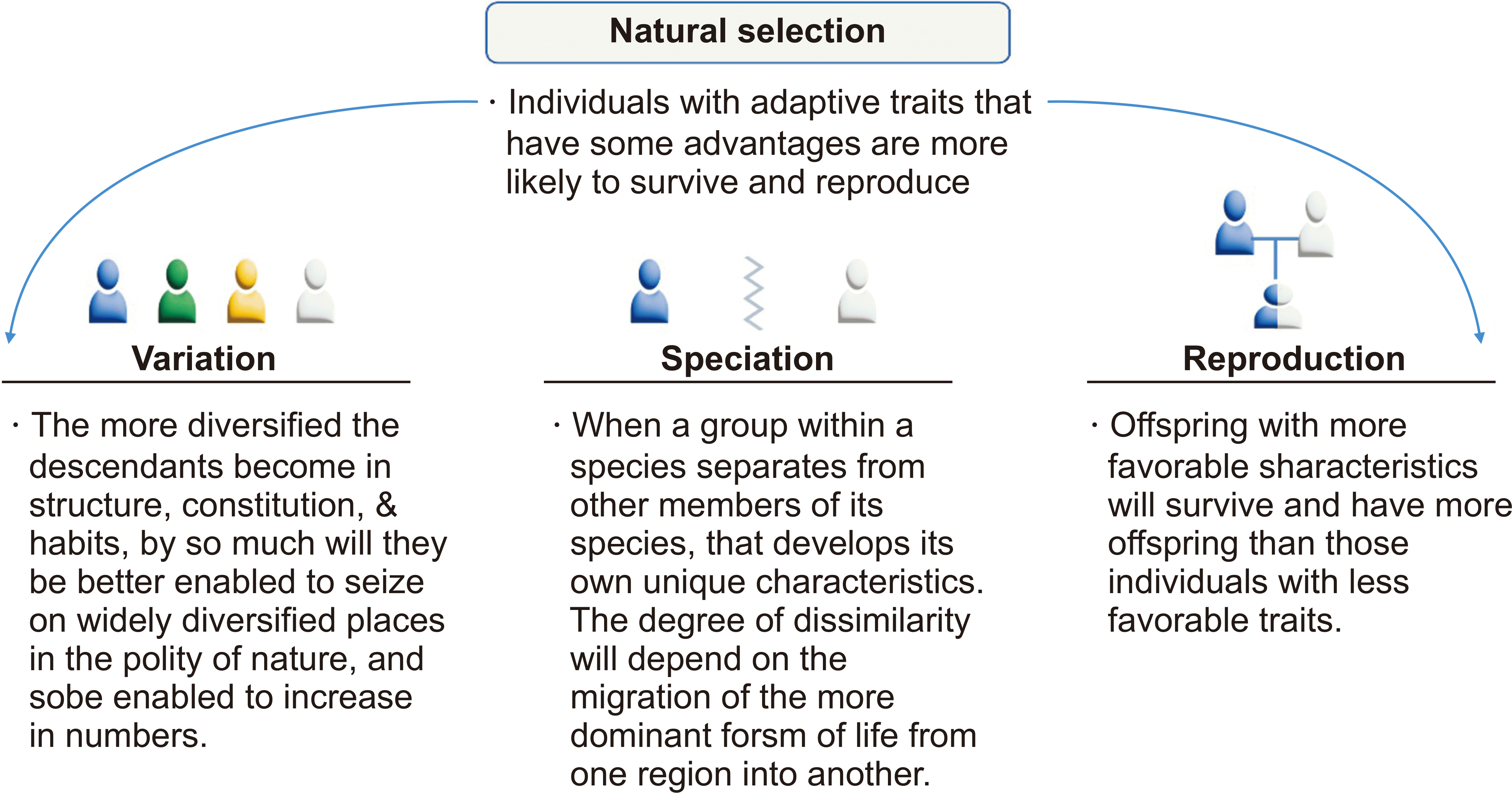
According to the natural selection theory, individuals who are more adapted to their environment are more likely to survive and pass on genes important to their survival (Figure 3) [94]. For this theory to be correct, genes associated with lupus should have been deleted. Furthermore, if genes associated with lupus have maintained under pathogen-driven positive selection, the risk of infection should be lower in lupus.
The theory of geographic distribution would make lupus patients different by region but similar in the region over time, meaning we could still manage the disease using designated medicines for a specific race or an approved region of the globe. However, these circumstances would not happen because genetic variation currently occurs more between individuals than ethnic groups.
The genetic risk load and clinical outcomes across different ethnicities could improve SLE management based on individual genetic risk profiles.
According to variation theory, lupus patients will become more genetically diverse in the future, comprehensive genetic studies should be continuously conducted to find out better targets, which will pave the way for personalized treatment for SLE patients.

SUPPLEMENTARY DATA

Supplementary data can be found with this article online at https://doi.org/10.4078/jrd.2024.0051

ACKNOWLEDGMENTS

None.

Notes

CONFLICT OF INTEREST

No potential conflict of interest relevant to this article was reported.

AUTHOR CONTRIBUTIONS

SYB and SCS contributed to the study conception , design, and data acquisition. All authors contributed to the analysis and/or interpretation of data. SYB and SCS contributed to drafting the manuscript. SYB and SCS contributed to revising the manuscript. All authors reviewed and approved the final version of the manuscript.

REFERENCES

1. Liu Z, Davidson A. 2012; Taming lupus-a new understanding of pathogenesis is leading to clinical advances. Nat Med. 18:871–82. DOI: 10.1038/nm.2752. PMID: 22674006. PMCID: PMC3607103.
2. Barber MRW, Drenkard C, Falasinnu T, Hoi A, Mak A, Kow NY, et al. 2021; Global epidemiology of systemic lupus erythematosus. Nat Rev Rheumatol. 17:515–32. DOI: 10.1038/s41584-021-00668-1. PMID: 34345022. PMCID: PMC8982275.
3. Izmirly PM, Wan I, Sahl S, Buyon JP, Belmont HM, Salmon JE, et al. 2017; The incidence and prevalence of systemic lupus erythematosus in New York County (Manhattan), New York: the Manhattan lupus surveillance program. Arthritis Rheumatol. 69:2006–17. DOI: 10.1002/art.40192. PMID: 28891252. PMCID: PMC11102806.
4. Lewis MJ, Jawad AS. 2017; The effect of ethnicity and genetic ancestry on the epidemiology, clinical features and outcome of systemic lupus erythematosus. Rheumatology (Oxford). 56(Suppl 1):i67–77. DOI: 10.1093/rheumatology/kew399. PMID: 27940583.
5. Askanase A, Khalili L, Tang W, Mertz P, Scherlinger M, Sebbag E, et al. 2023; New and future therapies: changes in the therapeutic armamentarium for SLE. Best Pract Res Clin Rheumatol. 37:101865. DOI: 10.1016/j.berh.2023.101865. PMID: 37633826.
6. Han Y, Liu L, Zang B, Liang R, Zhao X, Liu B. 2023; Advances in natural products and antibody drugs for SLE: new therapeutic ideas. Front Pharmacol. 14:1235440. DOI: 10.3389/fphar.2023.1235440. PMID: 37492083. PMCID: PMC10363611.
7. Appel GB, Contreras G, Dooley MA, Ginzler EM, Isenberg D, Jayne D, et al. Aspreva Lupus Management Study Group. 2009; Mycophenolate mofetil versus cyclophosphamide for induction treatment of lupus nephritis. J Am Soc Nephrol. 20:1103–12. DOI: 10.1681/ASN.2008101028. PMID: 19369404. PMCID: PMC2678035.
8. Isenberg D, Appel GB, Contreras G, Dooley MA, Ginzler EM, Jayne D, et al. 2010; Influence of race/ethnicity on response to lupus nephritis treatment: the ALMS study. Rheumatology (Oxford). 49:128–40. DOI: 10.1093/rheumatology/kep346. PMID: 19933596. PMCID: PMC2789586.
9. Merrill JT, Neuwelt CM, Wallace DJ, Shanahan JC, Latinis KM, Oates JC, et al. 2010; Efficacy and safety of rituximab in moderately-to-severely active systemic lupus erythematosus: the randomized, double-blind, phase II/III systemic lupus erythematosus evaluation of rituximab trial. Arthritis Rheum. 62:222–33. DOI: 10.1002/art.27233. PMID: 20039413. PMCID: PMC4548300.
10. Catalina MD, Bachali P, Yeo AE, Geraci NS, Petri MA, Grammer AC, et al. 2020; Patient ancestry significantly contributes to molecular heterogeneity of systemic lupus erythematosus. JCI Insight. 5:e140380. DOI: 10.1172/jci.insight.140380. PMID: 32759501. PMCID: PMC7455079.
11. Wolpoff MH, Hawks J, Frayer DW, Hunley K. 2001; Modern human ancestry at the peripheries: a test of the replacement theory. Science. 291:293–7. DOI: 10.1126/science.291.5502.293. PMID: 11209077.
12. Price M. 2020; Africans, too, carry Neanderthal genetic legacy. Science. 367:497. DOI: 10.1126/science.367.6477.497. PMID: 32001636.
13. Wolf AB, Akey JM. 2018; Outstanding questions in the study of archaic hominin admixture. PLoS Genet. 14:e1007349. DOI: 10.1371/journal.pgen.1007349. PMID: 29852022. PMCID: PMC5978786.
14. Hunter P. 2014; The genetics of human migrations: our ancestors migration out of Africa has left traces in our genomes that explain how they adapted to new environments. EMBO Rep. 15:1019–22. DOI: 10.15252/embr.201439469. PMID: 25216943. PMCID: PMC4253842.
15. Narbonne GM. 2005; The Ediacara biota: neoproterozoic origin of animals and their ecosystems. Annu Rev Earth Planet Sci. 33:421–42. DOI: 10.1146/annurev.earth.33.092203.122519.
16. Karlsson EK, Kwiatkowski DP, Sabeti PC. 2014; Natural selection and infectious disease in human populations. Nat Rev Genet. 15:379–93. DOI: 10.1038/nrg3734. PMID: 24776769. PMCID: PMC4912034.
17. Casanova JL, Abel L. 2005; Inborn errors of immunity to infection: the rule rather than the exception. J Exp Med. 202:197–201. DOI: 10.1084/jem.20050854. PMID: 16027233. PMCID: PMC2212996.
18. Allison AC. 1954; Protection afforded by sickle-cell trait against subtertian malarial infection. Br Med J. 1:290–4. DOI: 10.1136/bmj.1.4857.290.
19. Parkes M, Cortes A, Van Heel DA, Brown MA. 2013; Genetic insights into common pathways and complex relationships among immune-mediated diseases. Nat Rev Genet. 14:661–73. DOI: 10.1038/nrg3502. PMID: 23917628.
20. Abel L, Alcaïs A, Schurr E. 2014; The dissection of complex susceptibility to infectious disease: bacterial, viral and parasitic infections. Curr Opin Immunol. 30:72–8. DOI: 10.1016/j.coi.2014.07.002. PMID: 25083600.
21. Quintana-Murci L, Clark AG. 2013; Population genetic tools for dissecting innate immunity in humans. Nat Rev Immunol. 13:280–93. DOI: 10.1038/nri3421. PMID: 23470320. PMCID: PMC4015519.
22. Konstantinoudis G, Cameletti M, Gómez-Rubio V, Gómez IL, Pirani M, Baio G, et al. 2022; Regional excess mortality during the 2020 COVID-19 pandemic in five European countries. Nat Commun. 13:482. DOI: 10.1038/s41467-022-28157-3. PMID: 35079022. PMCID: PMC8789777.
23. Landoni G, Maimeri N, Fedrizzi M, Fresilli S, Kuzovlev A, Likhvantsev V, et al. 2021; Why are Asian countries outperforming the Western world in controlling COVID-19 pandemic? Pathog Glob Health. 115:70–2. DOI: 10.1080/20477724.2020.1850982. PMID: 33241776. PMCID: PMC7850376.
24. Fodil N, Langlais D, Gros P. 2016; Primary immunodeficiencies and inflammatory disease: a growing genetic intersection. Trends Immunol. 37:126–40. DOI: 10.1016/j.it.2015.12.006. PMID: 26791050. PMCID: PMC4738049.
25. Langefeld CD, Ainsworth HC, Cunninghame Graham DS, Kelly JA, Comeau ME, Marion MC, et al. 2017; Transancestral mapping and genetic load in systemic lupus erythematosus. Nat Commun. 8:16021. DOI: 10.1038/ncomms16021. PMID: 28714469. PMCID: PMC5520018.
26. Fumagalli M, Pozzoli U, Cagliani R, Comi GP, Riva S, Clerici M, et al. 2009; Parasites represent a major selective force for interleukin genes and shape the genetic predisposition to autoimmune conditions. J Exp Med. 206:1395–408. DOI: 10.1084/jem.20082779. PMID: 19468064. PMCID: PMC2715056.
27. Zhernakova A, Elbers CC, Ferwerda B, Romanos J, Trynka G, Dubois PC, et al. 2010; Evolutionary and functional analysis of celiac risk loci reveals SH2B3 as a protective factor against bacterial infection. Am J Hum Genet. 86:970–7. DOI: 10.1016/j.ajhg.2010.05.004. PMID: 20560212. PMCID: PMC3032060.
28. Jostins L, Ripke S, Weersma RK, Duerr RH, McGovern DP, Hui KY, et al. 2012; Host-microbe interactions have shaped the genetic architecture of inflammatory bowel disease. Nature. 491:119–24. DOI: 10.1038/nature11582. PMID: 23128233. PMCID: PMC3491803.
29. Prugnolle F, Manica A, Charpentier M, Guégan JF, Guernier V, Balloux F. 2005; Pathogen-driven selection and worldwide HLA class I diversity. Curr Biol. 15:1022–7. DOI: 10.1016/j.cub.2005.04.050. PMID: 15936272.
30. Ryder LP, Svejgaard A, Dausset J. 1981; Genetics of HLA disease association. Annu Rev Genet. 15:169–87. DOI: 10.1146/annurev.ge.15.120181.001125. PMID: 7039493.
31. Gough SC, Simmonds MJ. 2007; The HLA region and autoimmune disease: associations and mechanisms of action. Curr Genomics. 8:453–65. DOI: 10.2174/138920207783591690. PMID: 19412418. PMCID: PMC2647156.
32. Chen H, Hayashi G, Lai OY, Dilthey A, Kuebler PJ, Wong TV, et al. 2012; Psoriasis patients are enriched for genetic variants that protect against HIV-1 disease. PLoS Genet. 8:e1002514. DOI: 10.1371/journal.pgen.1002514. PMID: 22577363. PMCID: PMC3343879.
33. Matzaraki V, Kumar V, Wijmenga C, Zhernakova A. 2017; The MHC locus and genetic susceptibility to autoimmune and infectious diseases. Genome Biol. 18:76. DOI: 10.1186/s13059-017-1207-1. PMID: 28449694. PMCID: PMC5406920.
34. Ritari J, Koskela S, Hyvärinen K, FinnGen , Partanen J. 2022; HLA-disease association and pleiotropy landscape in over 235,000 Finns. Hum Immunol. 83:391–8. DOI: 10.1016/j.humimm.2022.02.003. PMID: 35221124.
35. Tsoi LC, Spain SL, Knight J, Ellinghaus E, Stuart PE, Capon F, et al. 2012; Identification of 15 new psoriasis susceptibility loci highlights the role of innate immunity. Nat Genet. 44:1341–8. DOI: 10.1038/ng.2467. PMID: 23143594. PMCID: PMC3510312.
36. Cortes A, Hadler J, Pointon JP, Robinson PC, Karaderi T, Leo P, et al. 2013; Identification of multiple risk variants for ankylosing spondylitis through high-density genotyping of immune-related loci. Nat Genet. 45:730–8. DOI: 10.1038/ng.2667. PMID: 23749187. PMCID: PMC3757343.
37. Diogo D, Bastarache L, Liao KP, Graham RR, Fulton RS, Greenberg JD, et al. 2015; TYK2 protein-coding variants protect against rheumatoid arthritis and autoimmunity, with no evidence of major pleiotropic effects on non-autoimmune complex traits. PloS one. 10:e0122271. DOI: 10.1371/journal.pone.0122271. PMID: 25849893. PMCID: PMC4388675.
38. Dendrou CA, Cortes A, Shipman L, Evans HG, Attfield KE, Jostins L, et al. 2016; Resolving TYK2 locus genotype-to-phenotype differences in autoimmunity. Sci Transl Med. 8:363ra149. DOI: 10.1126/scitranslmed.aag1974. PMID: 27807284. PMCID: PMC5737835.
39. Boisson-Dupuis S, Ramirez-Alejo N, Li Z, Patin E, Rao G, Kerner G, et al. 2018; Tuberculosis and impaired IL-23-dependent IFN-γ immunity in humans homozygous for a common TYK2 missense variant. Sci Immunol. 3:eaau8714. DOI: 10.3410/f.734676937.793572033. PMID: 30578352. PMCID: PMC6341984.
40. Kerner G, Ramirez-Alejo N, Seeleuthner Y, Yang R, Ogishi M, Cobat A, et al. 2019; Homozygosity for TYK2 P1104A underlies tuberculosis in about 1% of patients in a cohort of European ancestry. Proc Natl Acad Sci U S A. 116:10430–4. DOI: 10.1073/pnas.1903561116. PMID: 31068474. PMCID: PMC6534977.
41. Kerner G, Laval G, Patin E, Boisson-Dupuis S, Abel L, Casanova JL, et al. 2021; Human ancient DNA analyses reveal the high burden of tuberculosis in Europeans over the last 2,000 years. Am J Hum Genet. 108:517–24. DOI: 10.1016/j.ajhg.2021.02.009. PMID: 33667394. PMCID: PMC8008489.
42. Lindesmith L, Moe C, Marionneau S, Ruvoen N, Jiang X, Lindblad L, et al. 2003; Human susceptibility and resistance to Norwalk virus infection. Nat Med. 9:548–53. DOI: 10.1038/nm860. PMID: 12692541.
43. Panda D, Gjinaj E, Bachu M, Squire E, Novatt H, Ozato K, et al. 2019; IRF1 maintains optimal constitutive expression of antiviral genes and regulates the early antiviral response. Front Immunol. 10:1019. DOI: 10.3389/fimmu.2019.01019. PMID: 31156620. PMCID: PMC6529937.
44. Nunes-Santos CJ, Kuehn HS, Rosenzweig SD. 2020; IKAROS family zinc finger 1-associated diseases in primary immunodeficiency patients. Immunol Allergy Clin North Am. 40:461–70. DOI: 10.1016/j.iac.2020.04.004. PMID: 32654692. PMCID: PMC7394939.
45. Rascovan N, Sjögren KG, Kristiansen K, Nielsen R, Willerslev E, Desnues C, et al. 2019; Emergence and spread of basal lineages of Yersinia pestis during the Neolithic decline. Cell. 176:295–305.e10. DOI: 10.1016/j.cell.2018.11.005. PMID: 30528431.
46. Andrades Valtueña A, Neumann GU, Spyrou MA, Musralina L, Aron F, Beisenov A, et al. 2022; Stone Age Yersinia pestis genomes shed light on the early evolution, diversity, and ecology of plague. Proc Natl Acad Sci U S A. 119:e2116722119. DOI: 10.3410/f.742004133.793592836. PMID: 35412864. PMCID: PMC9169917.
47. Trowsdale J. 2011; The MHC, disease and selection. Immunol Lett. 137:1–8. DOI: 10.1016/j.imlet.2011.01.002. PMID: 21262263.
48. Spurgin LG, Richardson DS. 2010; How pathogens drive genetic diversity: MHC, mechanisms and misunderstandings. Proc Biol Sci. 277:979–88. DOI: 10.1098/rspb.2009.2084. PMID: 20071384. PMCID: PMC2842774.
49. Satta Y, O'HUigin C, Takahata N, Klein J. 1994; Intensity of natural selection at the major histocompatibility complex loci. Proc Natl Acad Sci U S A. 91:7184–8. DOI: 10.1073/pnas.91.15.7184. PMID: 8041766. PMCID: PMC44363.
50. Robinson J, Halliwell JA, Hayhurst JD, Flicek P, Parham P, Marsh SG. 2015; The IPD and IMGT/HLA database: allele variant databases. Nucleic Acids Res. 43:D423–31. DOI: 10.1093/nar/gku1161. PMID: 25414341. PMCID: PMC4383959.
51. Yasukochi Y, Satta Y. 2013; Current perspectives on the intensity of natural selection of MHC loci. Immunogenetics. 65:479–83. DOI: 10.1007/s00251-013-0693-x. PMID: 23549729. PMCID: PMC3651823.
52. Yasukochi Y, Satta Y. 2014; A human-specific allelic group of the MHC DRB1 gene in primates. J Physiol Anthropol. 33:14. DOI: 10.1186/1880-6805-33-14. PMID: 24928070. PMCID: PMC4072476.
53. Quintana-Murci L. 2016; Understanding rare and common diseases in the context of human evolution. Genome Biol. 17:225. DOI: 10.1186/s13059-016-1093-y. PMID: 27821149. PMCID: PMC5098287.
54. Fernández-Viña MA, Klein JP, Haagenson M, Spellman SR, Anasetti C, Noreen H, et al. 2013; Multiple mismatches at the low expression HLA loci DP, DQ, and DRB3/4/5 associate with adverse outcomes in hematopoietic stem cell transplantation. Blood. 121:4603–10. DOI: 10.1182/blood-2013-02-481945. PMID: 23596045. PMCID: PMC3668493.
55. Paul S, Weiskopf D, Angelo MA, Sidney J, Peters B, Sette A. 2013; HLA class I alleles are associated with peptide-binding repertoires of different size, affinity, and immunogenicity. J Immunol. 191:5831–9. DOI: 10.4049/jimmunol.1302101. PMID: 24190657. PMCID: PMC3872965.
56. Kaufman J. 2018; Generalists and specialists: a new view of how MHC class I molecules fight infectious pathogens. Trends Immunol. 39:367–79. DOI: 10.1016/j.it.2018.01.001. PMID: 29396014. PMCID: PMC5929564.
57. Manczinger M, Boross G, Kemény L, Müller V, Lenz TL, Papp B, et al. 2019; Pathogen diversity drives the evolution of generalist MHC-II alleles in human populations. PLoS Biol. 17:e3000131. DOI: 10.1371/journal.pbio.3000131. PMID: 30703088. PMCID: PMC6372212.
58. Crump JA, Luby SP, Mintz ED. 2004; The global burden of typhoid fever. Bull World Health Organ. 82:346–53. PMID: 15298225. PMCID: PMC2622843.
59. Sun H, Yang Z, Lin K, Liu S, Huang K, Wang X, et al. 2015; The adaptive change of HLA-DRB1 allele frequencies caused by natural selection in a Mongolian population that migrated to the South of China. PLoS One. 10:e0134334. DOI: 10.1371/journal.pone.0134334. PMID: 26230582. PMCID: PMC4521750.
60. Villesen P, Aagaard L, Wiuf C, Pedersen FS. 2004; Identification of endogenous retroviral reading frames in the human genome. Retrovirology. 1:32. DOI: 10.1186/1742-4690-1-32. PMID: 15476554. PMCID: PMC524368.
61. Belshaw R, Pereira V, Katzourakis A, Talbot G, Pačes J, Burt A, et al. 2004; Long-term reinfection of the human genome by endogenous retroviruses. Proc Natl Acad Sci U S A. 101:4894–9. DOI: 10.1073/pnas.0307800101. PMID: 15044706. PMCID: PMC387345.
62. Sekigawa I, Ogasawara H, Kaneko H, Hishikawa T, Hashimoto H. 2001; Retroviruses and autoimmunity. Intern Med. 40:80–6. DOI: 10.2169/internalmedicine.40.80. PMID: 11300167.
63. Stearrett N, Dawson T, Rahnavard A, Bachali P, Bendall ML, Zeng C, et al. 2021; Expression of human endogenous retroviruses in systemic lupus erythematosus: multiomic integration with gene expression. Front Immunol. 12:661437. DOI: 10.3389/fimmu.2021.661437. PMID: 33986751. PMCID: PMC8112243.
64. Adelman MK, Marchalonis JJ. 2002; Endogenous retroviruses in systemic lupus erythematosus: candidate lupus viruses. Clin Immunol. 102:107–16. DOI: 10.1006/clim.2001.5153. PMID: 11846452.
65. Katoh I, Kurata S. 2013; Association of endogenous retroviruses and long terminal repeats with human disorders. Front Oncol. 3:234. DOI: 10.3389/fonc.2013.00234. PMID: 24062987. PMCID: PMC3769647.
66. Mustelin T, Ukadike KC. 2020; How retroviruses and retrotransposons in our genome may contribute to autoimmunity in rheumatological conditions. Front Immunol. 11:593891. DOI: 10.3389/fimmu.2020.593891. PMID: 33281822. PMCID: PMC7691656.
67. Greenig M. 2019; HERVs, immunity, and autoimmunity: understanding the connection. PeerJ. 7:e6711. DOI: 10.7717/peerj.6711. PMID: 30984482. PMCID: PMC6452852.
68. Talotta R, Atzeni F, Laska MJ. 2020; Retroviruses in the pathogenesis of systemic lupus erythematosus: are they potential therapeutic targets? Autoimmunity. 53:177–91. DOI: 10.1080/08916934.2020.1755962. PMID: 32321325.
69. Perl A, Colombo E, Dai H, Agarwal R, Mark KA, Banki K, et al. 1995; Antibody reactivity to the HRES-1 endogenous retroviral element identifies a subset of patients with systemic lupus erythematosus and overlap syndromes. Correlation with antinuclear antibodies and HLA class II alleles. Arthritis Rheum. 38:1660–71. DOI: 10.1002/art.1780381119. PMID: 7488288.
70. Chen CJ, Lin KH, Lin SC, Tsai WC, Yen JH, Chang SJ, et al. 2005; High prevalence of immunoglobulin A antibody against Epstein-Barr virus capsid antigen in adult patients with lupus with disease flare: case control studies. J Rheumatol. 32:44–7. PMID: 15630723.
71. Marchini J, Howie B. 2010; Genotype imputation for genome-wide association studies. Nat Rev Genet. 11:499–511. DOI: 10.1038/nrg2796. PMID: 20517342.
72. Guerra SG, Vyse TJ, Cunninghame Graham DS. 2012; The genetics of lupus: a functional perspective. Arthritis Res Ther. 14:211. DOI: 10.1186/ar3844. PMID: 22640752. PMCID: PMC3446495.
73. Yin X, Kim K, Suetsugu H, Bang SY, Wen L, Koido M, et al. 2021; Meta-analysis of 208370 East Asians identifies 113 susceptibility loci for systemic lupus erythematosus. Ann Rheum Dis. 80:632–40. DOI: 10.1136/annrheumdis-2020-219209. PMID: 33272962. PMCID: PMC8053352.
74. Morris DL, Taylor KE, Fernando MM, Nititham J, Alarcón-Riquelme ME, Barcellos LF, et al. 2012; Unraveling multiple MHC gene associations with systemic lupus erythematosus: model choice indicates a role for HLA alleles and non-HLA genes in Europeans. Am J Hum Genet. 91:778–93. DOI: 10.1016/j.ajhg.2012.08.026. PMID: 23084292. PMCID: PMC3487133.
75. Kim K, Bang SY, Lee HS, Okada Y, Han B, Saw WY, et al. 2014; The HLA-DRβ1 amino acid positions 11-13-26 explain the majority of SLE-MHC associations. Nat Commun. 5:5902. DOI: 10.1038/ncomms6902. PMID: 25533202.
76. Bang SY, Choi JY, Park S, Choi J, Hong SJ, Lee HS, et al. 2016; Brief report: influence of HLA-DRB1 susceptibility alleles on the clinical subphenotypes of systemic lupus erythematosus in Koreans. Arthritis Rheumatol. 68:1190–6. DOI: 10.1002/art.39539. PMID: 26663868.
77. Sun C, Molineros JE, Looger LL, Zhou XJ, Kim K, Okada Y, et al. 2016; High-density genotyping of immune-related loci identifies new SLE risk variants in individuals with Asian ancestry. Nat Genet. 48:323–30. DOI: 10.1038/ng.3496. PMID: 26808113. PMCID: PMC4767573.
78. Gonzalez-Galarza FF, McCabe A, Santos EJMD, Jones J, Takeshita L, Ortega-Rivera ND, et al. 2020; Allele frequency net database (AFND) 2020 update: gold-standard data classification, open access genotype data and new query tools. Nucleic Acids Res. 48:D783–8. DOI: 10.1093/nar/gkz1029. PMID: 31722398. PMCID: PMC7145554.
79. Morris DL, Sheng Y, Zhang Y, Wang YF, Zhu Z, Tombleson P, et al. 2016; Genome-wide association meta-analysis in Chinese and European individuals identifies ten new loci associated with systemic lupus erythematosus. Nat Genet. 48:940–6. DOI: 10.1038/ng.3603. PMID: 27399966. PMCID: PMC4966635.
80. Lessard CJ, Sajuthi S, Zhao J, Kim K, Ice JA, Li H, et al. 2016; Identification of a systemic lupus erythematosus risk locus spanning ATG16L2, FCHSD2, and P2RY2 in Koreans. Arthritis Rheumatol. 68:1197–209. DOI: 10.1002/art.39548. PMID: 26663301. PMCID: PMC4981330.
81. Molineros JE, Looger LL, Kim K, Okada Y, Terao C, Sun C, et al. 2019; Amino acid signatures of HLA class-I and II molecules are strongly associated with SLE susceptibility and autoantibody production in Eastern Asians. PLoS Genet. 15:e1008092. DOI: 10.1371/journal.pgen.1008092. PMID: 31022184. PMCID: PMC6504188.
82. Geh D, Gordon C. 2018; Epratuzumab for the treatment of systemic lupus erythematosus. Expert Rev Clin Immunol. 14:245–58. DOI: 10.1080/1744666X.2018.1450141. PMID: 29542345.
83. Wallace DJ, Furie RA, Tanaka Y, Kalunian KC, Mosca M, Petri MA, et al. 2018; Baricitinib for systemic lupus erythematosus: a double-blind, randomised, placebo-controlled, phase 2 trial. Lancet. 392:222–31. DOI: 10.1016/S0140-6736(18)31363-1. PMID: 30043749.
84. He J, Zhang R, Shao M, Zhao X, Miao M, Chen J, et al. 2020; Efficacy and safety of low-dose IL-2 in the treatment of systemic lupus erythematosus: a randomised, double-blind, placebo-controlled trial. Ann Rheum Dis. 79:141–9. DOI: 10.1136/annrheumdis-2019-215396. PMID: 31537547. PMCID: PMC6937406.
85. Morand EF, Furie R, Tanaka Y, Bruce IN, Askanase AD, Richez C, et al. 2020; Trial of anifrolumab in active systemic lupus erythematosus. N Engl J Med. 382:211–21. DOI: 10.1056/NEJMoa1912196. PMID: 31851795.
86. van Vollenhoven RF, Hahn BH, Tsokos GC, Lipsky P, Fei K, Gordon RM, et al. 2020; Maintenance of efficacy and safety of ustekinumab through one year in a phase II multicenter, prospective, randomized, double-blind, placebo-controlled crossover trial of patients with active systemic lupus erythematosus. Arthritis Rheumatol. 72:761–8. DOI: 10.1002/art.41179. PMID: 31769212.
87. Isenberg D, Furie R, Jones NS, Guibord P, Galanter J, Lee C, et al. 2021; Efficacy, safety, and pharmacodynamic effects of the Bruton's tyrosine kinase inhibitor fenebrutinib (GDC-0853) in systemic lupus erythematosus: results of a phase II, randomized, double-blind, placebo-controlled trial. Arthritis Rheumatol. 73:1835–46. DOI: 10.1002/art.41811. PMID: 34042314.
88. Furie RA, Aroca G, Cascino MD, Garg JP, Rovin BH, Alvarez A, et al. 2022; B-cell depletion with obinutuzumab for the treatment of proliferative lupus nephritis: a randomised, double-blind, placebo-controlled trial. Ann Rheum Dis. 81:100–7. DOI: 10.1136/annrheumdis-2021-220920. PMID: 34615636. PMCID: PMC8762029.
89. Morand E, Pike M, Merrill JT, van Vollenhoven R, Werth VP, Hobar C, et al. 2023; Deucravacitinib, a tyrosine kinase 2 inhibitor, in systemic lupus erythematosus: a phase II, randomized, double-blind, placebo-controlled trial. Arthritis Rheumatol. 75:242–52. DOI: 10.1002/art.42391. PMID: 36369798. PMCID: PMC10100399.
90. Webb R, Kelly JA, Somers EC, Hughes T, Kaufman KM, Sanchez E, et al. 2011; Early disease onset is predicted by a higher genetic risk for lupus and is associated with a more severe phenotype in lupus patients. Ann Rheum Dis. 70:151–6. DOI: 10.1136/ard.2010.141697. PMID: 20881011. PMCID: PMC3034281.
91. Joo YB, Lim J, Tsao BP, Nath SK, Kim K, Bae SC. 2018; Genetic variants in systemic lupus erythematosus susceptibility loci, XKR6 and GLT1D1 are associated with childhood-onset SLE in a Korean cohort. Sci Rep. 8:9962. DOI: 10.1038/s41598-018-28128-z. PMID: 29967481. PMCID: PMC6028392.
92. Reid S, Alexsson A, Frodlund M, Morris D, Sandling JK, Bolin K, et al. 2020; High genetic risk score is associated with early disease onset, damage accrual and decreased survival in systemic lupus erythematosus. Ann Rheum Dis. 79:363–9. DOI: 10.1136/annrheumdis-2019-216227. PMID: 31826855. PMCID: PMC7034364.
93. Kwon YC, Ha E, Kwon HH, Park DJ, Shin JM, Joo YB, et al. 2023; Higher genetic risk loads confer more diverse manifestations and higher risk of lupus nephritis in systemic lupus erythematosus. Arthritis Rheumatol. 75:1566–72. DOI: 10.1002/art.42516. PMID: 37011055.
94. Darwin C. 1859. On the origin of species by means of natural selection, or the preservation of favored races in the struggle for life. John Murray;London: DOI: 10.5962/bhl.title.68064.
Figure 1
Early human migration throughout the history of the Earth. The Earth was created around 4.54 billion (B) years ago. The geological timeline represents deep time based on events throughout the Earth's history. The Precambrian is the earliest part of the Earth's history. Fossils of bacteria older than 3.46 B years have been found. Bacteria may have appeared 3 B years ago, followed by viruses 1.5 B years ago, with animals potentially appearing 1 B years ago. The succeeding eon is the current Phanerozoic Eon, which is divided into three eras: the Paleozoic, the Mesozoic, and the Cenozoic, which are split into three periods; the Paleogene, in which the HERV may have appeared, the Neogene, and the Quaternary, whose first epoch is the Pleistocene, which is divided into three sub-epochs; the Gelasian, from which the fossils of homo erectus were found, with its earliest occurrence about 2 million years ago, the Calabrian, and the Chibanian in which homo Neanderthalensis appeared around 300,000 years ago, followed by Homo sapiens who migrated from Africa to Europe and Asia where the fossils of homo Denisova were found. HERV: Human endogenous retrovirus.
jrd-31-4-200-f1.tif
Figure 2
Ancestry-driven pathways for SLE-risk SNP-associated genes. The ancestry-driven key signaling pathways in Asians, Europeans, and African Americans were analyzed by enrichr (https://maayanlab.cloud/Enrichr/#libraries) using non-HLA SNP-associated genes. SLE: systemic lupus erythematosus, SNP: single-nucleotide polymorphism, JAK–STAT: janus kinase–signal transducers and activators of transcription, IFN: interferon gamma.
jrd-31-4-200-f2.tif
Figure 3
Three ways to increase variation in natural evolution. Natural selection is a central mechanism responsible for the evolution of adaptive features. Darwin tried to establish the fundamental requirement for evolutionary change, which is variation among individuals. Speciation is the generation of distinct species by splitting a single lineage into two or more genetically independent ones. Geographic specification, also called allopatric specification, is a speciation mode that occurs when populations are geographically separated, i.e., where gene flow is disrupted. Traits that alter life expectancy are only evolutionarily relevant when they affect reproductive output. Therefore, characteristics that increase reproduction may increase frequency through generations even though they shorten overall life expectancy. The relative fitness of different traits depends on the current environment. Indeed, currently, fit traits may become unfit should the environment change. Adapted from the article of Darwin C. On the origin of species by means of natural selection, or the preservation of favored races in the struggle for life. London, John Murray, 1859 [94].
jrd-31-4-200-f3.tif
Table 1
Allele frequency of HLA-DRB1 in the general population according to ethnicity
HLA-DRB1* European African American Asian Korean
*01:01 0.0758 0.0260 0.0212 0.0648
*03:01 0.1067 0.0707 0.0437 0.0217
*04:01 0.0906 0.0229 0.0074 0.0073
*04:03 0.0061 0.0023 0.0186 0.0296
*04:04 0.0309 0.0069 0.0074 0.0138
*04:05 0.0035 0.0096 0.0494 0.0824
*04:06 0.0010 0.0006 0.0296 0.0485
*04:07 0.0105 0.0040 0.0017 0.0035
*04:08 0.0070 0.0006 0.0004 0.0011
*04:10 0.0000 0.0006 0.0027 0.0104
*07:01 0.1249 0.0977 0.0669 0.0680
*08:01 0.0285 0.0046 0.0004 0.0005
*08:02 0.0000 0.0010 0.0054 0.0240
*08:03 0.0000 0.0004 0.0794 0.0787
*09:01 0.0094 0.0316 0.1790 0.0934
*10:01 0.0082 0.0185 0.0161 0.0164
*11:01 0.0560 0.0871 0.0614 0.0465
*11:06 0.0000 0.0000 0.0001 0.0019
*12:01 0.0197 0.0395 0.0288 0.0429
*12:02 0.0002 0.0027 0.0841 0.0334
*13:01 0.0513 0.0555 0.0118 0.0201
*13:02 0.0418 0.0645 0.0311 0.0944
*14:02 0.0005 0.0006 0.0001 0.0002
*14:05 0.0000 0.0000 0.0209 0.0280
*15:01 0.1328 0.0293 0.1062 0.0747
*15:02 0.0000 0.0017 0.0194 0.0310
*16:02 0.0000 0.0154 0.0358 0.0111

Revised from the article of Gonzalez-Galarza et al. (Nucleic Acids Res 2020;48:D783-8) [78]. HLA: systemic lupus erythematosus.

Table 2
Amino acid positions 11, 13, and 26 of HLA-DRB1 associated with SLE
Amino acid positions 11, 13, and 26* OR Classical HLA-DRB1 alleles specific to amino acid General population
European African American Asian Korean
Asp–Phe–Tyr 1.23 *09:01 0.009 0.032 0.179 0.093
Gly–Tyr–Phe 1.51 *07:01 0.125 0.098 0.067 0.068
Leu–Phe–Leu 0.73 *01:01 0.076 0.026 0.021 0.065
Pro–Arg–Phe 1.54 *15:01, *15:02, *16:02 0.133 0.046 0.161 0.117
Ser–Gly–Phe 1.40 *08:01, *08:02, *08:03 0.029 0.006 0.085 0.103
Ser–Gly–Leu 0.70 *12:01, *12:02 0.020 0.042 0.113 0.076
Ser–Ser–Phe 0.69 *11:01, *11:03, *11:06, *11:08, *13:01, *13:02, *14:02 0.177 0.208 0.132 0.210
Ser–Ser–Tyr 1.45 *03:01 0.125 0.071 0.044 0.022
Val–Phe–Leu 0.84 *10:01 0.008 0.019 0.016 0.016
Val–His–Phe 0.71 *04:01, *04:03, *04:04, *04:05, *04:06, *04:07, *04:08, *04:10 0.146 0.048 0.117 0.197

HLA: systemic lupus erythematosus, SLE: systemic lupus erythematosus, OR: odds ratio. *HLA-DRB1 haplotypes are grouped based on the amino acid residues present within HLA-DRB1 at positions 11, 13, and 26. †Haplotype ORs have previously been reported to be associated with SLE. Revised from the article of Kim et al. (Nat Commun 2014;5:5902) [75]. ‡Revised from the article of Gonzalez-Galarza et al. (Nucleic Acids Res 2020;48:D783-8) [78].

Table 3
Clinical trials on SLE
Targeting Drug (mode of action) Trial phase Reference
B cell Obinutuzumab (anti-CD20 Ig) 3 Furie et al. [88]
Epratuzumab (anti-CD22 Ig) 3 Geh and Gordon [82]
Type-I interferon Anifrolumab (type-I interferon receptor inhibiting Ab) 3 Morand et al. [85]
Small molecule Baricitinib (JAK1/JAK2 inhibitor) 2 Wallace et al. [83]
Deucravacitinib (tyrosine kinase TYK2 inhibitor) 2 Morand et al. [89]
Fenebrutinib (Bruton’s tyrosine kinase inhibitor) 2 Isenberg et al. [87]
Interleukin Ustekinumab (anti-interleukin-12/23 p40 Ab) 2 van Vollenhoven et al. [86]
Low-dose Interleukin-2 2 He et al. [84]

SLE: systemic lupus erythematosus, JAK: janus kinase, TYK2: tyrosine kinase 2.

TOOLS
Similar articles