Abstract
Pneumatisation of the maxillary sinus (MS) is variable. The archived cone-beam computed tomography file of a 54-year-old female was retrospectively evaluated anatomically. Nasal or retrobullar recesses of the MSs (NRMS) were found. The MSs were bicameral. NRMSs extended from the postero-lateral chambers of the MSs into the lateral nasal walls. The right NRMS was reached superior to the middle turbinate and the ethmoidal bulla was applied on its anterior side. The left NRMS had two medial pouch-like ends, one beneath the ethmoidal bulla and the other on the anterior side of the basal lamella of the middle turbinate. Additional anatomical findings were the uncinate bulla, infraorbital recesses of the MS, maxillary recess of the sphenoidal sinus, and atypical posterior insertions of the superior nasal turbinates, maxillo-ethmoido-sphenoidal and ethmoido-sphenoidal. The NRMS is a novel finding and could lead to erroneous endoscopic corridors if not documented before the interventions.
There are four pairs of paranasal sinuses that communicate with the nasal fossae. The ethmoid and maxillary sinuses (MSs) are pneumatised at birth. The MS (Highmore’s antrum) may extend different recesses, alveolar, sphenoidal [1], zygomatic, palatal [2-5], infraorbital [6], or conchal [7].
Posterior to the ethmoidal bulla (EB) in the lateral nasal wall could be found retrobullar and suprabullar recesses or the basal lamella of the middle turbinate (BLMT). Recesses extending from the MS to the middle nasal meatus posteriorly to the EB were not reported despite the lateral nasal wall being a major surgical corridor.
The cone-beam computed tomography (CBCT) files of a 54-year-old female case were studied anatomically. The subject was scanned with an iCat CBCT machine (Imaging Sciences International) with the settings: resolution 0.250, field of view 130, and image matrix size 640×640, and was positioned according to the manufacturer’s. The CBCT data were analyzed with the Planmeca Romexis Viewer 3.5.0.R software. The patient has given written informed consent for all medical data to be used for if anonymised. The research was conducted ethically in accordance with the code of ethics of the World Medical Association (Declaration of Helsinki).
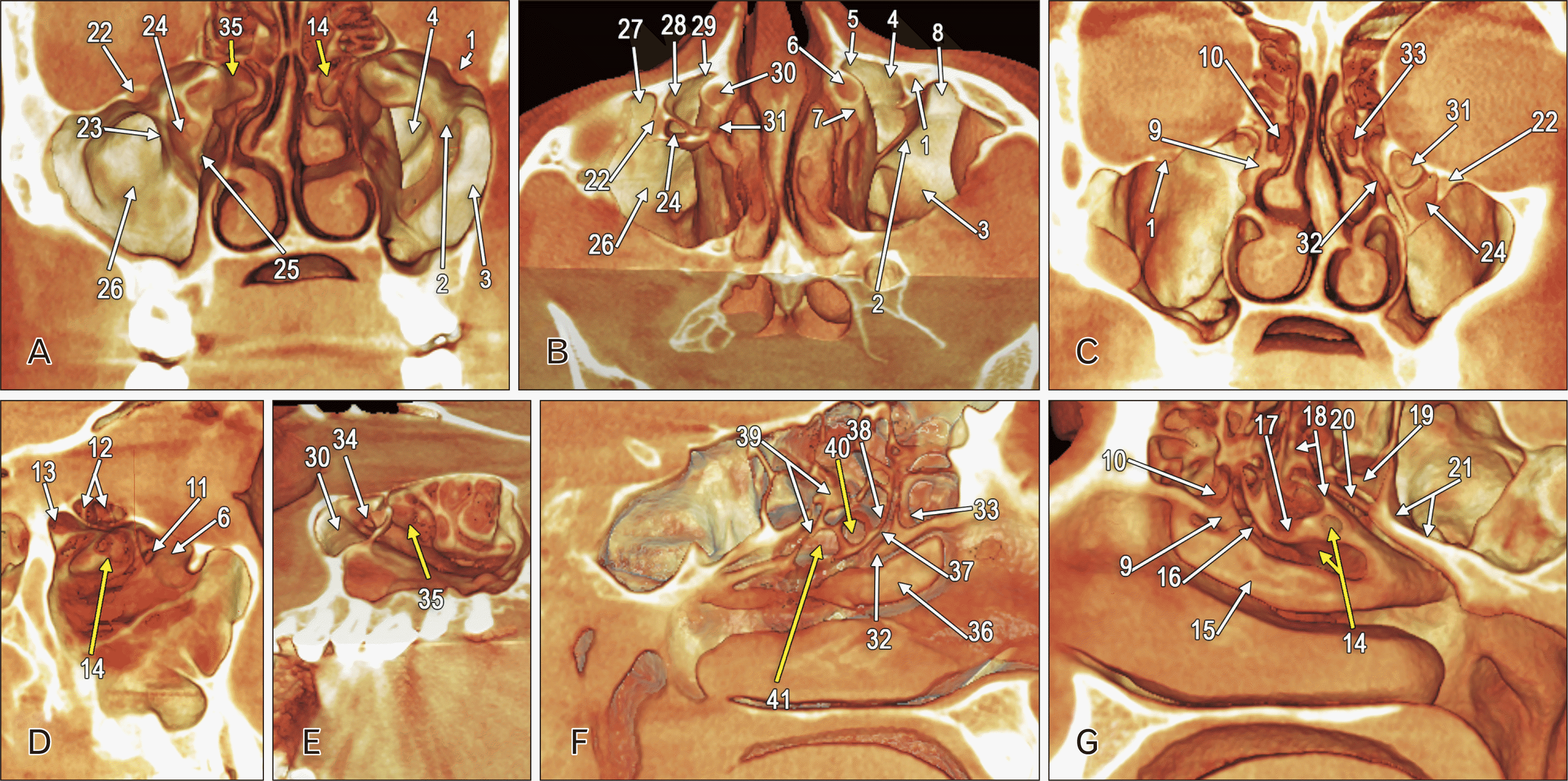
The height of the right MS in its midportion was 40.51 mm. It projected zygomatic, alveolar, and palatine recesses. Lateral and medial infraorbital recesses were found on the anterior sinus wall (Fig. 1A–C). The medial one extended in front of the lacrimonasal canal as a large prelacrimal recess and posterior to the canal as a narrow retrolacrimal recess (Fig. 1B, C). The infraorbital recesses were separated by an oblique septum inserted onto the anterior sinus wall at 3.26 mm lateral to the infraorbital foramen, onto the infraorbital canal in the sinus roof, and the sinus floor (Fig. 1A, B). The upper and lower postero-medial ends of that crescent-like septum were attached to the sinonasal wall. Immediately in front of the superior sinonasal insertion of that septum was the MS ostium (Fig. 1D). That septum incompletely divided the sinus cavity into two chambers, one smaller antero-medial and the other larger, postero-lateral (Fig. 1A, B).
The pterygopalatine angle of the right MS was elevated towards the sphenoidal sinus (SS), thus configuring a sphenoidal recess in front of the maxillary recess of the SS (Fig. 1D). The posterior ethmoid was inclined at 135.10° supero-medially, as referred to the MS roof. Therefore, on sagittal slices, the inferior posterior ethmoid air cells appeared as posterior infraorbital (Haller) cells (Fig. 1D).
A large medial or nasal recess of the postero-lateral chamber of the right maxillary sinus (NRMS) was found extended into the lateral nasal wall, inferior to the ethmoidal labyrinth, and superior to the middle and inferior turbinates (Fig. 1D, G). Its medial pouch-like end was narrowed posteriorly. The entrance into this NRMS was 25.25 mm high. The highest depth of the NRMS, as measured from the sinonasal wall, was 11.0 mm. The maximum right MS width at the level of the NRMS was 38.30 mm. The EB was applied on the anterior side of that NRMS (Fig. 1G). The tip of the NRMS reached within the BLMT (Fig. 1G). Postero-superiorly to the NRMS were the superior and Santorini’s supreme nasal turbinates (Fig. 1G). The NRMS was roofing the middle meatus posterior to the EB, and its infero-medial wall corresponded to the posterior fontanelle.
The maximum height of the left MS was 38.93 mm. It projected alveolar and zygomatic recesses. The sinus was almost wholly divided into antero-medial and postero-lateral chambers by an oblique septum inserted superiorly on the infraorbital canal (Fig. 1A). That septum contained an air cell opened into the antero-medial chamber (Fig. 1A–C) and septum separated medial and lateral infraorbital recesses (Fig. 1B). The medial one extended medially with equally sized prelacrimal and retrolacrimal recesses. The drainage ostium of the MS was in the retrolacrimal recess (Fig. 1E). Both MSs drained into the ethmoidal infundibulum on the outer side of the uncinate process (Fig. 1C). Each uncinate process, left and right, had pneumatised roots (uncinate bullae) (Fig. 1C, F, G).
A left NRMS extended from the postero-lateral chamber of the left MS was also found (Fig. 1E). The entrance into this NRMS was 38.25 mm high. The highest depth of the left NRMS, as measured from the sinonasal wall, was 12.5 mm. The maximum right MS width at the level of the NRMS was 26.39 mm. There were two successive medial pouch-like ends of the left NRMS (Fig. 1F). The anterior one reached beneath the EB, and the posterior one got posterior to it on the anterior side of the BLMT. The postero-superior side of the left NRMS faced the superior meatus and turbinate. It roofed inferiorly the middle meatus.
Both SSs were postsellar, extended pterygoid and maxillary recesses, pneumatised the optic struts, and drained into the sphenoethmoidal recesses.
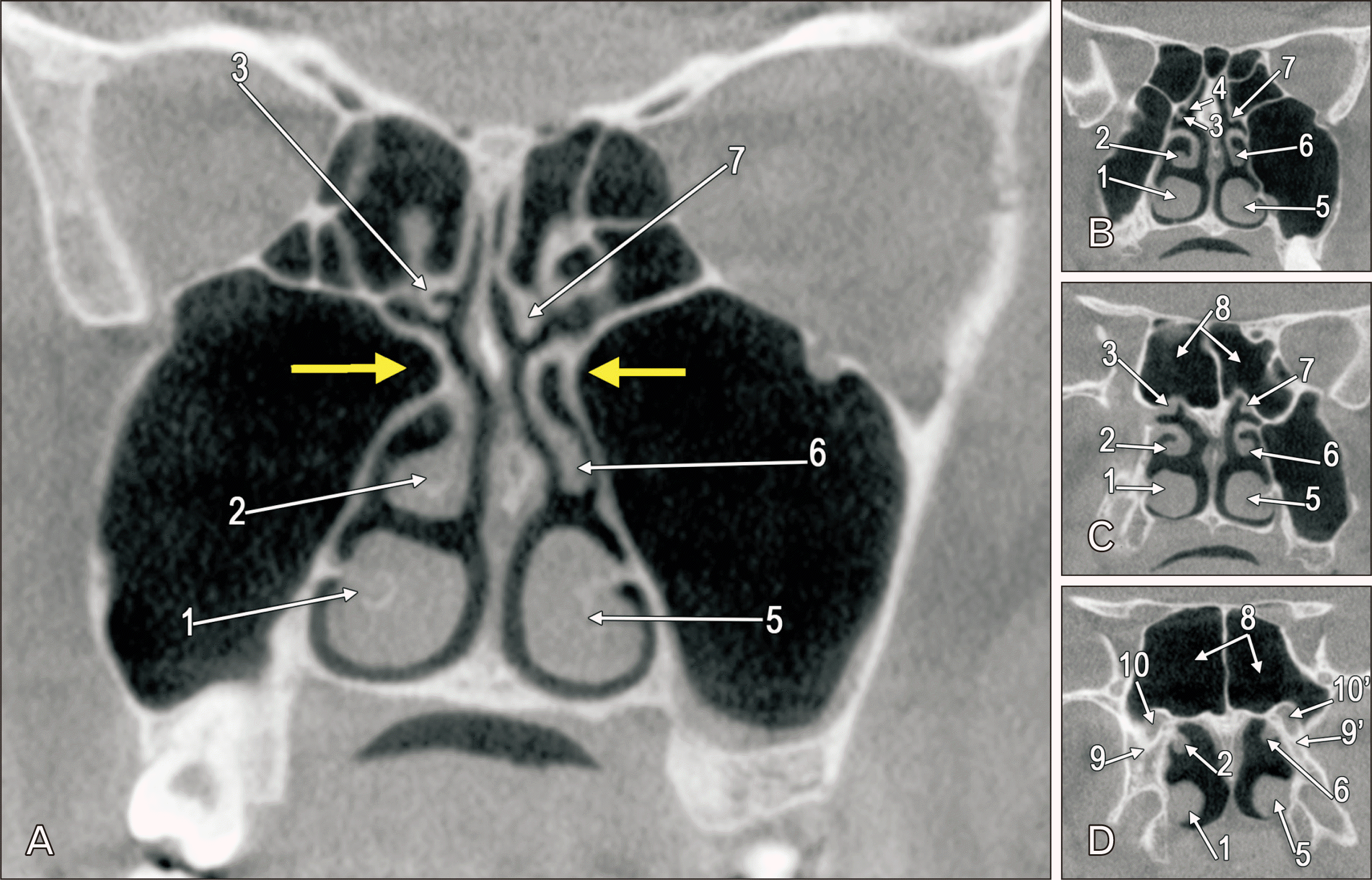
While the posterior ends of the SNTs were attached to the SSs’ walls, the tails of the middle turbinates reached the pterygoid roots. The intermediate segment of the right SNT was ethmoidal, while the anterior end was inserted on the MS wall. Thus, the right SNT’s insertion was maxillo-ethmoido-sphenoidal. That of the left SNT was just ethmoido-sphenoidal (Fig. 2). On each side, the upper margin of the middle nasal turbinate was pneumatised.
The ethmoid originates from the cartilaginous nasal capsule, but the other paranasal sinuses are extensions from the ethmoid into membranous bone. The EB appears in the lateral wall of the middle meatus by 12 weeks [8]. By 14 weeks, the primordial ethmoidal infundibulum and primordial MS develop between the uncinate process and the EB [8]. The anterior and middle ethmoid air cells develop from the EB [8]. These cells further tend to expand to occupy all available space [9]. As the anterior and middle ethmoid air cells and the MS have a common chronological and topographic embryologic origin, an imbalance of these processes could have led to the persistence of a NRMS within the ethmoid instead of an ethmoid air cell.
NRMSs were found extending in front of the BLMTs and beneath the EB. These were significant morphological changes in the lateral nasal walls. The MSs were bicameral and their drainage was associated with the antero-medial chambers. Additional findings were the uncinate bulla, infraorbital recesses of the MS, maxillary recess of the SS, and atypical posterior insertions of the nasal turbinates. Infraorbital recesses could be misjudged as infraorbital ethmoid air cells (Haller cells). The drainage pattern could help distinguish them.
Medial or conchal recesses of the MS may penetrate the inferior turbinate’s root [7]. These could not be misjudged as NRMS because they protrude into the inferior turbinate and not into the middle meatus. A sphenoidal recess of the MS is encountered in less than 10% of cases [1].
The highest mediolateral width of the MS was measured on axial views [10]. Values ranged from 26.82 mm to 28.01 mm [10]. No evidence of NRMS was reported then. The width of the right MS in the present case was over this range upper limit at the level of the NRMS, but on the left side, although the depth of the NRMS was included, the maximum width of the MS was lower than the inferior range limit in the study discussed here. The MS’ maximum width was determined on coronal CBCT slices [11]. The mean values were then 26.72±4.3 mm (right side) and 37.24±3.5 mm (left side) [11]. We got here 38.30 mm for the maximum width of the right MS and 26.39 mm for the left MS width, which is quite opposite to those results [11] that suggest the left MS is larger than the right one.
Intrasinus septa determine bicameral MSs. Only distinct and separate air spaces, each opening into the ethmoidal infundibulum, represent true antral duplication [12]. A MS was found divided by a bony vertical septum and had a non-draining antero-lateral compartment [13]. A double MS should be distinguished from a bicameral maxilla containing a hypoplastic MS in front of a huge extruded posterior ethmoid air cell [1]. Another much rarer type of duplication consists of two non-communicating air spaces separated by a coronally directed bony partition; the two cavities open separately into the middle meatus [12].
The NRMSs were retrobullar thus they could be also termed “retrobullar recesses” of the MSs. The nasal retrobullar recess is a cleft of the lateral nasal wall located postero-superiorly to the EB in front of the BLMT and could be found in >90% of cases [14]. When a NRMS and a nasal retrobullar recess occur concomitantly, the latter is shortened and lies above the former. When finding a closed air space inferior to such a retrobullar recess, surgeons should check so as not to open the MS into the nasal fossa accidentally.
The BLMT forms the EB’s posterior wall. A NRMS is an unexpected pneumatisation posterior to the EB. They should be distinguished between during endoscopic procedures. Different surgical corridors address the ethmoid and an essential landmark during endoscopic approaches is the BLMT that could be altered either by the individual expansion of ethmoid air cells or, rarely, by a NRMS.
We found maxillo-ethmoido-sphenoidal and, respectively, ethmoido-sphenoidal insertions of the SNTs. A previous study found maxillo-ethmoidal and ethmoido-sphenoidal insertions of SNTs modifying the surgical landmarks [15].
Therefore, the retrobullar recess of the MS may occur; thus, the origin of a retrobullar pneumatisation should be carefully documented when surgical approaches are designed.
Notes
References
1. Craiu C, Rusu MC, Hostiuc S, Săndulescu M, Derjac-Aramă AI. 2017; Anatomic variation in the pterygopalatine angle of the maxillary sinus and the maxillary bulla. Anat Sci Int. 92:98–106. DOI: 10.1007/s12565-015-0320-z. PMID: 26663153.
2. Chan HL, Monje A, Suarez F, Benavides E, Wang HL. 2013; Palatonasal recess on medial wall of the maxillary sinus and clinical implications for sinus augmentation via lateral window approach. J Periodontol. 84:1087–93. DOI: 10.1902/jop.2012.120371. PMID: 23106503.
3. Günaçar DN, Köse TE, Arsan B, Aydın EZ. 2022; Radioanatomic study of maxillary sinus palatal process pneumatization. Oral Radiol. 38:398–404. DOI: 10.1007/s11282-021-00569-9. PMID: 34554390.
4. Niu L, Wang J, Yu H, Qiu L. 2018; New classification of maxillary sinus contours and its relation to sinus floor elevation surgery. Clin Implant Dent Relat Res. 20:493–500. DOI: 10.1111/cid.12606. PMID: 29691967.
5. Serindere G, Serindere M, Gunduz K. 2023; Evaluation of maxillary palatal process pneumatization by cone-beam computed tomography. J Stomatol Oral Maxillofac Surg. 124:101432. DOI: 10.1016/j.jormas.2023.101432. PMID: 36921841.
6. Cârstocea L, Rusu MC, Mateşică DŞ, Săndulescu M. 2020; Air spaces neighbouring the infraorbital canal. Morphologie. 104:44–50. DOI: 10.1016/j.morpho.2019.07.002. PMID: 31492524.
7. Măru N, Rusu M, Săndulescu M. 2013; The conchal recess of the maxillary sinus: a 100 cases CB CT study. Rom J Funct Clin Macro Microsc Anat Anthropol. 12:271–5.
8. Wang RG, Jiang SC. 1997; The embryonic development of the human ethmoid labyrinth from 8-40 weeks. Acta Otolaryngol. 117:118–22. DOI: 10.3109/00016489709118002. PMID: 9039492.
9. Márquez S, Tessema B, Clement PA, Schaefer SD. 2008; Development of the ethmoid sinus and extramural migration: the anatomical basis of this paranasal sinus. Anat Rec (Hoboken). 291:1535–53. DOI: 10.1002/ar.20775. PMID: 18951481.
10. R SSS, Khan N, Parameswaran R, Boovaraghavan S, Nagi M. 2024; Evaluation of dimensional changes in maxillary and frontal sinus in adult patients with anterior open bite and normal overbite: a retrospective cone beam computed tomography (CBCT) study. Cureus. 16:e53710. DOI: 10.7759/cureus.53710. PMID: 38455800. PMCID: PMC10919753.
11. Aşantoğrol F, Coşgunarslan A. 2022; The effect of anatomical variations of the sinonasal region on maxillary sinus volume and dimensions: a three-dimensional study. Braz J Otorhinolaryngol. 88(Suppl 1):S118–27. DOI: 10.1016/j.bjorl.2021.05.001. PMID: 34053909. PMCID: PMC9734263.
12. Glass M. 1952; Duplication of the maxillary antrum symptomatology, diagnosis and treatment. S Afr Med J. 26:895–902. PMID: 13028765.
13. Yue V, Bleach NR, van Hasselt CA. 1994; Double maxillary antrum as a cause of maxillary sinus mucocele. Ear Nose Throat J. 73:839–41. DOI: 10.1177/014556139407301109. PMID: 7828478.
14. Bolger WE, Mawn CB. 2001; Analysis of the suprabullar and retrobullar recesses for endoscopic sinus surgery. Ann Otol Rhinol Laryngol Suppl. 186:3–14. DOI: 10.1177/00034894011100S501. PMID: 11372938.
15. Rusu MC, Hostiuc S, Motoc AGM, Mogoantă CA, Sava JC, Săndulescu M. 2020; The sphenoethmoidal sinus and the modified anatomy of the related structures. Rom J Morphol Embryol. 61:143–8. DOI: 10.47162/RJME.61.1.16. PMID: 32747905. PMCID: PMC7728111.
Fig. 1
Three-dimensional volume renderings. (A) Posterior view of the maxillary sinuses (MSs), posterior walls removed. (B) Superior view of the MSs, roofs removed. (C) Anterior view of the MSs, anterior walls removed. (D) Lateral view of the medial wall of the right MS. (E) Infero-lateral view of the medial wall of the left MS. (F) Medial view of the left lateral nasal wall. (G) Medial view of the right lateral nasal wall. Legends 1–21 indicate right-side landmarks. Legends 22–41 indicate left-side landmarks. 1. Infraorbital canal; 2. oblique intrasinus septum; 3. postero-lateral chamber of the MS; 4. medial infraorbital recess; 5. prelacrimal recess; 6. lacrimonasal canal; 7. retrolacrimal recess; 8. lateral infraorbital recess; 9. uncinate process; 10. uncinate bulla; 11. main drainage ostium of the right MS; 12. posterior Haller cells; 13. sphenoidal recess of the MS; 14. nasal recess of the MS; 15. middle nasal turbinate; 16. semilunar hiatus; 17. ethmoidal bulla; 18. basal lamella of the middle nasal turbinate; 19. Santorini’s supreme nasal turbinate; 20. superior nasal turbinate; 21. floor of the sphenoidal sinus; 22. infraorbital canal; 23. oblique intrasinus septum; 24. air cell of the intrasinus septum; 25. the slit-like passage between the chambers of the MS; 26. postero-lateral chamber of the MS; 27. lateral infraorbital recess; 28. medial infraorbital recess; 29. prelacrimal recess; 30. lacrimonasal canal; 31. retrolacrimal recess; 32. uncinate process; 33. uncinate bulla; 34. main drainage ostium of the left MS; 35. nasal recess of the MS; 36. middle nasal turbinate; 37. semilunar hiatus; 38. ethmoidal bulla; 39. basal lamella of the middle nasal turbinate; 40. anterior medial end of the nasal recess of the MS; 41. posterior medial end of the nasal recess of the MS.

Fig. 2
Anterior views of coronal two-dimensional anterior-to-posterior slices (A–D) through the nasal turbinates. The thick arrows indicate the nasal recesses of the maxillary sinuses. 1. right inferior turbinate; 2. right middle turbinate; 3. right superior turbinate; 4. Santorini’s supreme turbinate; 5. left inferior turbinate; 6. left middle turbinate; 7. left superior turbinate; 8. sphenoidal sinuses; 9, 9’. non-lamellar base of the pterygoid process; 10, 10’. vidian canal.


 PDF
PDF Citation
 Print
Print


 XML Download