Introduction
The transversus abdominis (TA) muscle, the deepest of the anterolateral abdominal wall muscles ends anteriorly in aponeurosis. The aponeurosis passes medially and blends at the linea alba. The medial edges of the TA muscles are closest near the xiphoid process, when traced caudally they diverge laterally and are furthest from the lateral edge of rectus sheath at the umbilicus. Near the xiphoid process, the muscular part of TA extends behind the rectus abdominis into the posterior layer of the rectus sheath [
1]. In the traditional teaching of rectus sheath formation, all the three anterior abdominal wall muscles become aponeurotic at linea semilunaris
i.e., the lateral border of rectus abdominis [
2,
3]. The Gray’s Anatomy mentions that the TA muscle gets incorporated into the posterior rectus sheath above the umbilicus [
1].
Novitsky et al. [
4], in 2012, first described the transversus abdominis release (TAR) as a novel approach for surgical repair of large complex ventral hernias. In TAR, following creation of retro-rectus space, the posterior rectus sheath is incised medial to the lateral edge of rectus abdominis muscle (linea semilunaris) to expose the medial edge of the TA muscle. The TA muscle is then divided longitudinally parallel to lateral margin of rectus sheath, to enter the preperitoneal space, separating the peritoneum away from the anterior abdominal wall. This myofascial release allows adequate medial mobilization of the posterior rectus sheath, while maintaining the neurovascular supply, and offers wider space for mesh placement [
5]. The mesh is placed in the preperitoneal or retromuscular space following approximation of the medial edges of posterior rectus sheath [
4]. The TAR has removed the boundary of linea semilunaris as a limitation to the performance of retro-muscular repairs [
6]. Punekar et al. [
7] (2016) studied 100 computed tomography (CT) abdomen images and presented a revised understanding that TA muscle is incorporated in the posterior rectus sheath, above the level of umbilicus. Hence, we sought to describe the medial margin of TA muscle at different anatomic landmarks, the extent of its incorporation in the posterior rectus sheath and its relation to the inferior epigastric vessels, to improve our understanding of anatomy relevant to TAR.
Materials and Methods
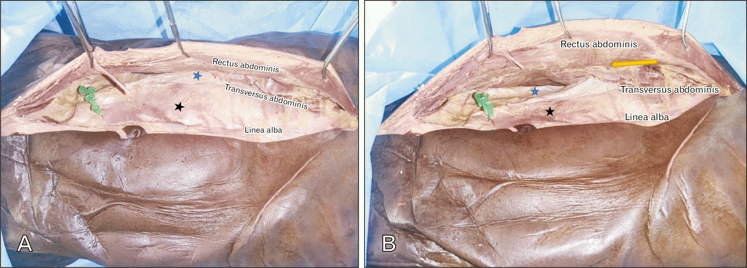
Ethical approval for this study was taken from the Institutional Review Board of Christian Medical College (IRB no. 13674). Ten conventional formalin fixed cadavers (5 males, 5 females), which are donated to the Department of Anatomy for teaching and research purposes were dissected, these cadavers were obtained after getting the consent from the donor families. The age of cadavers ranged from 35 to 70 years, with no prior midline hernial defects, or laparotomy scars. The authors followed the same dissection steps as done in TAR surgeries (
Fig. 1A). A midline skin incision was made from xiphisternum to pubic symphysis. The anterior rectus sheath was incised on either side of linea alba (approximately 1 cm lateral). The retro-rectus space was developed towards the linea semilunaris on either side, visualizing the junction between the posterior and anterior rectus sheath, ending just medial to the neurovascular bundle penetrating the posterior rectus sheath. The posterior rectus sheath was incised 0.5 cm medial to the neurovascular bundle, to expose the underlying TA muscle (
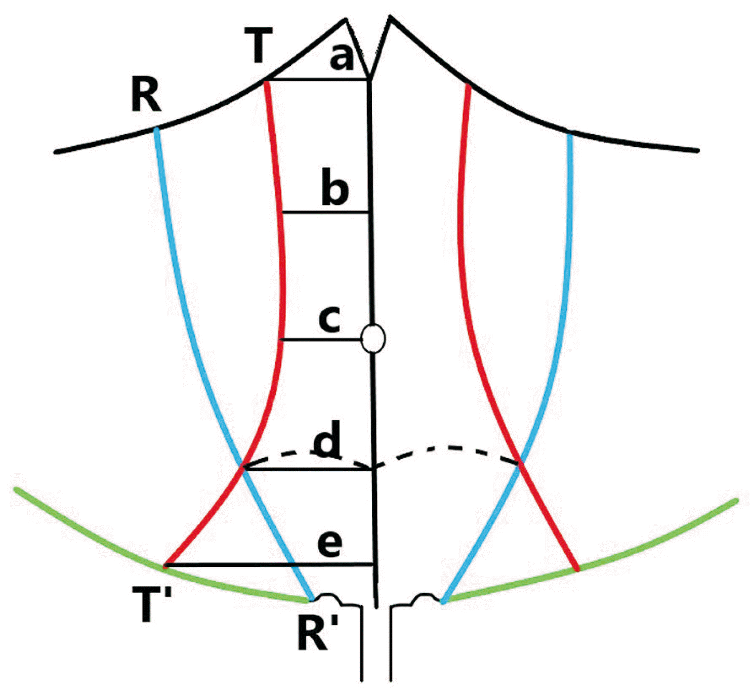
Fig. 1B). Once exposed, the measurements of the medial edge of muscular part of TA in the posterior rectus sheath to the linea alba (TL) and the distance from the lateral margin of rectus abdominis to the linea alba (RL) was measured at different anatomic landmarks, such as 1) xiphisternum, 2) midway between the xiphisternum and the umbilicus, 3) level of umbilicus, 4) arcuate line, and 5) midway between arcuate line and pubic symphysis, by using a vernier caliper/measuring tape in mm by two observers (
Fig. 2). The mean value of the observers was taken for each landmark. The mean TA distance was calculated by RL–TL. The distance between the inferior epigastric vessels and the medial border of TA muscle was also measured.
Fig. 1
(A) Anterior view of the anterior abdominal wall, showing transversus abdominis release procedure on a cadaver. Black asterisk, posterior rectus sheath; blue asterisk, fascia transversalis (plane developed beneath the transversus abdominis); green pins, arcuate line. (B) Showing the lateral view of the anterior abdominal wall, yellow arrow pointing at the neurovascular structures piercing the posterior rectus sheath.


Fig. 2
Schematic diagram showing the measurements of transversus abdominis muscle. TT’, medial margin of transversus abdominis muscle; RR’, lateral margin of rectus abdominis; dotted line, arcuate line; measurements were taken at (a) the level of xiphisternum; (b) the midway between xiphisternum and umbilicus; (c) the level of umbilicus; (d) the level of arcuate line; (e) the midway between arcuate line and pubic symphysis.


Results
The distance from the linea alba to the medial border of TA and the lateral border of rectus abdominis was measured at 5 levels, as described in the methodology and tabulated in
Table 1.
Table 1
Measurements of transversus abdominis muscle taken at different anatomical landmarks
|
Xiphisternum |
Minimum |
|
Maximum |
|
Mean±SD |
|
Median |
|
IQR |
P-value |
|
R |
L |
R |
L |
R |
L |
R |
L |
R |
L |
|
Distance between medial margin of TA to LA (mm) |
11.31 |
10.6 |
|
24.72 |
20.9 |
|
17.0±4.7 |
15.6±3.2 |
|
16.9 |
15.7 |
|
20.6–12.8 |
17.9–12.9 |
0.40 |
|
Distance between the lateral border of RA to LA (mm) |
54.1 |
56.9 |
|
92.7 |
83.4 |
|
75.9±12.4 |
69.8±8.5 |
|
82.6 |
68.6 |
|
84.8–64.4 |
77.6–62.2 |
0.02*
|
|
TA distance (RL–TL) |
41.3 |
39.7 |
|
80.6 |
80.2 |
|
61.6±11.8 |
58.9±11.9 |
|
61.3 |
60.6 |
|
70.5–52.6 |
67.7–52.1 |
0.37 |
|
Midway between xiphisternum and umbilicus |
|
|
|
Distance between medial margin of TA to LA (mm) |
20.5 |
21.25 |
|
72.71 |
51.77 |
|
49.4±16.0 |
42.8±9.3 |
|
49.5 |
47.1 |
|
63.6–37.2 |
64.4–49.5 |
0.53 |
|
Distance between the lateral border of RA to LA (mm) |
48.2 |
48.3 |
|
92.7 |
83.4 |
|
74.8±12.7 |
69.9±9.4 |
|
77.2 |
71.3 |
|
84.6–66.5 |
77.1–65.5 |
0.05 |
|
TA distance (RL–TL) |
12.84 |
15.5 |
|
39.1 |
36.6 |
|
25.4±9.0 |
27.1±5.9 |
|
27.4 |
27.8 |
|
32.8–15.9 |
32.1–15.2 |
0.29 |
|
Umbilicus |
|
|
|
Distance between medial margin of TA to LA (mm) |
58.6 |
57.8 |
|
125.6 |
114.4 |
|
93.8±19.2 |
91.0±17.9 |
|
98.3 |
95.6 |
|
102.6–96.4 |
106.8–81.1 |
0.12 |
|
Distance between the lateral border of RA to LA (mm) |
51.1 |
56.2 |
|
85.7 |
89.8 |
|
69.2±10.3 |
69.6±9.9 |
|
68.5 |
69.4 |
|
74.3–61.2 |
76.6–60.6 |
0.80 |
|
TA distance (RL–TL) |
–40 |
–38.8 |
|
–67 |
–1.7 |
|
–24.6±11.9 |
–21.4±12.4 |
|
–27.4 |
–21.3 |
|
–19.6 to –34.8 |
–8.9 to –33.3 |
0.26 |
|
Arcuate line |
|
|
|
|
|
|
|
|
|
|
|
|
|
|
|
|
Distance between medial margin of TA to LA (mm) |
58.4 |
48.2 |
|
107 |
97.7 |
|
88.7±13.5 |
83.3±14.7 |
|
92.4 |
87.4 |
|
95.9–87.2 |
93.9–79.9 |
0.15 |
|
Distance between the lateral border of RA to LA (mm) |
53.7 |
52.8 |
|
82.4 |
77 |
|
63.7±7.7 |
63.3±6.2 |
|
63.3 |
61.6 |
|
66.9–57.7 |
67.3–59.6 |
0.70 |
|
TA distance (RL–TL) |
–36.3 |
–37.2 |
|
–4.1 |
11.6 |
|
–24.9±8.6 |
–19.9±14.4 |
|
–27 |
–23 |
|
–23.4 to –30.2 |
–10.5 to –32.1 |
0.18 |
|
Midway between arcuate line and symphysis pubis |
|
|
|
Distance between medial margin of TA to LA (mm) |
56.2 |
58.5 |
|
82.6 |
73.4 |
|
73.2±6.9 |
67.0±4.7 |
|
76 |
67.3 |
|
77.2–72.4 |
71.2–63.4 |
0.01*
|
|
Distance between the lateral border of RA to LA (mm) |
41.5 |
39.5 |
|
68.2 |
67.3 |
|
50.4±6.9 |
48.1±8.7 |
|
49.3 |
45.2 |
|
53.5–45.9 |
49.7–41.3 |
0.34 |
|
TA distance (RL–TL) |
–30.9 |
–26.5 |
|
–6 |
–5.7 |
|
–22.9±6.5 |
–18.9±6.0 |
|
–24.1 |
–20.4 |
|
–17.3 to –28.1 |
–15.2 to –23.3 |
0.11 |

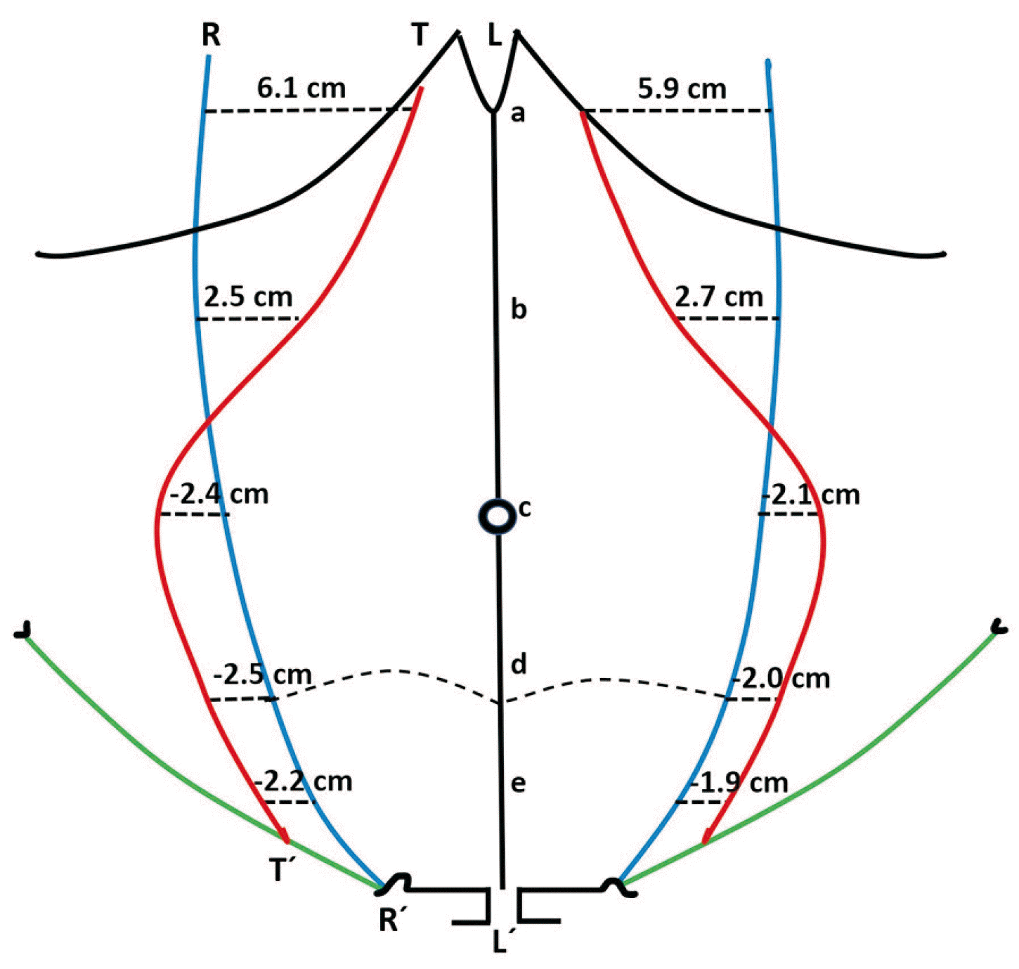
The proportion of the TA muscle being incorporated in the posterior rectus sheath differed at each level. At the level of xiphisternum, the TA muscle resides within the posterior rectus sheath, such that the mean overlap of the two muscles (rectus abdominis and TA) was 61.6 mm on right side and 58.9 mm on the left side. The mean overlap of these two muscles midway between the umbilicus and xiphisternum was 25.4 mm on the right side and 27.1 mm on the left. Between this level and the umbilicus, the TA muscle exited the posterior rectus sheath. The mean distance between the two muscles on the right side at the level of the umbilicus, arcuate line and midway between arcuate line and pubic symphysis were –24.6, –24.9, and –22.9 mm respectively. On the left side these values were –21.4, –19.9, and –18.9 mm respectively (
Table 1,
Fig. 3,
Supplementary Fig. 1).
Fig. 3
Depiction of the outlines of the medial margin of transversus abdominis muscle (TT’, red line); lateral margin of rectus abdominis muscle (RR’, blue line); LL’, linea alba; RL, distance between lateral margin of rectus abdominis and linea alba; TL, distance between the medial margin of transversus abdominis and linea alba. The black dotted lines represent the mean TA distance (RL–TL) at various levels (a, xiphisternum; b, midway between xiphisternum and umbilicus; c, umbilicus; d, arcuate line; e, midway between arcuate line and pubic symphysis). The positive distances indicate overlap and negative distances indicate lack of overlap. The distances in mm are converted to cm for the sake of ease and clarity.


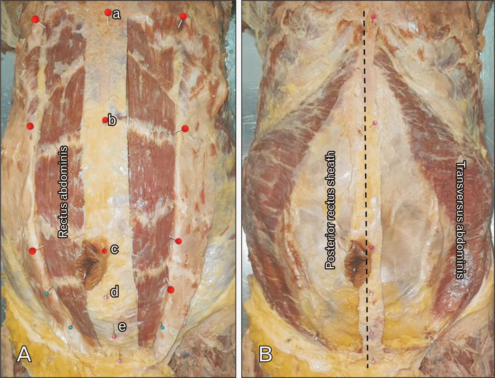
The TA muscle was present within the posterior rectus sheath at the level of xiphisternum, and midway between the xiphisternum and the umbilicus in all the ten cadavers (100%) (
Fig. 4).
Fig. 4
Dissection of anterior abdominal wall demonstrating the rectus abdominis muscle, transversus abdominis (TA), and posterior layer of rectus sheath (A, B). Measurements were taken at (a) xiphisternum, (b) midway between xiphisternum and umbilicus, (c) umbilicus, (d) arcuate line, and (e) midway between arcuate line and pubic symphysis. Dotted line indicates linea alba. Note that the TA completely exits the posterior rectus sheath at the level of umbilicus.


The mean distance between the medial border of TA muscle and the inferior epigastric vessels just above the pubic symphysis was 18.9 mm on the right and, and 17.2 mm on the left (
Table 2). There was no divarication of the rectus abdominis in any of the cadavers.
Table 2
Measurements of transversus abdominis muscle in relation to the inferior epigastric vessels
|
Minimum |
|
Maximum |
|
Mean±SD |
|
Median |
|
IQR |
P-value |
|
R |
L |
R |
L |
R |
L |
R |
L |
R |
L |
|
Distance between medial margin of TA to inferior epigastric vessels (mm) |
13.7 |
9.3 |
|
27.4 |
25.9 |
|
18.9±4.6 |
17.2±5.5 |
|
19.0 |
16.9 |
|
14.4–22.1 |
12.8–21.5 |
0.34 |

Discussion
The anterior abdominal wall is considered to have two parts: anterolateral and middle. The anterolateral portion is composed of the external oblique, the internal oblique, and the TA muscles. The middle portion is composed of the rectus abdominis and pyramidalis muscles. From various origin, the TA muscle fibres pass medially, the uppermost fibres become aponeurotic near the midline, posterior to the rectus abdominis. At the level of the umbilicus, the aponeurotic fibers begin lateral to the rectus abdominis muscle [
8]. Traditional teaching describes the formation of posterior rectus sheath by the posterior lamina of internal oblique aponeurosis and the aponeurosis of the TA, from the costal margin to a point midway between the umbilicus and the pubis [
3]. This was challenged by Novitsky et al. [
4] in 2012, while describing TAR procedure as a modified approach to posterior component separation (PCS). Punekar et al. [
7] (2016), after studying 100 CT abdomen images presented a revised understanding of the components of the posterior rectus sheath, showing the incorporation of TA muscle in the posterior rectus sheath above the umbilicus. Below the umbilicus, the TA muscle exited the posterior rectus sheath and retreated laterally behind the internal oblique muscle in the lower abdomen [
7]. This updated knowledge on TA anatomy is important, it forms the basis for PCS with TAR which helps cover large hernial defects in the ventral abdominal wall.
The key step to the PCS with TAR procedure is the release of the TA muscle itself. In conjunction with the internal oblique muscle, the TA muscle serves as a corset which provides hoop tension around the abdomen. The theory behind TAR is that by dividing this muscle, circumferential tension will be released, providing significant medial advancement of the rectus muscle and its enveloping sheaths, thus allowing re-approximation of the anterior rectus sheath in the midline. TAR affords 8 cm to 12 cm advancement per side in most patients [
9]. In a cadaveric model, posterior sheath advancement of over 11 cm was obtained, at the end of the TAR, allowing for restoration of the visceral sac. This confirms the suitability of PCS with TAR procedure for reliable abdominal wall reconstructions for very large ventral abdominal defects [
10]. The TAR has several advantages which include significant medial mobilization of the posterior rectus sheath that allows for extensive lateral dissection in a potentially unlimited space between the TA muscle and the underlying transversalis fascia. It avoids disruption of the neurovascular supply to the rectus abdominis muscle and anterolateral abdominal wall skin thereby avoiding muscle weakness, paresthesia, numbness, and skin necrosis. Even though the exact clinical significance of preserving these neurovascular bundles remains debated, it has been suggested that damage to these bundles may predispose to abdominal wall bulging and laxity [
11].
The present study demonstrates the significant incorporation of the TA muscle within the posterior rectus sheath above the level of umbilicus, which was quite different from the traditional teaching on rectus sheath formation. It also highlights that the TA muscle exits the posterior rectus sheath below a point midway between the umbilicus and the xiphisternum. This contrasts the study done by Punekar et al. [
7] in American population, where the TA left the posterior rectus sheath at the level of umbilicus. These findings are very important if one needs to release the transverse abdominis muscle to get a formidable advancement of the posterior rectus sheath to cover large defects. This study suggests that functional transfer of abdominal wall components can help in the reconstruction of large ventral abdominal defects, without the need for muscle flaps like tensor fascia lata flap. This is based on the fact that contractile dynamic muscular tissues resist strain and stress well [
12]. This procedure avoids disruption of the neurovascular supply to the rectus abdominis and anterolateral abdominal wall muscles, as the plane of dissection is created between the TA and the fascia transversalis.
In the present study, the distance between the medial border of TA and the inferior epigastric vessels just above the level of pubic symphysis was recorded. Inferior epigastric artery (IEA) false aneurysms are recognized complications following abdominal surgery or trauma. If left untreated potential complications include painful persistent swelling, abscess formation or rupture [
13]. Abdominal port placement and dissection of the muscles are two steps which can potentially injure the epigastric vessels [
14]. The IEA traverses the posterior wall of rectus sheath at the level of arcuate line between 4 to 8 cm from the midline [
14]. Creating the retro-rectus muscle plane and further dissection of the medial margin of the TA muscle are risk factors for inferior epigastric vessel damage. There are a few case reports on the injury of the IEA during the abdominal wall reconstruction and further placement of sublay mesh [
13-
15]. The distance between the medial edge of TA muscle and the inferior epigastric vessels was less than 2 cm, in the present study.
In conclusion, the present study demonstrated the presence of muscular part of TA within the posterior rectus sheath above the umbilicus, its complete exit from the rectus sheath at the level of umbilicus, and its lateral retreat posterior to the internal oblique below the level of umbilicus. This is opposed to the traditional teaching on posterior rectus sheath formation. This knowledge of updated anatomy of the posterior rectus sheath helps in understanding the TAR for ventral abdominal hernia repairs. The limitation of the study include the shrinkage of specimens caused by formalin fixation.


 PDF
PDF Citation
 Print
Print


 XML Download