Introduction
Celiac trunk, the artery of foregut, and the 3 branches originating from it (common hepatic, splenic and left gastric) are supplying the alimentary canal down to the opening of bile duct, and the derivatives, spleen, liver, and pancreas. It arose from the front of the aorta, between crura of diaphragm, opposite the body of T
12 vertebra. It is a short wide trunk, flanked by the celiac group of the preaortic lymph nodes. The celiac ganglion of the sympathetic system lies one on each side and sends nerves to the artery which are carried along all its branches. Common hepatic artery (CHA) leaves celiac trunk and runs forward to continue between layers of lesser omentum anteriorly to the portal vein. it gives a branch, gastroduodenal and becomes the proper hepatic, that divides into right hepatic and left hepatic branches [
1,
2].
Celiac trunk direction is inclined by the hepatic artery origin and by the topography of neck of the pancreas. The hepatic artery is responsible for pulling celiac trunk to the right and against adults, the hepatic artery diameter in younger children appear larger than diameter of the splenic artery. In newborn, celiac artery has different course and directed usually to the left [
3].
Noah et al. [
4] mentioned that angiography forms the basis for effective operative planning and the clinician is often challenged by arterial variants. These variations that are hardly mentioned in anatomical textbooks must be carefully taken into consideration by surgeons. The authors noticed variations in 25.5% from a total of 204 angiographies. Cavdar et al. [
5] mentioned that the anatomical variations in celiac trunk are due to changes in the development of ventral splanchnic arteries and these variations should be kept in mind during surgical or non-surgical evaluation. Covey et al. [
6] added that radiologists who perform hepatic artery embolization should be aware of the hepatic artery variations, as failure to diagnose any aberrant artery may lead to imperfect embolization to liver tumors.
Before liver resections or transplantations, angiography to celiac trunk and superior mesenteric arteries is routinely done [
7]. In mixed population of organ donors, anatomy of hepatic artery was anomalous in 42.22% of patients [
8]. Although anatomical variations of hepatic arteries are common, most can be safely managed. Because of their small caliber, reconstruction of these vessels during transplantation is difficult. Also, they are very close to the other hilar structures. The creation of practical techniques for these anastomoses can simplify the surgery and minimize the incidence of vascular complications [
9,
10].
Since anatomic understanding is the starting point of medical knowledge [
11], it was determined to do research in Egyptian individuals to evaluate and describe the prevalence of celiac and superior mesenteric arterial variants, and to supply angiographic evidence for the classic arterial anatomy.
Go to :

Results
In this research, 389 angiographs were selected, and the data were recorded (
Table 1). Of the available 389 angiograms, 286 patients (73.52%) had the standard anatomy of celiac trunk. The celiac trunk was divided into 3 branches: left gastric artery (LGA), splenic a. and CHA. Then CHA bifurcated to GDA and PHA. PHA branched to RHA and LHA arteries (
Fig. 1). On the other hand, 103 patients (26.47%) had a single or multiple vessel variation. So, the total variations in these patients reached 145 (37.27%).
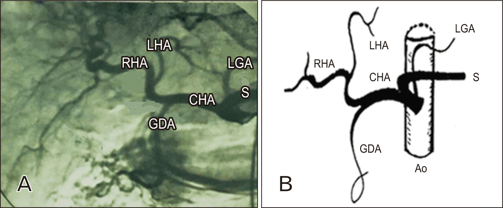 | Fig. 1Anteroposterior angiogram (A) showing the standard anatomy of the celiac axis. The celiac trunk gives splenic artery (S), the CHA and the LGA. Notice the GDA, proper hepatic artery that divides into RHA and LHA. Sub image (B) is a schematic diagram showing the standard anatomy of the celiac axis. Ao, aorta; LGA, left gastric artery; GDA, gastroduodenal artery; RHA, right hepatic artery; LHA, left hepatic artery; CHA, common hepatic artery. 
|
Table 1
Anatomic variations in celiac trunk and superior mesenteric artery
|
Anatomic variations |
Number of cases (%) |
|
Total number of patients |
389 (100) |
|
Standard arterial anatomy |
286 (73.52) |
|
Patients carrying arterial variations |
103 (26.47) |
|
Absent celiac trunk |
2 (0.51) |
|
Replaced LHA to LGA |
22 (5.65) |
|
Replaced LHA to celiac trunk |
15 (3.85) |
|
Total variations of LHA |
37 (9.51) |
|
Accessory RHA from LHA |
5 (1.28) |
|
Accessory RHA from SMA |
17 (4.37) |
|
Total accessory RHAs |
22 (5.65) |
|
Replaced RHA to aorta |
5 (1.28) |
|
Replaced RHA to SMA |
15 (3.85) |
|
Replaced RHA to celiac trunk |
13 (3.34) |
|
Total replaced RHAs |
33 (8.48) |
|
Total variations of RHA |
55 (14.13) |
|
Origin of GDA from the celiac trunk |
7 (1.79) |
|
Origin of GDA from RHA |
9 (2.31) |
|
Origin of GDA from LHA |
11 (2.82) |
|
Origin of LGA from splenic artery |
2 (0.51) |
|
Replaced PHA to SMA |
1 (0.25) |
|
Hepatomesentric trunk |
1 (0.25) |
|
Quadrification of celiac trunk |
2 (0.51) |
|
Quadrification of CHA |
1 (0.25) |
|
Origin of splenic artery from aorta |
2 (0.51) |
|
Origin of Inf phrenic artery from celiac trunk |
8 (2.05) |
|
Double hepatic artery |
7 (1.79) |
|
Total variants |
145 (37.27) |

Sometimes, the right or the left inferior phrenic arteries originated from celiac trunk. This finding was seen in 8 patients (2.05%) of this study (
Fig. 2). Also, celiac trunk quadrifurcation was noticed in 2 cases (0.51%) (
Fig. 3).
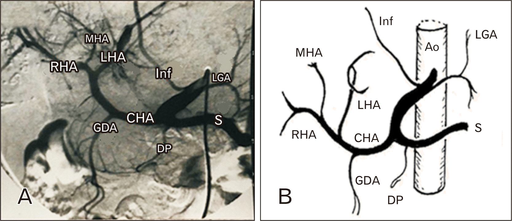 | Fig. 2(A) Anteroposterior angiogram of celiac axis showing the splenic artery (S), the CHA, the LGA, and the right inferior phrenic artery (Inf) branching from celiac trunk. Notice the GDA, the dorsal pancreatic branch (DP) of the splenic artery and an MHA branching from the RHA. Sub image (B) is a schematic diagram showing the anatomical variants. CHA, common hepatic artery; LGA, left gastric artery; GDA, gastroduodenal artery; MHA, middle hepatic artery; RHA, right hepatic artery; LHA, left hepatic artery; Ao, aorta. 
|
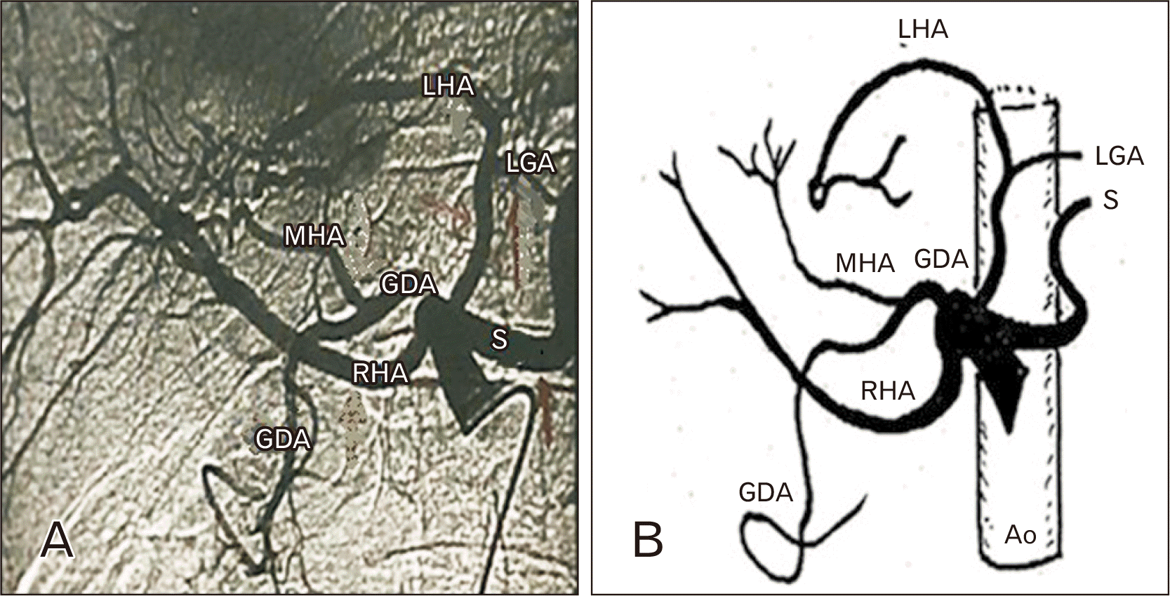 | Fig. 3(A) Anteroposterior angiogram of the celiac axis showing quadrifurcation of the celiac trunk into RHA, common origin of LHA, LGA, GDA, and splenic artery (S). Notice the MHA originating from the GDA. Sub image (B) is a schematic diagram showing the anatomical variants. RHA, right hepatic artery; LHA, left hepatic artery; LGA, left gastric artery; GDA, gastroduodenal artery; MHA, middle hepatic artery; Ao, aorta. 
|
Although absence of the celiac trunk is a very rare condition, this arrangement was found in 2 cases (0.51%) of our series. In 1 case, splenic artery arose directly from aorta (
Fig. 4A), and the RHA was replaced to the SMA (
Fig. 4B). In the other case, splenic artery originated from the aorta (
Fig. 5A), while the CHA and SMA appeared fused together in a common trunk, the hepatomesenteric trunk that was found in only 1 patient (0.25%) of this study (
Fig. 5B). Another unusual case was the division of the CHA into GDA, RHA, LHA, and middle hepatic to form what is called quadrifurcation of the CHA. This was noticed in only 1 case (0.25%) of the present work (
Fig. 5C). Also, middle hepatic artery (MHA) was visualized in 7 (1.79%) patients in this study. It arose from the RHA in 2 patients (
Fig. 2), from the GDA in 4 patients (
Fig. 3) and from CHA artery in only 1 (
Fig. 5C).
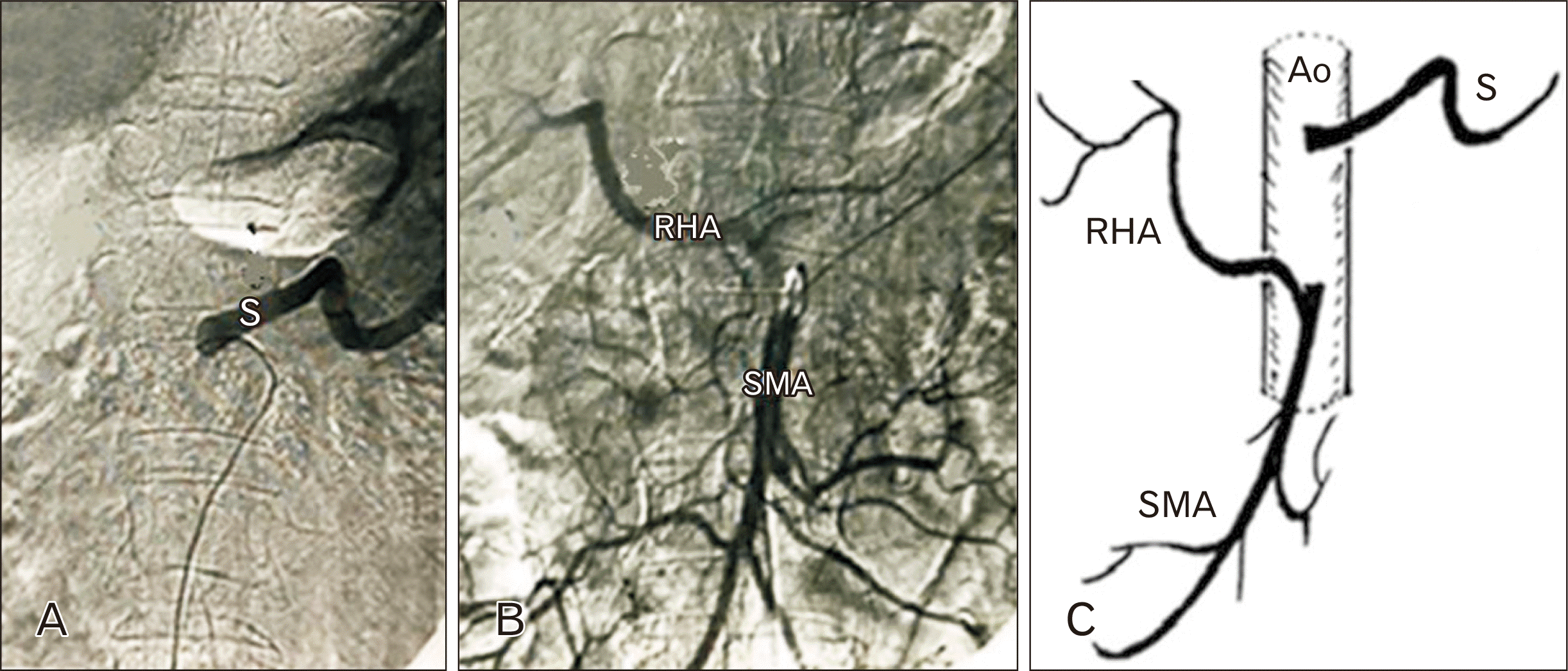 | Fig. 4Anteroposterior angiograms of the same patient showing absence of celiac trunk with separate origin of the splenic artery (S) from aorta (A), and replaced RHA to the SMA (B). Sub image (C) is a schematic diagram showing the anatomical variants. RHA, right hepatic artery; SMA, superior mesenteric artery; Ao, aorta. 
|
 | Fig. 5(A) Anteroposterior angiograms of the same patient showing separate origin of splenic artery (S) from aorta, the common origin of CHA and SMA forming a hepatomesenteric trunk (B); and quadrifurcation of the CHA into RHA, LHA, MHA, and GDA (C). Sub image (D) is a schematic diagram showing the anatomical variants. CHA, common hepatic artery; SMA, superior mesenteric artery; RHA, right hepatic artery; LHA, left hepatic artery; MHA, middle hepatic artery; GDA, gastroduodenal artery; Ao, aorta. 
|
The splenic artery was usually the most voluminous branch of the celiac trunk. Two patients (0.51%) had an abnormal origin of splenic a. from abdominal aorta. In 1 case, the RHA was replaced to the SMA (
Fig. 4A, B), while in the other case was associated with a hepatomesenteric trunk (
Fig. 5A, B). In the 2 cases, no typical celiac trunk was noticed.
Variations in PHA were observed in only 1 case (0.25%) of this study where it is replaced to the SMA and gave right & left hepatic arteries (
Fig. 6A), while the GDA arose from the celiac trunk (
Fig. 6B).
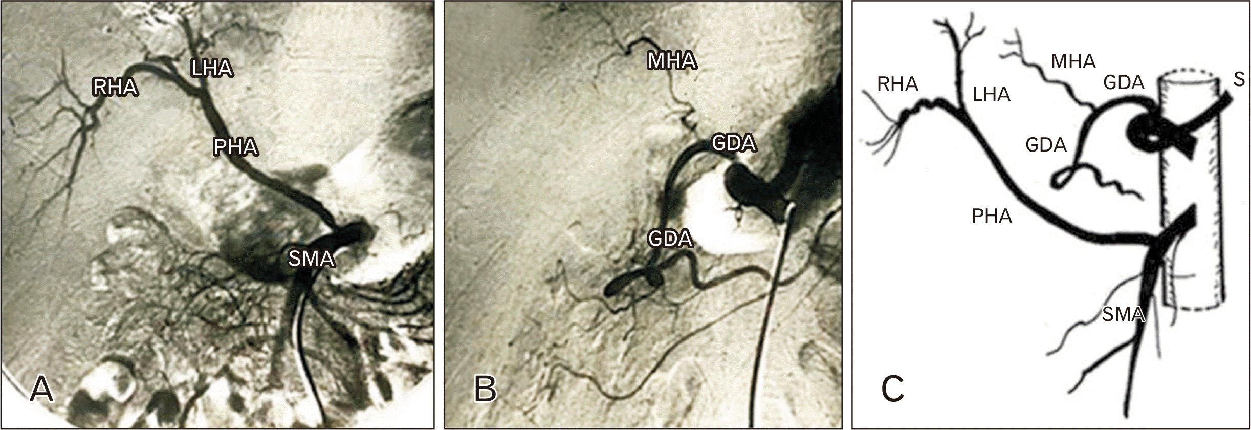 | Fig. 6(A) Anteroposterior angiograms of the same patient showing replacement of the PHA to the SMA and its division into RHA and LHA. The celiac axis gives GDA from which a small MHA arises (B). Sub image (C) is a schematic diagram showing the anatomical variants. PHA, proper hepatic artery; SMA, superior mesenteric artery; RHA, right hepatic artery; LHA, left hepatic artery; GDA, gastroduodenal artery; MHA, middle hepatic artery; S, splenic artery. 
|
Variant anatomy of the LHA was seen in 37 patients (9.51%) of our series and included only 2 different origins, LGA (22 patients) and celiac trunk (15 patients). When the LHA was replaced to LGA (5.65%), the right hepatic a. appeared to arise from celiac trunk directly and gave GDA (
Fig. 7), or the GDA arose from celiac trunk and RHA is replaced to the SMA (
Fig. 8A, B). The second abnormal origin of LHA was from celiac trunk and occurred in 15 cases (3.85%) of this series. Some cases showed that GDA arose from the LHA with replacement of the RHA to the aorta (
Fig. 9A, B). Other cases were associated with what is called double hepatic arteries, where the 2 hepatic arteries arise directly from celiac trunk, and GDA arises either from right or left hepatic arteries (
Figs. 10,
11A). Double hepatic arteries were noticed in 7 cases (1.79%) of the present study.
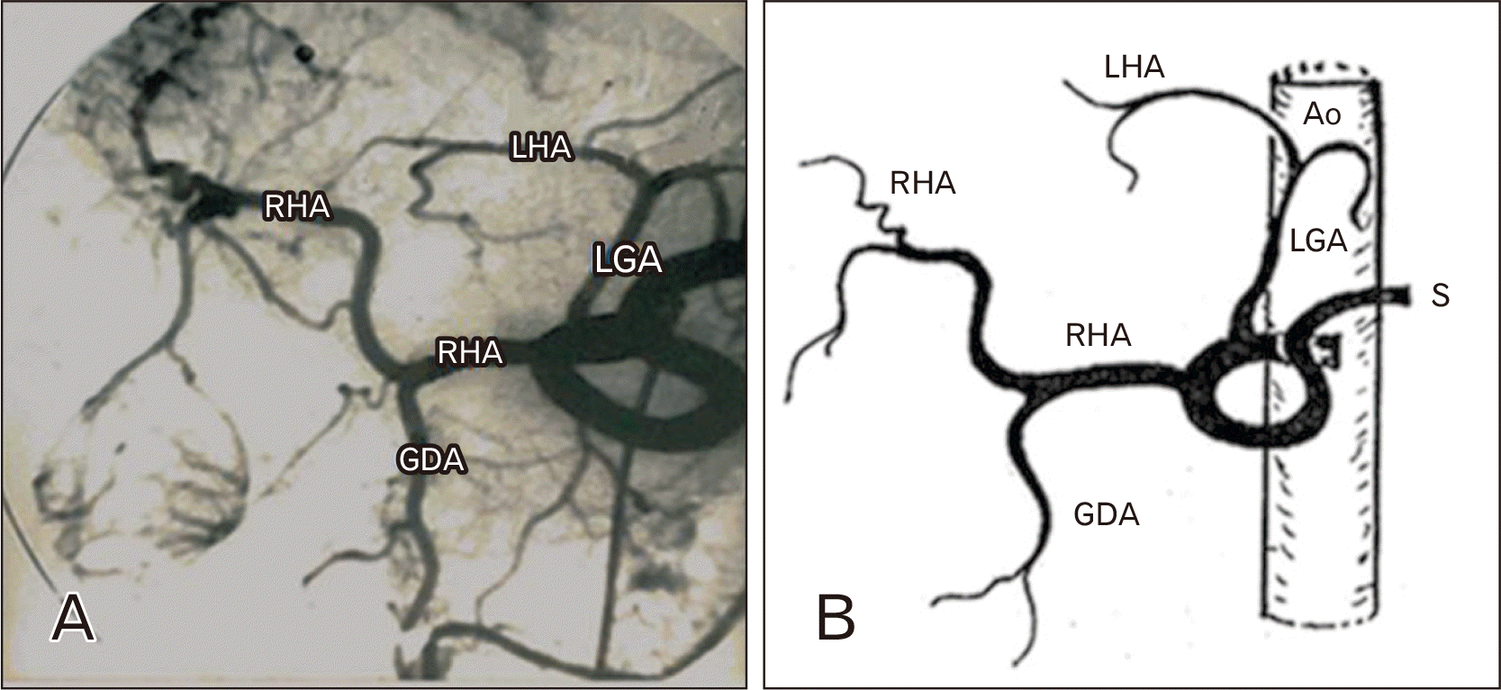 | Fig. 7(A) Anteroposterior angiogram of the celiac axis showing replacement of the LHA to the LGA. The RHA arises from the celiac trunk and gives the GDA branch. Sub image (B) is a schematic diagram showing the anatomical variants. LHA, left hepatic artery; LGA, left gastric artery; RHA, right hepatic artery; GDA, gastroduodenal artery; Ao, aorta; S, splenic artery. 
|
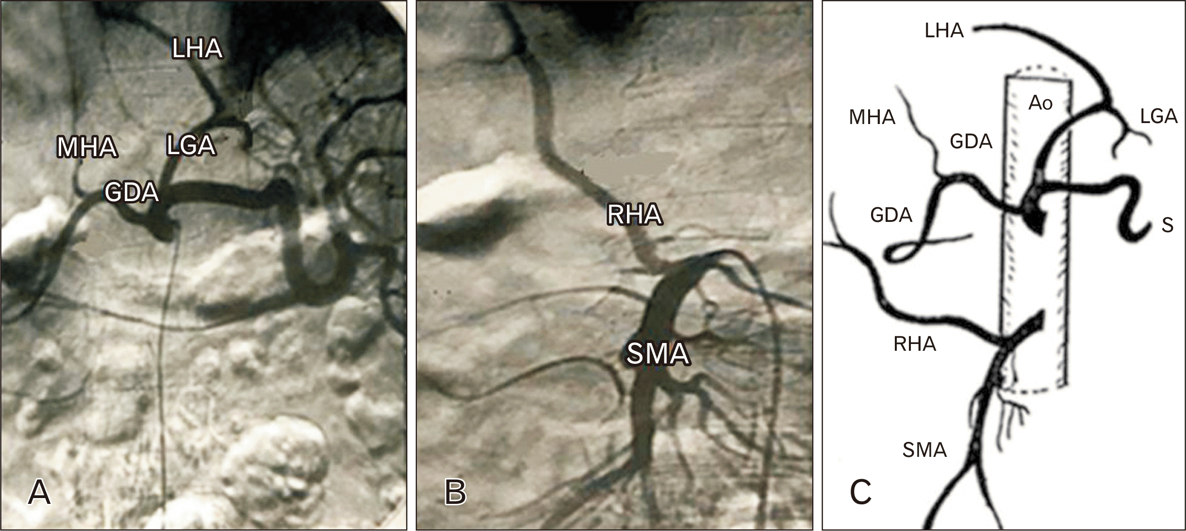 | Fig. 8(A) Anteroposterior angiograms of the same patient showing replacement of the LHA to the LGA with the GDA arising from the celiac trunk and giving the MHA. Replacement of the RHA to the SMA is also seen (B). Sub image (C) is a schematic diagram showing the anatomical variants. LHA, left hepatic artery; LGA, left gastric artery; GDA, gastroduodenal artery; MHA, middle hepatic artery; RHA, right hepatic artery; SMA, superior mesenteric artery; Ao, aorta; S, splenic artery. 
|
 | Fig. 9(A) Anteroposterior angiograms of the same patient showing the LHA originating from the celiac trunk and giving GDA branch, while the RHA is replaced to the aorta (B). Sub image (C) is a schematic diagram showing the anatomical variants. LHA, left hepatic artery; GDA, gastroduodenal artery; RHA, right hepatic artery; LGA, left gastric artery; Ao, aorta; S, splenic artery. 
|
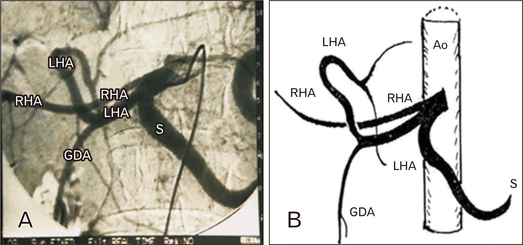 | Fig. 10(A) Anteroposterior angiogram showing the RHA and the LHA originating from celiac trunk (double hepatic artery). Notice the GDA branching from the LHA. Sub image (B) is a schematic diagram showing the anatomical variants. RHA, right hepatic artery; LHA, left hepatic artery; GDA, gastroduodenal artery; Ao, aorta; S, splenic artery. 
|
 | Fig. 11(A) Anteroposterior angiograms of the same patient showing RHA and the LHA originating from the celiac trunk (double hepatic) with the GDA arising from the RHA. An accessory RHA is seen originating from the SMA (B). Sub image (C) is a schematic diagram showing the anatomical variants. RHA, right hepatic artery; LHA, left hepatic artery; GDA, gastroduodenal artery; SMA, superior mesenteric artery; S, splenic artery. 
|
Anatomical variants in the RHA in this study reached 55 variations (14.13%). The abnormal origins of RHA included the SMA, the aorta, LHA, and celiac trunk. Accessory RHAs appeared in 22 (5.65%) cases: 17 accessory RHAs (4.37%) originated from the SMA (
Figs. 11B,
12B) and 5 cases (1.28%) originated from the LHA (
Fig. 12A). Replaced RHA was seen in 33 patients (8.48%) of our series. In 5 cases (1.28%), the RHA was replaced to the aorta (
Fig. 9B). In another 13 cases, it was replaced to the celiac trunk (
Fig. 7). In the remaining 15 cases, the RHA was replaced to the SMA (
Figs. 4B,
8B,
13B).
 | Fig. 12(A) Anteroposterior angiograms of the same patient showing the LHA giving GDA and a small accessory RHA. Another accessory RHA is seen originating from the SMA (B). Sub image (C) is a schematic diagram showing the anatomical variants. LHA, left hepatic artery; GDA, gastroduodenal artery; RHA, right hepatic artery; SMA, superior mesenteric artery. 
|
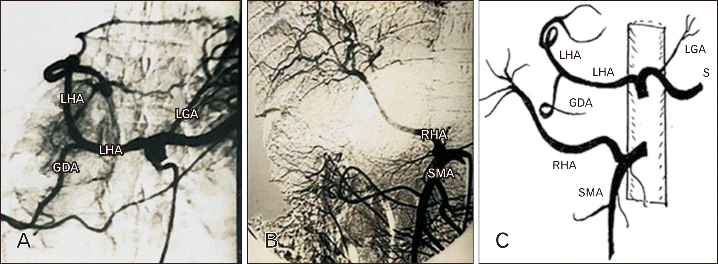 | Fig. 13Anteroposterior angiograms of the same patient showing the LHA arising from the celiac trunk and giving the GDA. The LGA appeared arising from the splenic artery (S) while the RHA was not depicted (A). The RHA was replaced to the SMA (B). Sub image (C) is a schematic diagram showing the anatomical variants. LHA, left hepatic artery; GDA, gastroduodenal artery; LGA, left gastric artery; RHA, right hepatic artery; SMA, superior mesenteric artery. 
|
The LGA was the smallest of the 3 branches of celiac trunk; however, its apparent diameter was always exceeding that of the right gastric branch. It was noticed that the diameter of LGA increases when it produces the aberrant LHA. The LGA always arises from celiac trunk in this work. In only 2 cases (0.51%), it arose from the splenic artery (
Fig. 13A).
GDA arose normally from the common hepatic a., however, the following abnormal origins appeared in this study: 7 cases (1.79%) from the celiac trunk (
Figs. 3,
6B,
8A), 11 cases (2.82%) from the LHA (
Figs. 9A,
10,
13A) and 9 cases (2.31%) from the RHA (
Figs. 7,
11A). In the latter, GDAs may arise from the RHA just after division of the common hepatic a. and this condition is called the late origin of GDA (
Fig. 14). The late origin of the GDA occurred in 4 cases (1.02%) and is differentiated from double hepatic arteries by the presence of a CHA. There is no CHA in cases of a double hepatic arteries, and in this case, the GDA originates from right or left hepatic arteries.
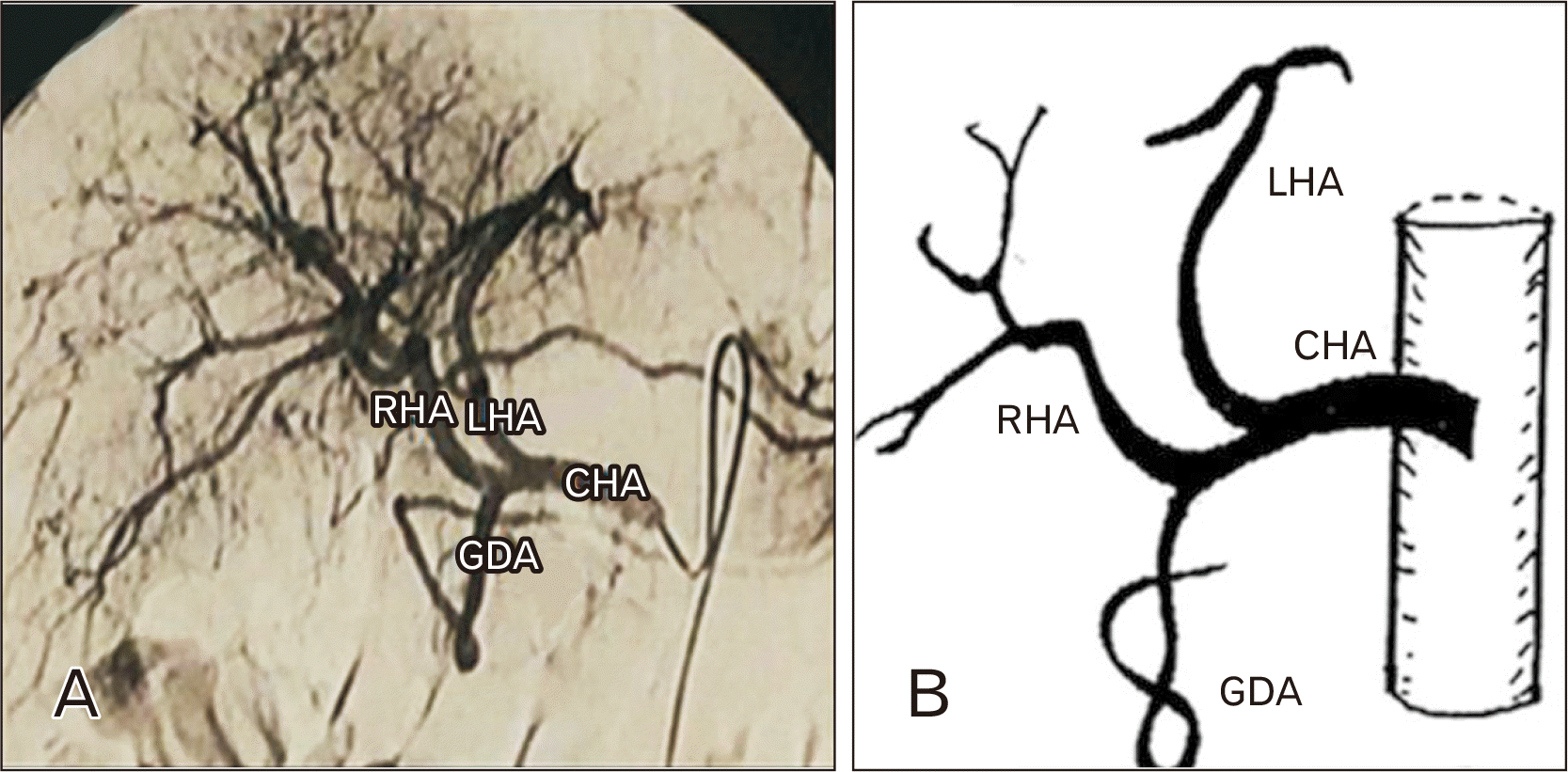 | Fig. 14(A) Anteroposterior angiogram showing the late origin of GDA arising just distal to the origin of LHA. Sub image (B) is a schematic diagram showing the anatomical variants. RHA, right hepatic artery; GDA, gastroduodenal artery; LHA, left hepatic artery; CHA, common hepatic artery. 
|
Go to :

Discussion
In this work, there were 389 available angiograms from which 103 patients (26.47%) had a single or multiple vessel variation. So, the total variations in these patients were 145 (37.27%). Previous angiographic and cadaveric studies reported similar results, Noah et al. [
4] found 69 (33.82%) vessel variations in their angiographic study on 204 patients. Farghadani et al. [
12] mentioned that 63.9% of cases had classic arterial anatomy while 36.1% had variant types. Santos et al. [
13] stated that normal anatomical pattern was the most prevalent in most studies (75.0%). In our study the word accessory means that 1 branch which supplies one side of the liver ectopically arises while the remaining blood supply comes from the typical site. Some authors use the word aberrant instead of accessory or use both terms interchangeably. The term replaced may also refer to the cases in which the whole arterial blood supply of one side of the liver originates from atypical site.
In the present study, the 3 classical branches (trifurcation) occurred in 286 patients (73.52%). Similarly, Juszczak et al. [
14] found the celiac trunk consists of left gastric, common hepatic and splenic arteries in 82% of cadavers. This standard anatomy of celiac trunk has been reported in approximately 84% of patients [
15] and 89.8% [
16]. However, quadrifurcation of celiac trunk was noticed in only 2 cases (0.51%) of the current study. This finding represents an exceptional arterial variation in the upper abdomen as was suggested by Vandamme and Bonte [
3]. They added the confirmation that, although trifurcation of celiac trunk is still considered to be the normal appearance, quadrifurcation and even a division into 5 branches were also noticed. Santos et al. [
13] observed quadrifurcation in 8.33% of cases.
Vandamme and Bonte [
3], mentioned that the celiac trunk can be easily considered as it consists of 3 main stems; hepatic, splenic and left gastric arteries, while the other branches are less important collaterals. Some celiac trunks give one or more collaterals. The commonest collateral was the double or single inferior phrenic. Our data reflect similar results as the origin of inferior phrenic artery from celiac trunk was noticed in 8 patients (2.05%) of our series. On the other hand, Juszczak et al. [
14] observed another origin of the left inferior phrenic from the left gastric in 2% of cases. Also, middle hepatic a. was visualized in 1.8% of patients of this study, it might arise from the CHA, RHA or GDA. This was in favor with Michels [
17], who recorded one MHA arose from the left or right hepatic arteries, and Covey et al. [
6] who confirmed the presence of MHA and considered it normal. On the other hand, Ghosh [
18] mentioned that the MHA was the dominant artery in each specimen which supplied the quadrate lobe of the liver and arose as a sub-branch of LHA in 59 specimens (47.2%) and as a sub-branch of RHA in 66 specimens (52.8%).
Absent celiac trunk is found in 2 cases (0.51%) of our series. The splenic artery originates directly from aorta while the RHA is replaced to the SMA in 1 case and the other case was associated with a hepatomesenteric trunk. Higashi and Hirai [
19] reported similar results but the splenic, the left gastric, and the common hepatic originate independently from aorta in this order. The superior mesenteric, in their study, divided from anterior aspect of the aorta below origin of the common hepatic a. and gave off an accessory right hepatic. Another case of agenesis (absent) celiac trunk was also described by Başar et al. [
20] who mentioned that instead of coeliac trunk, another artery arises from abdominal aorta opposite 1st lumbar vertebra and supplies the branches of celiac and superior mesenteric arteries. It gives splenic, jejunal, ileal, pancreatico-duodenal, proper hepatic and left gastric, successively. Santos et al. [
13] stated that celiac trunk was absent in 41.7% of the findings.
The various anatomy of the CHA was described by many authors, Shoumura et al. [
21] reported 2 cases with CHA that arise from abdominal aorta. Sponza et al. [
22] noticed that in 2% of cases, CHA was detected arising from the SMA. They added that in that anatomical variant, the vessel was running posterior to portal vein and not anterior to it. Farghadani et al. [
12] mentioned that variation in the CHA origin is detected in 2.6% of patients and its trifurcation to gastroduodenal, RHA, and LHA was detected in 1.8% of patients. In our study, the only abnormal origin of the CHA, was its common origin with the superior mesenteric from the aorta by a common hepatomesenteric trunk. It was observed in only 1 patient (0.25%). Different incidences of hepatomesenteric trunk have also been reported: Lippert and Pabst [
15] 3%, Kadir [
23] 0.4%, Kahraman et al. [
24] 2% and Wang et al. [
16] (4.47%). Based on these data, Kahraman et al. [
24] suggested that the whole arterial supply of duodenum originates from the SMA in the presence of a hepatomesenteric trunk. CHA relation to head of pancreas may cause difficulties during resection in pancreas. Unpanned division or ligation of the CHA in pancreatic surgery may lead to duodenal and hepatic ischemia or necrosis. So, it is very important to make a selective angiography to define the variants in the celiac axis before doing any elective resection in pancreas.
In the current study, quadrifurcation of the CHA into GDA, RHA, LHA, and MHA were seen in only 1 patient (0.25%). Coinciding with our results, Covey et al. [
6] reported quadrifurcation of the CHA in 0.5% of patients and added that, while not considered as a variant by many authors, it has important surgical significance, especially in patients with pumps introduced for hepatic infusion of chemo-therapeutic materials.
Covey et al. [
6] reported that the PHA is quite rare in literature. it was replaced to the SMA in 0.3% of patients and GDA arose separately from the aorta. Similarly, the PHA is replaced to the SMA in only 0.25% of patients of our series. Another uncommon but important variant was the double hepatic artery, where the right & left hepatic arteries originated separately from the celiac trunk that was found in 1.79% of cases of the present study. This finding was noticed by Fasel et al. [
7] in 3.7% of patients and became evident through the report of Blumgart and Fong [
25] who added that double hepatic artery is of practical importance in patients that are evaluated before hepatic arterial infusion and pump placement. In the patients with standard anatomy, the catheters should be introduced in GDA in a backward direction and the catheter tip in the proximal part of GDA or in the PHA to perfuse both right and left hepatic arteries. This is in case of surgical catheterization but in per-cutaneous route, catheters are typically introduced to the PHA for infuse both hepatic branches.
the LHA is replaced by LGA in 5.65% of patients and to the celiac trunk in 3.85% of patients. accessory LHA was not seen in our series. However, Kadir [
23] mentioned that in 23% of individuals, either a branch of the LHA or the entire LHA arises from the LGA. Also, Noah et al. [
4] found that the LHA takes origin from the LGA in 10.3% of patients as an accessory artery. Farghadani et al. [
12] found the LHA originating from the LGA in 6.9% of cases. Lastly, Covey et al. [
6] stated that the accessory LHA originates from the LGA in 10.7% of patients, while in 2.5% of patients, it takes origin from the celiac axis. It should be noted that our data was limited to the replaced LHA to the LGA as there were no accessory LHAs, while the variants reported by other authors included both accessory and replaced LHA to the LGA.
A variant source of the RHA was found in 14.13% of cases in our study. Of these, accessory RHAs were found in 5.65% of patients of which 4.37% originated from the SMA and 1.25% branched from the LHA. Replaced RHA was seen in 8.48% of patients. It was replaced to the aorta (1.25%), celiac trunk (3.34%), and SMA (3.85%). Going in line with these results, Shoumura et al. [
21] found 2 cases with accessory RHA originating from SMA in 184 Japanese cadavers. Sponza et al. [
22] mentioned that in 19% of the cases, RHA is seen to arise from SMA. Noah et al. [
4] mentioned that in 9.3% of patients, RHA takes origin only from the SMA and the incidence of both accessory and replaced RHA to the SMA was 14.2%. Oran et al. [
26] described a case of RHA that arose from the aorta independently. They added that anatomical variants of celiac trunk and SMA are frequent, so, the data about these deviations is vital before conducting surgical or radiological actions. Farghadani et al. [
12] reported the origin of the RHA from the SMA in 9.6%, from the celiac axis in 1.8% and from the aorta in 1.3% of patients. Covey et al. [
6], concluded that in 12.2% of cases, the RHA is replaced to the SMA. Accessory RHAs were noticed in 2.5% of cases: accessory RHAs arose from SMA in 1.5% of cases and originate from the celiac axis and GDA in only 1 case for each.
Variants of celiac and superior mesenteric arteries can be explained embryologically. They were linked longitudinally by primitive anastomosis, from which left gastric, splenic, and hepatic arteries originate. Partial absence or persistence of anastomosis may clarify the existence of partial or complete replacement of celiac trunk branches by the SMA. Moreover, in primitive liver, the RHA originated from the SMA, and the CHA was arising from the celiac artery or from the aorta directly while the LHA arose from the vessels that supply the stomach [
27]. Kahraman et al. [
24] suggested that partial persistance of the longitudial anastomosis propably form the hepatomesenteric trunk. Accordingly, based on our data, both right and left hepatic arteries or one of them may persist originating from superior mesenteric or left gastric arteries. Wang et al. [
16] concluded that displacement, persistence, and interruption of the longitudinal anastomosis might be the embryological mechanism of the different origins of superior mesenteric, celiac arteries and their branches.
The LGA supplies the liver with an aberrant LHA in 5.65% of cases in this series adding to its importance anatomically and surgically. The LGA showed an abnormal source from the splenic artery in only 2 cases (0.51%) of our series. However, Kadir [
23] reported that the LGA was a branch of the celiac trunk in 90% of individuals but in 2.5% it originates directly from aorta, and in the remainder, it arises from splenic artery.
In their work on 156 abdominal preparations explored both angiography and by dissection, Vandamme and Bonte [
3] described the unchanging origin of GDA from the CHA opposite the junction between the first and second parts of the duodenum. However, Kadir [
23], mentioned that in 89% of individuals, the GDA arises from the celiac trunk. This was more frequent than the ratio observed in our study (1.79%). Our data include other important variants, as the GDA originated from other sources: in addition to celiac trunk, it originated from LHA in 2.82% of cases, and from the RHA in 2.31% of patients from which 1.02% described as late origin of GDA. This late origin of GDA from the RHA was also noticed by Covey et al. [
6] in 3.7% of patients. In view of this relatively high prevalence of celiac and SMA variations (26.47% in this work), we suggested that surgeons and interventional radiologists working in this area should keep in their mind the high possibility of its occurrence and the threats that may exist.
Limitations
This study was conducted in a single radiological center. But the participating liver institute is committed to providing free medical services to a high flow of patients and serving great areas in the country.
Go to :


 PDF
PDF Citation
 Print
Print


 XML Download