Journal List > Korean J Orthod > v.54(5) > 1516088485

Satapathy, Das, Barik, Mohanty, Rath, and Mishra: Effectiveness of autologous leukocyte–platelet-rich fibrin on the rate of maxillary canine retraction, rotation, pain, and soft tissue healing: A split-mouth randomized controlled trial

Abstract

Objective

To assess the effectiveness of leukocyte–platelet-rich fibrin (L-PRF) compared with conventional treatment on canine retraction, rotation, pain, and soft tissue healing.

Methods

Sixteen adult patients aged 18–25 years (10 females, and 6 males; mean age 22.25 ± 2.26 years) with Class I bimaxillary protrusion and Class II div 1 malocclusion participated in this single-center, split-mouth randomized controlled trial at the Orthodontics Department of a single hospital in SCB Dental College and Hospital, Cuttack, India. Randomization was performed using a computer-assisted function with a 11 allocation ratio. The intervention included the placement of L-PRF on the experimental side and follow-up for 90 days. The primary outcome measures were canine retraction, rotation, pain, and soft tissue healing. The range of tooth movement was evaluated at 15-day intervals 0th day (T0), 15th day (T1), 30th day (T2), 45th day (T3), 60th day (T4), 75th day (T5), and 90th day (T6). Canine rotation was assessed at T0 and T6, and pain and soft tissue healing were evaluated on the 3rd, 7th, and 15th days of the treatment.

Results

Cumulatively, the L-PRF group demonstrated a significantly greater tooth movement as compared to conventional treatment group (P < 0.001). Overall, canine retraction was 1.5 times greater on the L-PRF side than on the control side. Canine rotation showed no significant relationship, whereas pain and soft tissue healing were significantly better on the L-PRF side than on the control side.

Conclusions

Local administration of L-PRF amplifies canine retraction while improving pain and soft tissue repair.

INTRODUCTION

The orthodontic profession has witnessed a growing interest in accelerated orthodontic tooth movement (OTM), driven by patient preferences and the desire to mitigate the risks associated with prolonged treatment durations, including root resorption, decalcification, and periodontal problems.1 OTM occurs because of remodeling processes within the alveolar bone and periodontal ligament (PDL). This process is characterized by bone resorption on the compression side of the tooth and bone deposition on the tension side.
Orthodontic forces trigger disturbances in the microcirculation of the PDL, leading to an initial inflammatory response characterized by increased vascular permeability and leukocyte migration. Subsequently, the migrated cells release inflammatory cytokines and pro-inflammatory lymphocytic growth and chemotactic factors.
Therefore, increased cytokine expression during bone remodeling can accelerate tooth movement.2,3
Numerous methods have been suggested to expedite tooth movement, ranging from invasive surgical techniques, such as osteotomies and corticotomy, to noninvasive approaches, such as pulsed electromagnetic fields, mechanical vibration, and low-level laser therapy.4,5 Among the noninvasive methods, biomaterials, such as platelet-rich plasma (PRP) and platelet-rich fibrin (PRF), have been recommended as promising alternatives for expediting OTM while minimizing the risk of bone and periodontal loss.6,7 The platelet concentration is a source of autologous growth factors and secretory cytokines, which play a significant role in angiogenesis, wound healing, and bone regeneration.8
In recent years, leukocytes have been included in PRP compositions, known as leukocyte–PRP (L-PRP), and are gaining increased attention for their therapeutic potential in tissue regeneration. L-PRP can enhance bone regeneration by upregulating cellular proliferation, viability, migration, angiogenesis, and osteogenesis.9,10 Furthermore, the presence of leukocytes in PRP correlates with elevated levels of catabolic cytokines, such as interleukin-1 (IL-1), interleukin-6 (IL-6), and tumor necrosis factor-alpha, leading to the production of catabolic metalloproteinases.11,12 Moreover, the bone turnover efficiency of PRF is higher than that of PRP because of its extended period of activity.13 Overall, leukocyte–PRF (L-PRF) has demonstrated better persistent release of pro-inflammatory factors from its thin and flexible fibrin matrix than PRP and PRF.14
Although existing literature has addressed the effects of L-PRF on OTM in extraction sockets, its influence on soft tissue healing and pain remains unexplored. This randomized controlled trial aimed to fill this knowledge gap by investigating the effects of L-PRF on maxillary canine retraction and rotation and its role in soft tissue healing and pain in individuals with Class I bimax protrusion and Class II div I malocclusion by comparing these effects to conventional solutions.

MATERIALS AND METHODS

Trial design

This was a single-center, randomized split-mouth clinical trial employing a 1:1 allocation ratio. The methodology used remained unchanged after trial completion.

Participants, eligibility, and study setting

This study spanned a period of 3 months, from October to December 2022, and was conducted within the Orthodontics and Dentofacial Orthopaedics Department at SCB Dental College and Hospital, Cuttack, India. This study was approved by the Institutional Ethics Committee of SCB Dental College and Hospital, Cuttack, India (IES/SCBDCH/092/2021) and registered in the Clinical Trial Registry of India (CTRI) (registration number: CTRI/2023/01/048932). Although the registration application was submitted before the trial commenced, registration confirmation was received after the trial concluded in January 2023. The protocol is accessible on the CTRI website (https://ctri.nic.in/clinicaltrials/login.php). Participants of both sexes, aged between 18 and 25 years, were included based on predefined inclusion and exclusion criteria. All owners of eligible participants provided informed consent after receiving a comprehensive explanation of the study procedures.
Inclusion criteria:
  • Class I bimaxillary protrusion requiring first premolar extraction

  • Class II div 1 malocclusion requiring maxillary first premolar extraction

  • Moderate crowding or spacing (2–5 mm) in the maxillary and mandibular arches

Exclusion criteria:
  • Long-term corticosteroid therapy or bisphosphonate and nonsteroidal anti-inflammatory drug use and a comprehensive history of orthodontic treatment

  • Presence of a pacemaker or ankylosed tooth

  • Pregnancy

Sample size derivation

The methods of Pandis15 (standard for split-mouth design) were used for sample size estimation:
n=f(α,β)×σ2/(μ1μ2)2
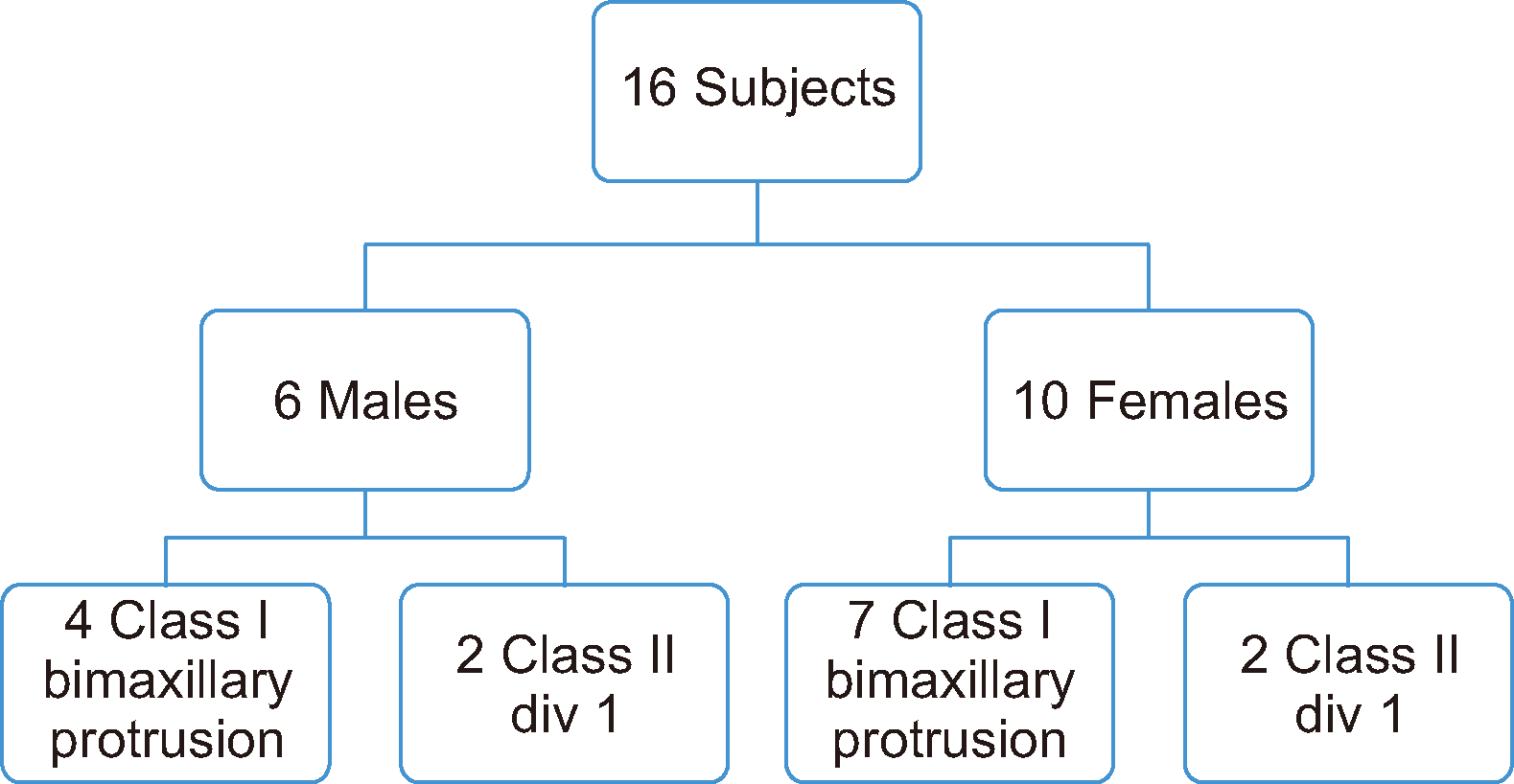
where σ pertains to the standard deviation, (µ1–µ2) intra-person difference, and ƒ (α, β) a function of significance level and power. With a difference between sides of 0.5 mm and canine distalization movement of 1 mm per month and a standard deviation of the difference of 0.7 mm at a 5% significance level (95% confidence interval) and 80% power, the computed sample was rounded up to 16 cases. Power analysis was performed for all outcomes. The distribution of malocclusions is shown in Figure 1.

Interim analyses and stopping guidelines

This study did not involve any interim analyses, and no specific stopping guidelines were established.

Randomization

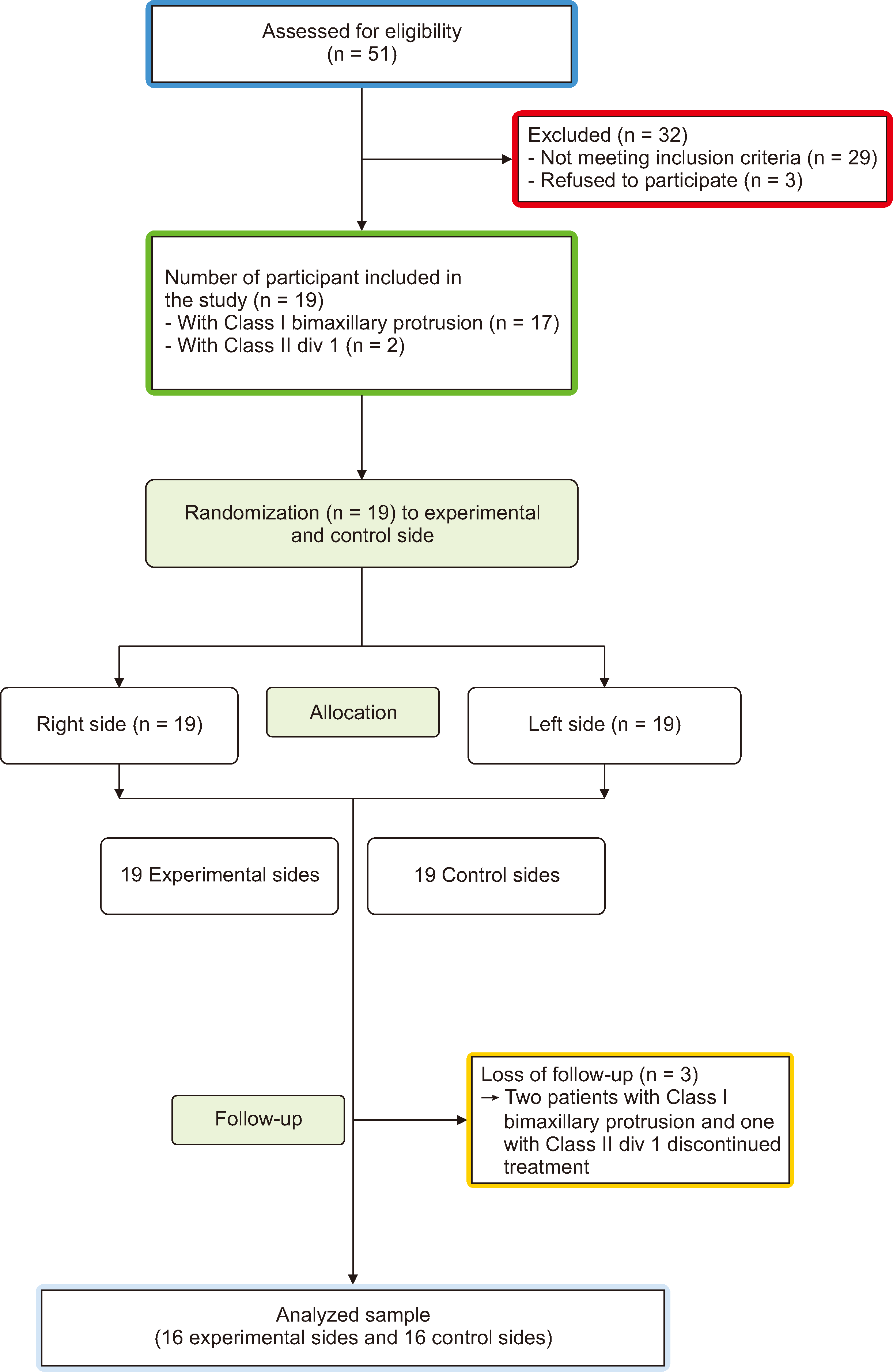
Random allocation was performed using a computer-assisted function that generated random numbers (Microsoft Office 2022, Microsoft, Random, Redmond, WA, USA). The Consolidated Standards of Reporting Trials statement was used as a guide (Figure 2).16 One extraction side was randomly assigned for L-PRF insertion, whereas the contralateral extraction side of the same patient was allocated for the conventional method. Sequentially numbered opaque envelopes were prepared, each containing information about the corresponding group based on the generated order. These envelopes remain sealed in a secure location until they were opened by the main researcher (SKS). The randomization process was overseen by an individual who was not directly involved in the study, thus ensuring impartial management.

Allocation concealment

The evaluator obtained each randomized allocation via a centralized, secure, web-based interface (RandoWeb, Assistance Publique-Hopitaux de Paris [AP-HP], Paris, France). This sequence was masked until the intervention was assigned.

Blinding

Immediately following the extraction, the researcher responsible for applying L-PRF opened one envelope and performed the specified procedure (L-PRF or conventional treatment). Only the main researcher (SKS) was aware of the intervention used for each patient. All other investigators, including SKD, involved in recruitment, and AKB, handling data collection/outcomes, were blinded to the intervention. The allocation details were not disclosed to the participants during the study. SKS ensured the confidentiality of personal data for potential and enrolled participants through the secure collection, sharing, and maintenance of protocols. This involved using a computer without Internet access, implementing password protection, and encrypting data stored on a USB drive.

Interventions

Pre-surgical orthodontic preparation

All 16 participants underwent orthodontic treatment performed by a single orthodontist. Pre-adjusted edgewise Mclaughlin–Bennett–Trevisi brackets of 0.018 inch (in) slot with –70 torque for maxillary canines (Leone SpA, Firenze, Italy) were bonded to all teeth. In contrast, triple-slot molar tubes were connected or banded to the molars. Bonding was performed using Transbond XT (3M Unitek, Monrovia, CA, USA) and cured using a light-emitting diode curing light (Coltene/Whaledent Inc., Cuyahoga Falls, OH, USA). The bands were luted with a glass ionomer luting cement (GC Fuji I; GC Corporation, Tokyo, Japan). Alignment and leveling were initiated using light wires, that is, 0.012-in nickel-titanium (NiTi), 0.014-in NiTi, and 0.016-in round (NiTi) arch wire, and eventually progressed to stiffer wires, such as 0.016 × 0.022-in rectangular NiTi, 0.016 × 0.022-in rectangle stainless steel (SS), and 0.017 × 0.025-in rectangular SS arch wires. Complete alignment of the dentition was performed before the initiation of canine retraction.

Micro-implant placement

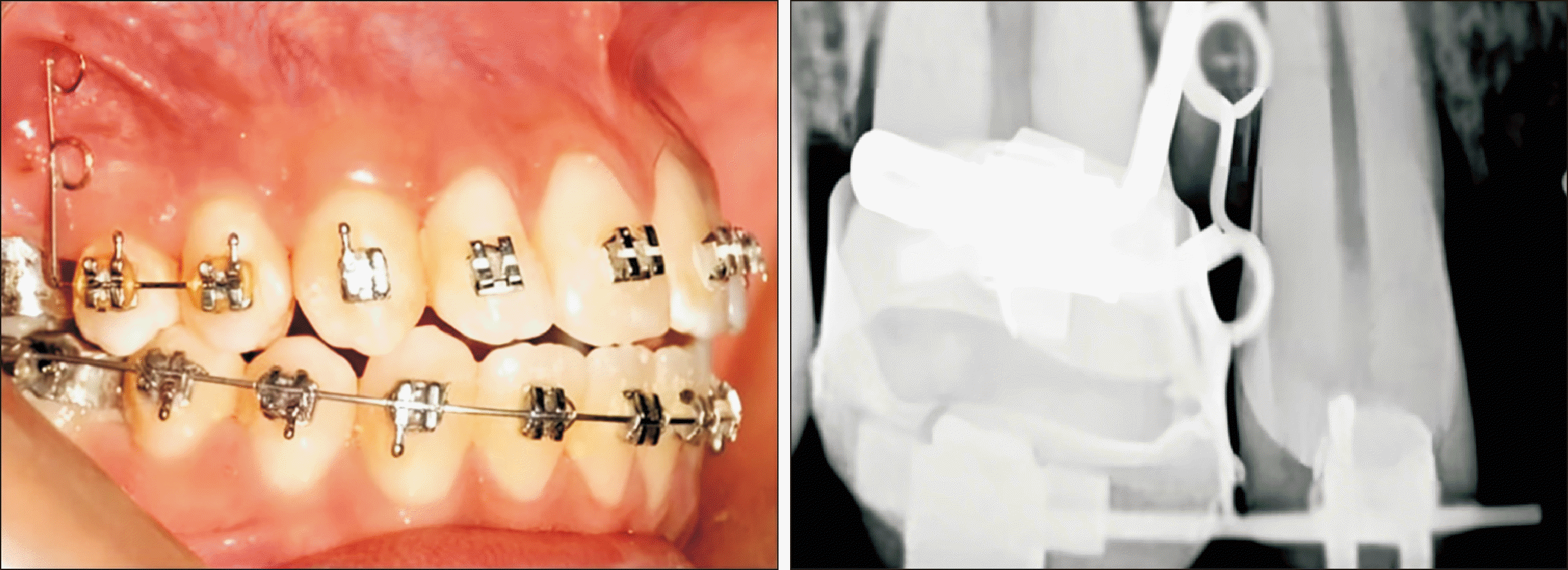
After alignment and leveling, a guiding template fabricated from a 0.017 × 0.025-in rectangular SS wire was used to correct the positioning of the micro-implant during its placement. After placing one of the free arms in the molar tube slot, intraoral periapical radiographs were obtained to determine the implant placement site. Micro-implants of 1.5 mm width and 8 mm length (FavAnchor® microimplant color code: pink, S. H. Pitkar Orthotools Ltd., Pune, India) were inserted under 2% local infiltration of lignocaine between the first maxillary first molar and second premolar on either side at a height of 5 mm from the alveolar crest (Figure 3). After leveling and alignment, atraumatic extraction of the maxillary premolars was performed under local anesthesia with 2% lignocaine diluted to 1:200,000.

Preparation and placement of leukocyte–platelet-rich fibrin plug

Using specific guidelines17 Choukroun’s PRF was used for preparing LPRF were used to prepare and place L-PRF on the experimental side before extracting the first premolars. Approximately 10 mL of venous blood (Figure 4A) was centrifuged without anticoagulants for 12 minutes at 2,700 rpm. At the apex of the tube, a layer of plasma devoid of platelets was observed, and an RBC clog was formed at the base (Figure 4B). Using sterile tweezers, the middle portion of the L-PRF plug was extracted and dissected using a BP No. 15 blade, approximately 2 mm below the intersection between the middle and lower layers (Figure 4C). After carefully inserting the L-PRF plug into the socket (Figure 4D), a 3-0 silk suture was used to close the socket. Similarly, the contralateral first premolar was gently extracted, and the socket was covered with a sterile gauze pack. All participants received post-extraction instructions and appropriate medications. The interventions were concurrently provided to each patient.

Orthodontic intervention for individual canine retraction

Individual canine retraction after extraction was accomplished using sliding mechanics of a 0.016 × 0.022-in SS arch wire with the help of a microimplant placed between the first molar and second premolar and a closed NiTi coil spring to exert a constant force of 150 g, which was measured through a Dontrix gauge at the sites.

Study records and outcome measure assessment

Pre-treatment records, such as lateral cephalograms, orthopantomogram, plaster models, and intra- and extra-oral photographs, were obtained.

Outcome measures

Primary outcome

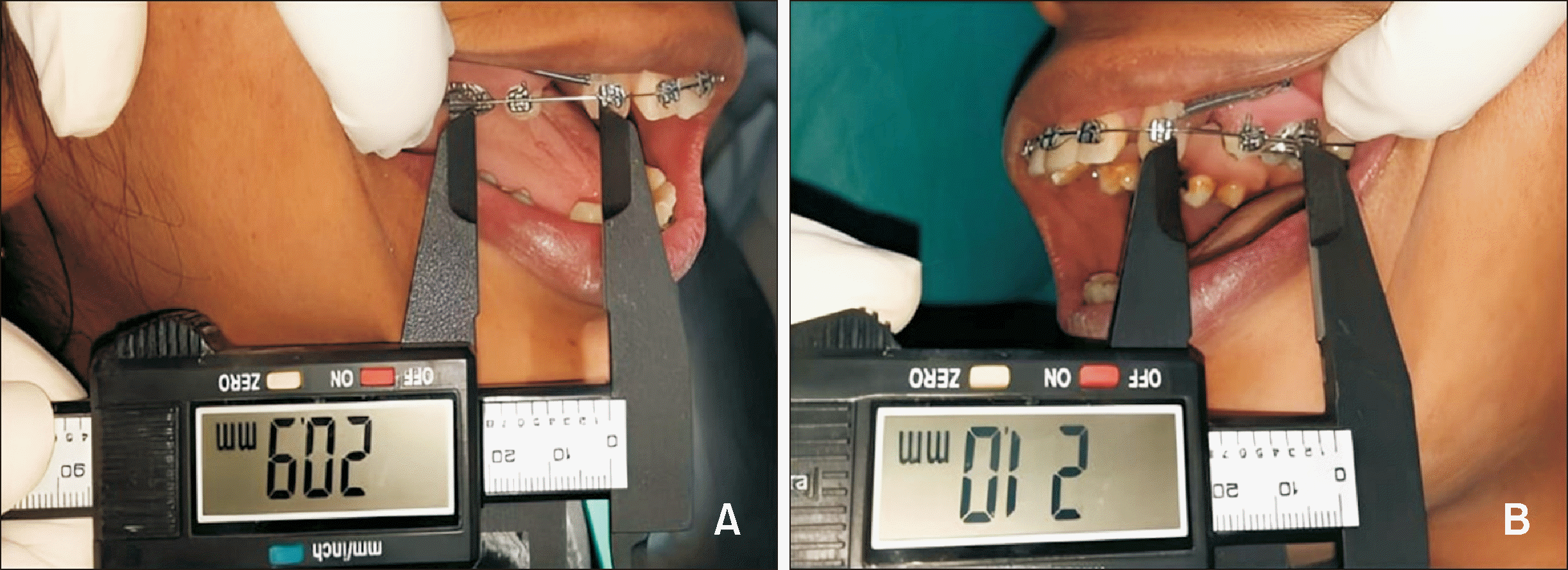
Canine distalization or retraction, which was intraorally measured using a digital Vernier caliper (Absolute Digimatic Caliper, Mitutoyo, Kawasaki, Japan) as the distance between the distal aspect of the maxillary canine bracket and the mesial aspect of the molar tube slot (Figure 5A and 5B), as the primary outcome. The measurements were recorded every 2 weeks (six time points, after placement of L-PRF): on the day of L-PRF placement (T0), 15th day (T1), 30th day (T2), 45th day (T3), 60th day (T4), 75th day (T5), and 90th day (T6) for 3 months between L-PRF and control sites.

Secondary outcomes

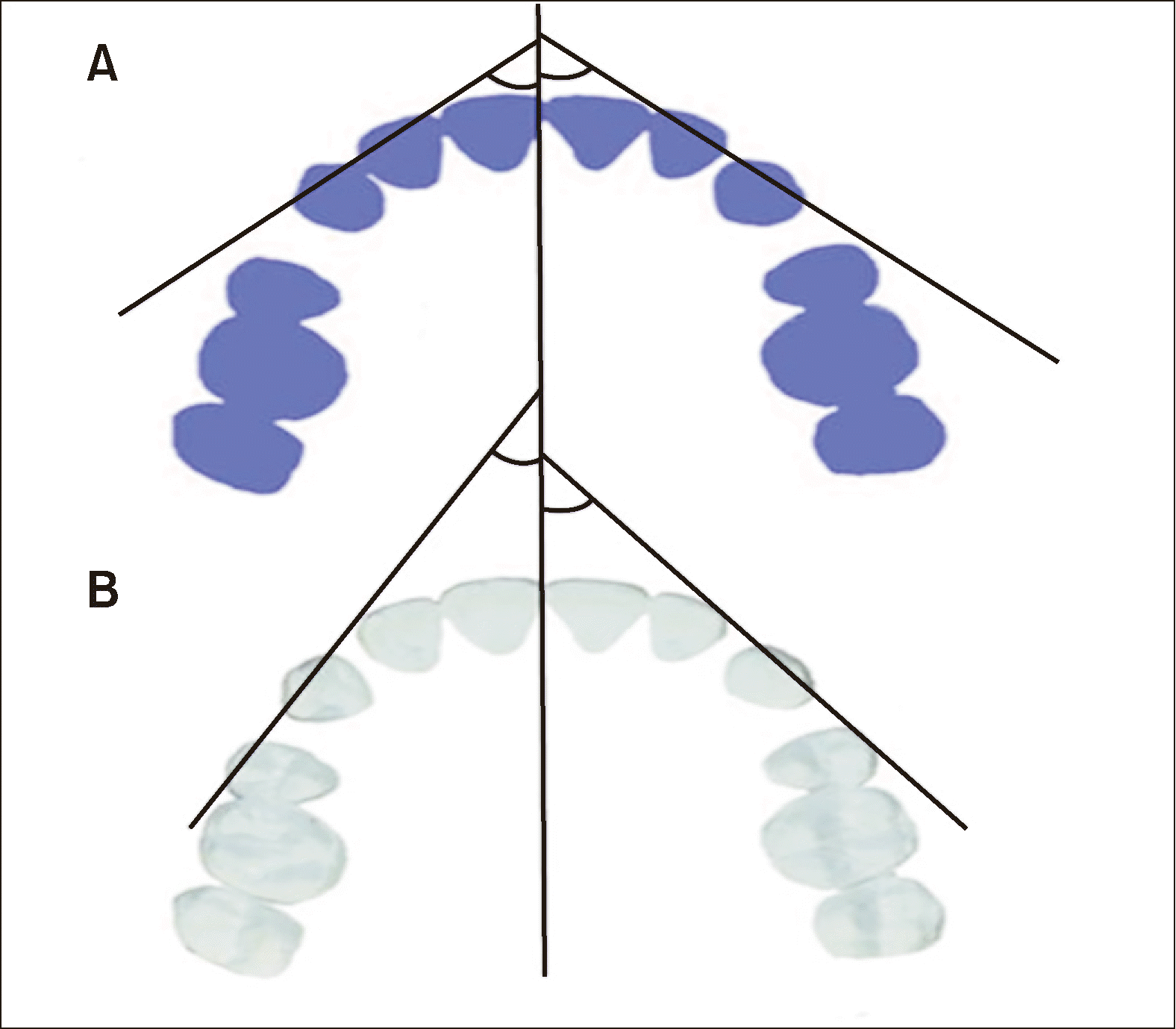
Canine rotation: Occlusograms were used to measure the rotation of canines using the method described by Ziegler and Inglervall.18 The angle formed by the line running through the mid-palatal raphe and the line connecting the canine’s mesial and distal contact sites was measured. The change in angle indicated the amount of rotation that occurred after the completion of canine retraction. The recordings were performed at the start (T0) and end of the study period (T6) (Figure 6).
Soft tissue healing and pain: The Healing Index by Landry et al.19 and the numerical pain rating scale by McCaffery and Beebe20 were used to evaluate soft tissue response and pain in the extraction sockets of L-PRF and control sites on the 3rd, 7th, and 15th days (Figure 7). No changes in trial outcomes were made after the trial commenced.

Statistical analyses

Statistical Package for the Social Sciences (SPSS version 21; IBM Corp., Armonk, NY, USA) was used for statistical analyses. Data normality was assessed using the Shapiro–Wilk test. For intragroup comparisons among different study visits, repeated measures analysis of variance was used. The intraclass correlation coefficient was used to evaluate examiner reliability. Unpaired Student’s t test was used to compare the L-PRF and control groups. A paired t test was used after summing the data at the cluster level to account for the clustering effects. The effect estimates were calculated by dividing the difference between the two groups (treatment group mean minus control group mean) by the standard deviation of one of the groups. Pearson’s correlation coefficients were also calculated, and the chi-squared test was used to determine whether there was a significant difference between categorical variables. For all statistical inferences, P < 0.05 was considered significant.

RESULTS

Participant flow

This study included 16 participants and 32 sides, with 16 participants each randomly assigned to the L-PRF and the control groups. All participants were tracked until the end of the study period (Figures 1 and 2). The intraclass correlation coefficient was r = 0.84, demonstrating high evaluator reliability.

Baseline data

Age was categorized into two subgroups: ≤ 20 and > 20 years. Most of the study participants (62.5%) belonged to the age group of > 20 years. The mean age of the participants was 22.25 ± 2.26 years. Most study participants (75%) were female (Figure 1).

Primary outcome

Canine distalization

Table 1 shows the degree of canine retraction or distalization on the L-PRF and control sides at various time intervals.
The mean changes in canine distalization between the L-PRF and the control groups were compared from T0 to T6 at monthly intervals and overall (Table 2). At each of the three time points, T0–T2 (1.24 ± 0.13 mm vs. 0.68 ± 0.12 mm, P < 0.001), T2–T4 (1.55 ± 0.10 mm vs. 0.77 ± 0.08 mm, P < 0.001), and T4–T6 (0.83 ± 0.06 mm vs. 0.77 ± 0.07 mm, P = 0.002), the difference between the L-PRF and the control side was statistically significant, indicating higher canine movement on the L-PRF side compared with the control side.
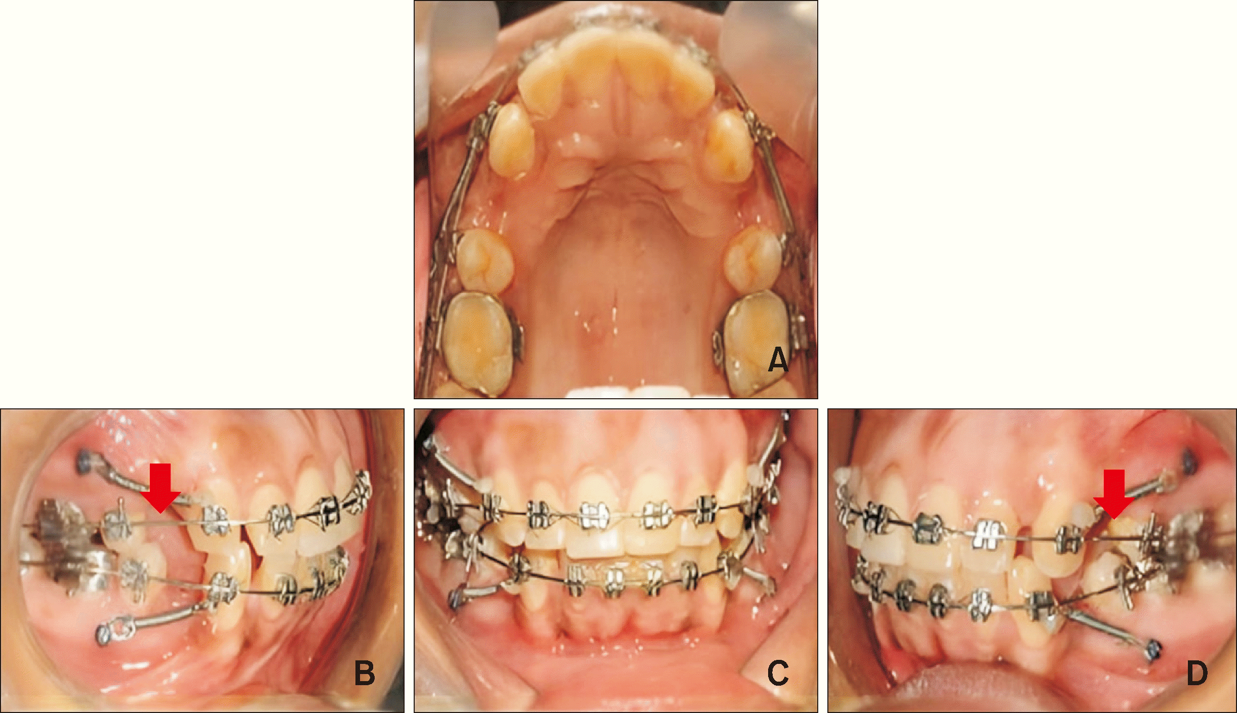
Overall, from T0 to T6, the distalization of the canine at the L-PRF side was higher (3.66 ± 0.24 mm) compared with that of the control side (2.18 ± 0.13 mm), the difference of which was statistically significant (P < 0.001) (Table 2). This indicates that L-PRF has the potential to induce rapid tooth movement when applied to an extraction socket. Figure 7 shows intraoral images of canine retraction 30 days post-treatment.

Secondary outcomes

Canine rotation

Inter-group comparison (Table 3) showed that the difference between the L-PRF and control sides at both T0 (33.99 ± 9.31° vs. 35.01 ± 5.30°, P = 0.705) and T6 (27.07 ± 9.20° vs. 30.12 ± 5.30°, P = 0.259) was nonsignificant, which signifies that L-PRF has little role in canine rotational movement.
Intra-group comparisons at T0 and T6 (Table 3) showed a significant difference in the mean for both the L-PRF (33.99 ± 9.31° vs. 27.07 ± 9.20°, P < 0.0001) and control (35.01 ± 5.30° vs. 30.12 ± 5.30°, P < 0.0001) sides. The fact that SS ligatures were used to tie the canines on both sides during retraction might have contributed to the nonsignificant differences between the two sides.

Healing and pain index

Inter-group comparison (Table 4) of both healing and pain index scores showed a significant difference in the mean for both the L-PRF and control sides on the 3rd, 7th, and 15th days, indicating that L-PRF aids in faster healing and pain modulation.
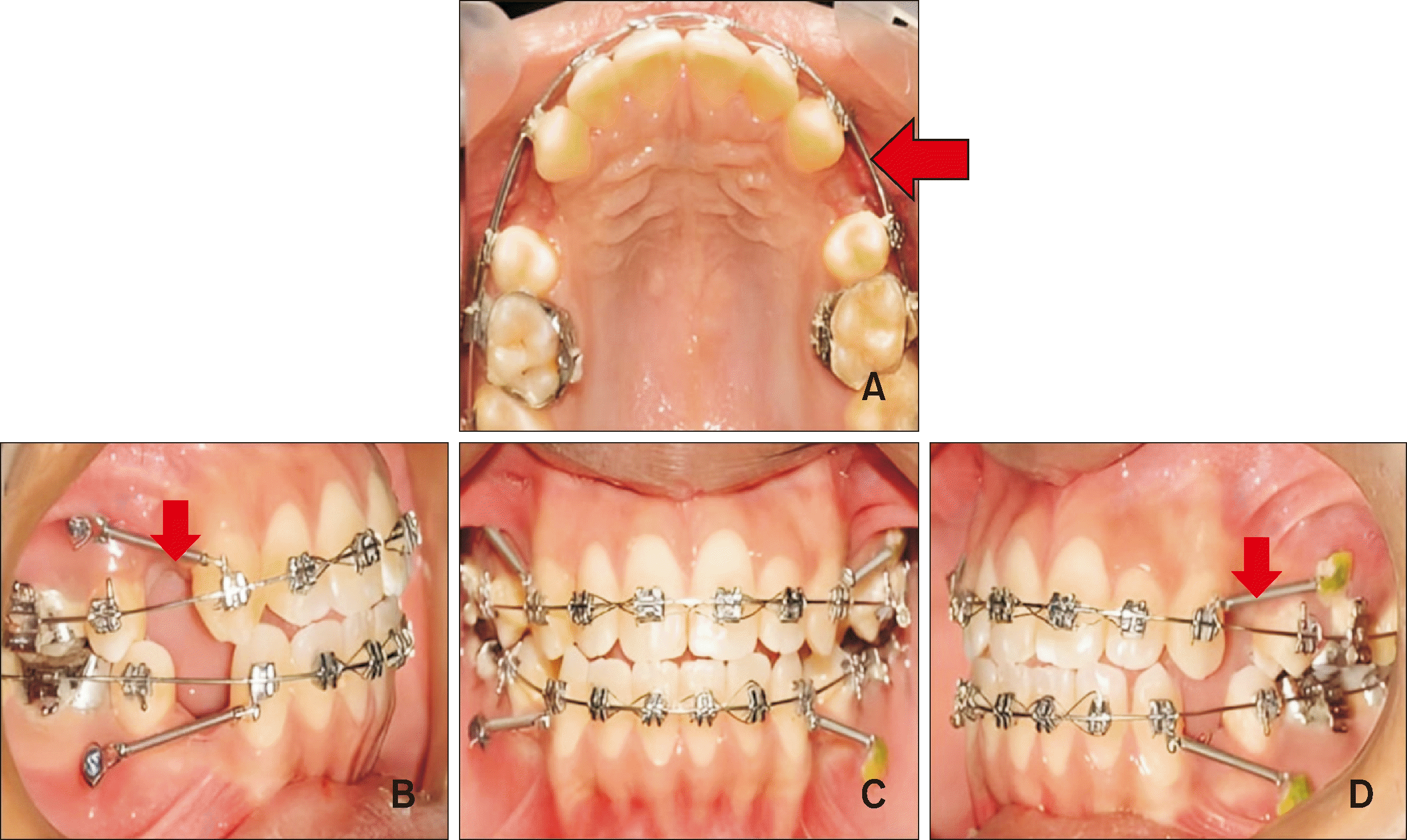
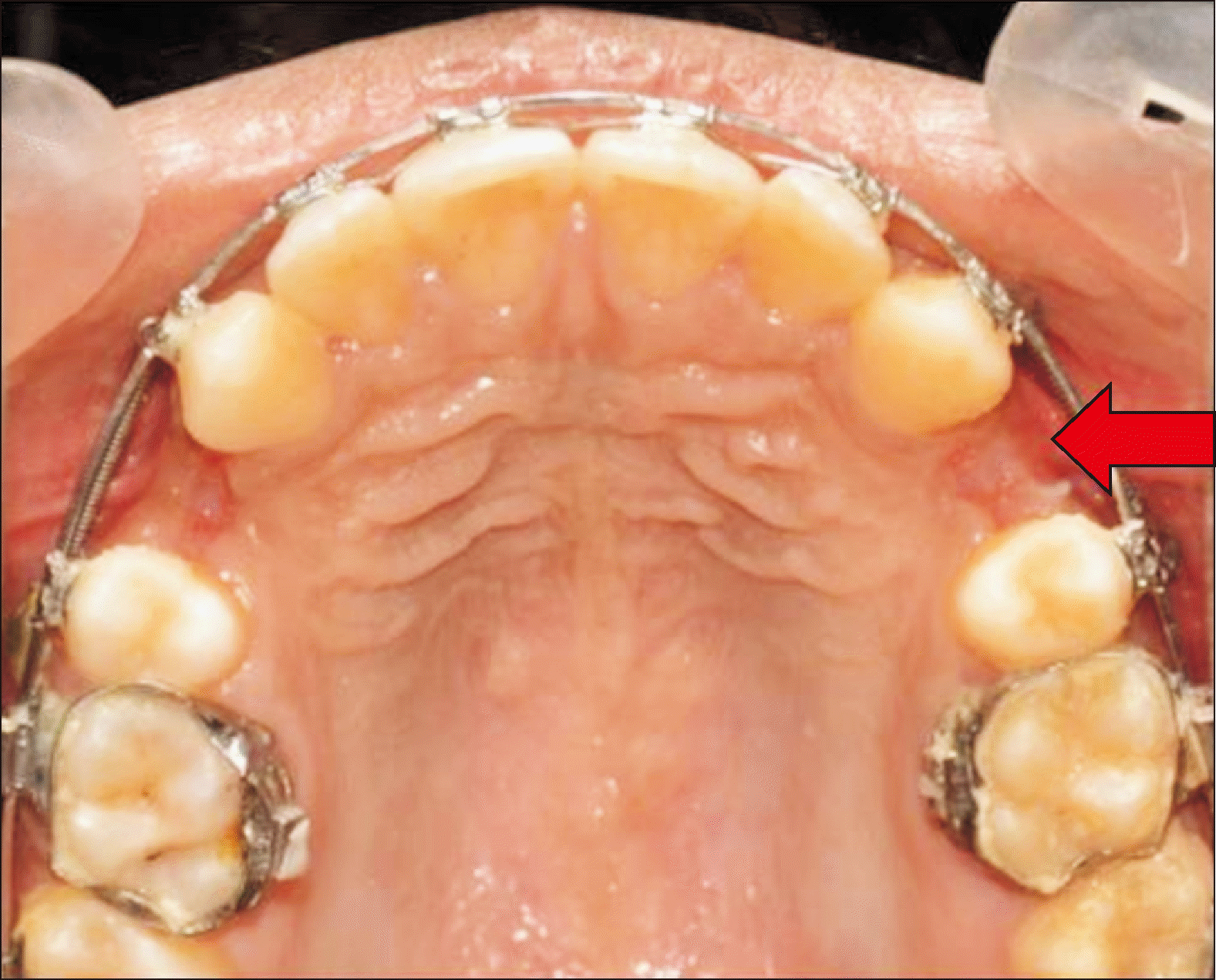
Similarly, the intra-group comparison (Table 5) of both index scores on the 3rd, 7th, and 15th days resulted in a significant difference between the L-PRF and control sides. Figures 8 and 9 (enhanced Figure 8A) show the intraoral images of soft tissue healing 7 days post-treatment.

Harms

No adverse reaction was noted during the trial.

DISCUSSION

Numerous surgical and nonsurgical approaches have been suggested to hasten tooth movement and yield diverse outcomes. Surgical techniques, such as tooth extraction and periodontally accelerated osteogenic orthodontics, induce a regional acceleratory phenomenon (RAP),21,22 reducing the duration of orthodontic treatment. Despite their apparent success, these methods require comprehensive long-term evaluations.23,24
Minimally invasive techniques have come to the forefront in the pursuit of less invasive approaches to expedite OTM. PRP has emerged as a recent addition to noninvasive methods aimed at accelerating tooth movement,7 offering the dual benefit of preventing alveolar bone loss while triggering a RAP-like effect and fostering alveolar bone regeneration.25
Animal studies have indicated that L-PRF results in increased tooth mobility, with a 1.7-fold increase observed in rat models and a 2.13-fold increase in dog models.6,7 A recently published systematic review suggested that platelet-rich concentrates might play a role in accelerating OTM in animal models.26

Effects of L-PRF on canine distalization

Except at T6, the current study found a statistically significant retraction on the L-PRF side compared with the control side at all time points (T1, T2, T3, T4, and T5). Biweekly canine retraction changes on the L-PRF side were 1.5 times more at T1, 2.5 times more at T2 and T3, almost twice at T4, and nearly similar at T5 and T6 compared with that on the control side.
Studies have presented divergent perspectives regarding the use of L-PRF in canine retraction. These conflicting findings may stem from variations in the intervention procedures, including distribution methods (membrane, injection, or plugs), dosage, concentrate presentation (L-PRF, PRF, or PRP), and observation periods.27-30
Studies investigating the placement of the L-PRF clot in extraction sockets have yielded mixed results. In some instances, canine retraction was less pronounced on the control side compared with that on the L-PRF side when canine distalization commenced immediately after the extraction of the first premolar.27,28
Retraction rates of 0.32 (control side) and 0.52 (L-PRF side) mm/month were reported by Nemtoi et al.28 (P = 0.006). Tehranchi et al.27 observed that throughout all subsequent visits (biweekly for 4 months), the teeth migrated faster on the L-PRF side than on the control side (P = 0.006). In contrast to the above, a study by Reyes Pacheco et al.31 found a considerably superior canine distalization rate (0.23 mm/month) on the control side, although they began canine retraction 15 days post-initial premolar extraction (L-PRF side: 0.67 mm/month, P = 0.004; control side: 0.90 mm/month). An 8-week study recently published by Barhate et al.32 demonstrated that when L-PRF was inserted into the extraction socket, canine retraction was significantly higher on the L-PRF side for the first 4 weeks and comparable for the remaining 4 weeks. Thus, canine distalization after L-PRF insertion may be predominantly caused by normal alveolar bone remodeling in response to a constant and optimal force magnitude. When injectable PRF was used, other studies22,30,33-35 revealed an increase in the canine retraction rate.
It is not desirable to draw conclusions by merely synthesizing the final output of each incorporated study, owing to their different trial times. Nevertheless, we may acquire a deeper understanding of the PRF effect by performing a time-dependent evaluation of each study’s data. For the first 3 months, PRF succeeded in accelerating OTM, with the most significant rate of acceleration occurring around the interval during the second month. The current study showed an accelerating effect of L-PRF during the first 3 months of placement, which is consistent with other studies reported in the literature.27,30,33,34 However, three of them27,30,34 showed a maximal effect during the second month after L-PRF application, implying that L-PRF exerts the most significant effect on OTM acceleration after the first month.
Although the causal mechanism remains unknown, we can presume that (1) immediately following L-PRF application, there is increased production of cytokines, which produce bone remodeling and then accelerate OTM,30 and (2) after a defined period, as L-PRF dissociates and there is a concomitant decrease in growth factors and cytokines, there may be some decrease in OTM via a negative feedback mechanism.29 However, the natural effect and mechanism of L-PRF and PRP must be elucidated by additional studies using yardstick protocols and PRP/L-PRF parameters.

Rotational effect of L-PRF

The mean change in canine rotation was not statistically significant between the L-PRF and control sides at T0 and T6. This contradicts the findings of Reyes Pacheco et al.,31 who demonstrated that the mean canine rotations were 5.80° on the test side and 8.50° on the control side (P = 0.001). The L-PRF side showed higher retroclination at the end of the distalization period. In contrast to the study by Reyes Pacheco et al.,31 three studies in the literature29,33,34 found statistically nonsignificant variations in canine rotation between the control and L-PRF, which is consistent with the result of the current study.

Effects of L-PRF on soft tissue healing and postoperative pain

In the current study, both indices showed a gradual improvement in pain and healing from the 3rd day to the 15th day of assessment, which is in line with the observation by de Almeida Barros Mourão et al.36 However, a study by El-Timamy et al.22 found no such positive correlation of L-PRF.

Study generalizability and limitations

This study was a split-mouth trial. Compared with parallel-group trials, this design requires fewer patients, which may result in non-normal distributions and affect the results. Although the sample size was determined using standard methods, a larger sample size would have resulted in a more foreseeable outcome and provided more value to the current study. The second limitation is derived from the fact that OTM is multifactorial and involves multiple cytokines and growth factors. Because such factors have not been considered, the effect cannot be fully generalized. In addition, as the measurement was performed using standard instruments, some degree of inaccuracy could creep in, as it was performed on the chair side in the patient’s mouth. Further studies considering other OTM-associated biomarkers and growth factors can provide a broader perspective and improve our understanding of tooth movement.

CONCLUSIONS

Cumulatively, the L-PRF group demonstrated a significantly greater tooth movement as compared to conventional treatment group (P < 0.001); however, there was no significant difference thereafter. There was also a significant improvement in healing and pain reduction on the L-PRF side compared with that on the control side.

Notes

AUTHOR CONTRIBUTIONS

Conceptualization: SKS, SKD. Data curation: SKD, SKR. Formal analysis: SKS, AKB. Investigation: SKS, DM. Methodology: SKS, SKD, DM. Project administration: SKD, AKB. Resources: AKB, DM. Software: SKR. Supervision: SKD, AKB, DM, SKR. Validation: SKD, AKB, DM. Visualization: SKD, MM. Writing–original draft: SKS. Writing–review & editing: SKD, AKB, DM, SKR, MM.

CONFLICTS OF INTEREST

No potential conflict of interest relevant to this article was reported.

FUNDING

None to declare.

REFERENCES

1. Segal GR, Schiffman PH, Tuncay OC. 2004; Meta analysis of the treatment-related factors of external apical root resorption. Orthod Craniofac Res. 7:71–8. https://doi.org/10.1111/j.1601-6343.2004.00286.x. DOI: 10.1111/j.1601-6343.2004.00286.x.
2. Teixeira CC, Khoo E, Tran J, Chartres I, Liu Y, Thant LM, et al. 2010; Cytokine expression and accelerated tooth movement. J Dent Res. 89:1135–41. https://doi.org/10.1177/0022034510373764. DOI: 10.1177/0022034510373764. PMID: 20639508. PMCID: PMC3318047.
3. Andrade I Jr, Taddei SRA, Souza PEA. 2012; Inflammation and tooth movement: the role of cytokines, chemokines, and growth factors. Semin Orthod. 18:257–69. https://doi.org/10.1053/j.sodo.2012.06.004. DOI: 10.1053/j.sodo.2012.06.004.
4. Nishimura M, Chiba M, Ohashi T, Sato M, Shimizu Y, Igarashi K, et al. 2008; Periodontal tissue activation by vibration: intermittent stimulation by resonance vibration accelerates experimental tooth movement in rats. Am J Orthod Dentofacial Orthop. 133:572–83. https://doi.org/10.1016/j.ajodo.2006.01.046. DOI: 10.1016/j.ajodo.2006.01.046. PMID: 18405822.
5. Wilcko MT, Wilcko WM, Bissada NF. 2008; An evidence-based analysis of periodontally accelerated orthodontic and osteogenic techniques: a synthesis of scientific perspectives. Semin Orthod. 14:305–16. https://doi.org/10.1053/j.sodo.2008.07.007. DOI: 10.1053/j.sodo.2008.07.007.
6. Güleç A, Bakkalbaşı BÇ, Cumbul A, Uslu Ü, Alev B, Yarat A. 2017; Effects of local platelet-rich plasma injection on the rate of orthodontic tooth movement in a rat model: a histomorphometric study. Am J Orthod Dentofacial Orthop. 151:92–104. https://doi.org/10.1016/j.ajodo.2016.05.016. DOI: 10.1016/j.ajodo.2016.05.016.
7. Rashid A, ElSharaby FA, Nassef EM, Mehanni S, Mostafa YA. 2017; Effect of platelet-rich plasma on orthodontic tooth movement in dogs. Orthod Craniofac Res. 20:102–10. https://doi.org/10.1111/ocr.12146. DOI: 10.1111/ocr.12146.
8. Del Corso M, Vervelle A, Simonpieri A, Jimbo R, Inchingolo F, Sammartino G, et al. 2012; Current knowledge and perspectives for the use of platelet-rich plasma (PRP) and platelet-rich fibrin (PRF) in oral and maxillofacial surgery part 1: periodontal and dentoalveolar surgery. Curr Pharm Biotechnol. 13:1207–30. https://doi.org/10.2174/138920112800624391. DOI: 10.2174/138920112800624391.
9. Yuan T, Guo SC, Han P, Zhang CQ, Zeng BF. 2012; Applications of leukocyte- and platelet-rich plasma (L-PRP) in trauma surgery. Curr Pharm Biotechnol. 13:1173–84. 20112800624445. DOI: 10.2174/138920112800624445. PMID: 21740374.
10. Schär MO, Diaz-Romero J, Kohl S, Zumstein MA, Nesic D. 2015; Platelet-rich concentrates differentially release growth factors and induce cell migration in vitro. Clin Orthop Relat Res. 473:1635–43. https://doi.org/10.1007/s11999-015-4192-2. DOI: 10.1007/s11999-015-4192-2. PMID: 25690170. PMCID: PMC4385378.
11. Anitua E, Zalduendo M, Troya M, Padilla S, Orive G. 2015; Leukocyte inclusion within a platelet rich plasma-derived fibrin scaffold stimulates a more pro-inflammatory environment and alters fibrin properties. PLoS One. 10:e0121713. https://doi.org/10.1371/journal.pone.0121713. DOI: 10.1371/journal.pone.0121713. PMID: 25823008. PMCID: PMC4379078. PMID: 1487ebf3db6e4948981f50c38ce0e938.
12. Pifer MA, Maerz T, Baker KC, Anderson K. 2014; Matrix metalloproteinase content and activity in low-platelet, low-leukocyte and high-platelet, high-leukocyte platelet rich plasma (PRP) and the biologic response to PRP by human ligament fibroblasts. Am J Sports Med. 42:1211–8. https://doi.org/10.1177/0363546514524710. DOI: 10.1177/0363546514524710. PMID: 24627579.
13. Baeyens W, Glineur R, Evrard L. 2010; [The use of platelet concentrates: platelet-rich plasma (PRP) and platelet-rich fibrin (PRF) in bone reconstruction prior to dental implant surgery]. Rev Med Brux. 31:521–7. French.
14. Dohan Ehrenfest DM, Bielecki T, Jimbo R, Barbé G, Del Corso M, Inchingolo F, et al. 2012; Do the fibrin architecture and leukocyte content influence the growth factor release of platelet concentrates? An evidence-based answer comparing a pure platelet-rich plasma (P-PRP) gel and a leukocyte- and platelet-rich fibrin (L-PRF). Curr Pharm Biotechnol. 13:1145–52. https://doi.org/10.2174/138920112800624382. DOI: 10.2174/138920112800624382.
15. Pandis N. 2012; Sample calculation for split-mouth designs. Am J Orthod Dentofacial Orthop. 141:818–9. https://doi.org/10.1016/j.ajodo.2012.03.015. DOI: 10.1016/j.ajodo.2012.03.015. PMID: 22640684.
16. Schulz KF, Altman DG, Moher D. 2010; CONSORT 2010 statement: updated guidelines for reporting parallel group randomised trials. J Pharmacol Pharmacother. 1:100–7. https://doi.org/10.4103/0976-500X.72352. DOI: 10.4103/0976-500X.72352. PMID: 21350618. PMCID: PMC3043330.
17. Dohan Ehrenfest DM, Rasmusson L, Albrektsson T. 2009; Classification of platelet concentrates: from pure platelet-rich plasma (P-PRP) to leucocyte- and platelet-rich fibrin (L-PRF). Trends Biotechnol. 27:158–67. https://doi.org/10.1016/j.tibtech.2008.11.009. DOI: 10.1016/j.tibtech.2008.11.009.
18. Ziegler P, Ingervall B. 1989; A clinical study of maxillary canine retraction with a retraction spring and with sliding mechanics. Am J Orthod Dentofacial Orthop. 95:99–106. https://doi.org/10.1016/0889-5406(89)90388-0. DOI: 10.1016/0889-5406(89)90388-0.
20. McCaffery M, Beebe A. 1989. Pain: clinical manual for nursing practice. Mosby;St. Louis: https://books.google.co.kr/books/about/Pain.html?id=GlBwnQAACAAJ&redir_esc=y. DOI: 10.1016/0304-3959(89)90226-1.
21. Frost HM. 1983; The regional acceleratory phenomenon: a review. Henry Ford Hosp Med J. 31:3–9. https://pubmed.ncbi.nlm.nih.gov/6345475/. DOI: 10.1159/000351897.
22. El-Timamy A, El Sharaby F, Eid F, El Dakroury A, Mostafa Y, Shaker O. 2020; Effect of platelet-rich plasma on the rate of orthodontic tooth movement. Angle Orthod. 90:354–61. https://doi.org/10.2319/072119-483.1. DOI: 10.2319/072119-483.1.
23. Gkantidis N, Mistakidis I, Kouskoura T, Pandis N. 2014; Effectiveness of non-conventional methods for accelerated orthodontic tooth movement: a systematic review and meta-analysis. J Dent. 42:1300–19. https://doi.org/10.1016/j.jdent.2014.07.013. DOI: 10.1016/j.jdent.2014.07.013. PMID: 25072362.
24. Hoffmann S, Papadopoulos N, Visel D, Visel T, Jost-Brinkmann PG, Präger TM. 2017; Influence of piezotomy and osteoperforation of the alveolar process on the rate of orthodontic tooth movement: a systematic review. J Orofac Orthop. 78:301–11. https://doi.org/10.1007/s00056-017-0085-1. DOI: 10.1007/s00056-017-0085-1.
25. Liou EJ. 2016; The development of submucosal injection of platelet rich plasma for accelerating orthodontic tooth movement and preserving pressure side alveolar bone. APOS Trends Orthod. 6:5–11. https://doi.org/10.4103/2321-1407.173725. DOI: 10.4103/2321-1407.173725.
26. Li Z, Zhou J, Chen S. 2021; The effectiveness of locally injected platelet-rich plasma on orthodontic tooth movement acceleration. Angle Orthod. 91:391–8. https://doi.org/10.2319/061320-544.1. DOI: 10.2319/061320-544.1.
27. Tehranchi A, Behnia H, Pourdanesh F, Behnia P, Pinto N, Younessian F. 2018; The effect of autologous leukocyte platelet rich fibrin on the rate of orthodontic tooth movement: a prospective randomized clinical trial. Eur J Dent. 12:350–7. https://doi.org/10.4103/ejd.ejd_424_17. DOI: 10.4103/ejd.ejd_424_17. PMID: 30147398. PMCID: PMC6089057.
28. Nemtoi A, Sirghe A, Nemtoi A, Haba D. 2018; The effect of a plasma with platelet-rich fibrin in bone regeneration and on rate of orthodontic tooth movement in adolescents. Rev Chim. 69:3727–30. https://doi.org/10.37358/RC.18.12.6829. DOI: 10.37358/RC.18.12.6829.
29. Erdur EA, Karakaslı K, Oncu E, Ozturk B, Hakkı S. 2021; Effect of injectable platelet-rich fibrin (i-PRF) on the rate of tooth movement. Angle Orthod. 91:285–92. https://doi.org/10.2319/060320-508.1. DOI: 10.2319/060320-508.1.
30. Nakornnoi T, Leethanakul C, Samruajbenjakun B. 2019; The influence of leukocyte-platelet-rich plasma on accelerated orthodontic tooth movement in rabbits. Korean J Orthod. 49:372–80. https://doi.org/10.4041/kjod.2019.49.6.372. DOI: 10.4041/kjod.2019.49.6.372. PMID: 31815105. PMCID: PMC6883214.
31. Reyes Pacheco AA, Collins JR, Contreras N, Lantigua A, Pithon MM, Tanaka OM. 2020; Distalization rate of maxillary canines in an alveolus filled with leukocyte-platelet-rich fibrin in adults: a randomized controlled clinical split-mouth trial. Am J Orthod Dentofacial Orthop. 158:182–91. https://doi.org/10.1016/j.ajodo.2020.03.020. DOI: 10.1016/j.ajodo.2020.03.020.
32. Barhate UH, Duggal I, Mangaraj M, Sharan J, Duggal R, Jena AK. 2022; Effects of autologous leukocyte-platelet rich fibrin (L-PRF) on the rate of maxillary canine retraction and various biomarkers in gingival crevicular fluid (GCF): a split mouth randomized controlled trial. Int Orthod. 20:100681. https://doi.org/10.1016/j.ortho.2022.100681. DOI: 10.1016/j.ortho.2022.100681. PMID: 36151016.
33. Çağlı Karcı İ, Baka ZM. 2021; Assessment of the effects of local platelet-rich fibrin injection and piezocision on orthodontic tooth movement during canine distalization. Am J Orthod Dentofacial Orthop. 160:29–40. https://doi.org/10.1016/j.ajodo.2020.03.029. DOI: 10.1016/j.ajodo.2020.03.029.
34. Zeitounlouian TS, Zeno KG, Brad BA, Haddad RA. 2021; Effect of injectable platelet-rich fibrin (i-PRF) in accelerating orthodontic tooth movement: a randomized split-mouth-controlled trial. J Orofac Orthop. 82:268–77. https://doi.org/10.1007/s00056-020-00275-x. DOI: 10.1007/s00056-020-00275-x.
35. Zeitounlouian TS, Zeno KG, Brad BA, Haddad RA. 2021; Three-dimensional evaluation of the effects of injectable platelet rich fibrin (i-PRF) on alveolar bone and root length during orthodontic treatment: a randomized split mouth trial. BMC Oral Health. 21:92. https://doi.org/10.1186/s12903-021-01456-9. DOI: 10.1186/s12903-021-01456-9. PMID: 33653326. PMCID: PMC7971145. PMID: b6d0a221c58c43a9962c172673cf8714.
36. de Almeida Barros Mourão CF, de Mello-Machado RC, Javid K, Moraschini V. 2020; The use of leukocyte- and platelet-rich fibrin in the management of soft tissue healing and pain in post-extraction sockets: a randomized clinical trial. J Craniomaxillofac Surg. 48:452–7. https://doi.org/10.1016/j.jcms.2020.02.020. DOI: 10.1016/j.jcms.2020.02.020.

Figure 1
Distribution of malocclusion among the 16 study participants.
kjod-54-5-303-f1.tif
Figure 2
Patient selection and leukocyte–platelet-rich fibrin allocation (Consolidated Standards of Reporting Trials diagram).16
kjod-54-5-303-f2.tif
Figure 3
Intraoral and radiographic views of a guiding template for the safe placement of micro-implant.
kjod-54-5-303-f3.tif
Figure 4
Preparation and placement of leukocyte–platelet-rich fibrin (L-PRF). A, Drawing of venous blood. B, Three layers of the venous blood post centrifugation. C, Dissected section of L-PRF. D, Placement of L-PRF in the extraction socket.
kjod-54-5-303-f4.tif
Figure 5
Measurement of canine distalization/retraction using a digital Vernier caliper (the distance between the distal aspect of the maxillary canine bracket and the mesial aspect of the molar tube slot). A, Measurement in the maxillary right quadrant. B, Measurement in the maxillary left quadrant.
kjod-54-5-303-f5.tif
Figure 6
Measurement of canine rotation. A, Occlusogram showing the angle between the mid-palatal raphe and a line joining the mesial and distal contact points canine on day 0. B, Occlusogram showing the angle between the mid-palatal raphe and a line joining the mesial and distal contact points of the canine after 3 months of retraction.
kjod-54-5-303-f6.tif
Figure 7
Intraoral images of canine retraction after 30 days of canine retraction. A, The extraction space of the maxillary right quadrant denotes the control side, and the maxillary left denotes the experimental side with leukocyte–platelet-rich fibrin (L-PRF). B, Maxillary right quadrant without L-PRF. The red arrow shows less distalization on the control side. C, Frontal view of both arches in occlusion. D, Maxillary left quadrant with L-PRF. The red arrow shows more distalization on the L-PRF side than on the control side.
kjod-54-5-303-f7.tif
Figure 8
Intraoral images of soft tissue healing after 7 days of leukocyte–platelet-rich fibrin (L-PRF) placement. A, Extraction space of the maxillary right quadrant denotes the control side, and the maxillary left indicates the experimental side with L-PRF. The red arrow shows better soft tissue healing on left side of maxillary arch with L-PRF placement. B, Maxillary right quadrant without L-PRF. The red arrow shows less healing on the control side. The red arrow indicates right side extraction socket without L-PRF, i.e., control side with less soft tissue healing. C, Frontal view of both arches in occlusion. D, Maxillary left quadrant with L-PRF. The red arrow shows better healing on the L-PRF side than on the control side.
kjod-54-5-303-f8.tif
Figure 9
Enhanced image of Figure 8A showing superior soft tissue healing. The red arrow denotes better soft tissue healing in the maxillary left quadrant with the leukocyte–platelet-rich fibrin side compared with that on the control side.
kjod-54-5-303-f9.tif
Table 1
Mean measurement in canine distalization between the experimental and control groups from T0 to T6
Time period Experimental group (mm) Control group (mm) t value P value Effect size Mean difference CI
T0 21.38 ± 0.49 21.15 ± 0.48 1.32 0.196 0.466 0.467 −0.23 to 1.16
T1 20.92 ± 0.44 20.79 ± 0.44 0.75 0.454 0.258 0.268 −0.42 to 0.96
T2 20.13 ± 0.50 20.46 ± 0.43 −1.97 0.057 −0.653 −0.698 −1.41 to 0.01
T3 19.30 ± 0.53 20.07 ± 0.44 −4.42 0.000 −1.440 −1.565 −2.35 to −0.77
T4 18.58 ± 0.53 19.69 ± 0.44 −6.38 < 0.0001**** −2.078 −2.256 −3.14 to −1.36
T5 18.10 ± 0.55 19.28 ± 0.46 −6.67 < 0.0001**** −2.152 −2.360 −3.26 to −1.45
T6 17.71 ± 0.57 18.97 ± 0.43 −6.81 < 0.0001**** −2.189 −2.410 −3.32 to −1.49

T0, 0th day; T1, 15th day; T2, 30th day; T3, 45th day; T4, 60th day; T5, 75th day; T6, 90th day; CI, confidence interval.

Statistically significant, ****P < 0.0001.

Unpaired t test.

Table 2
Mean change in canine distalization between the experimental and control sides from T0 to T6
Time period Experimental group (mm) Control Group (mm) t value P value Effect size Mean difference CI
T0–T2 1.24 ± 0.13 0.68 ± 0.12 12.48 < 0.001*** 4.30 4.476 3.17–5.77
T2–T4 1.55 ± 0.10 0.77 ± 0.08 23.29 < 0.001*** 7.80 8.613 6.39–10.83
T4–T6 0.83 ± 0.06 0.77 ± 0.07 3.26 0.002** 0.87 1.133 0.38–1.88
T0–T6 3.66 ± 0.24 2.18 ± 0.13 20.99 < 0.001*** 6.16 7.668 5.66–9.67

T0, 0th day; T2, 30th day; T4, 60th day; T6, 90th day; CI, confidence interval.

Statistically significant, **P < 0.01, ***P < 0.001.

Unpaired t test.

Table 3
Inter- and intra-group comparison of mean change of canine rotation between the experimental and control groups from T0 to T6
T0 (°) T6 (°) P value
Inter-group
Experimental group 33.99 ± 9.31 35.01 ± 5.30 0.705
Control group 29.81 ± 9.20 30.12 ± 5.30 0.259
Intra-group
Experimental group 33.99 ± 9.31 35.01 ± 5.30 < 0.0001****
Control group 29.81 ± 9.20 30.12 ± 5.30 < 0.0001****

T0, 0th day; T6, 90th day.

****P < 0.0001.

Statistically significant (P < 0.05) by student t test.

Statistically significant (P < 0.05) by paired t test.

Table 4
Inter-group comparison of mean change of healing and pain index score between the experimental and control groups from 3rd, 7th, and 15th days
Variable: healing index
Time period Experimental group Control group P value
3rd day 4.00 ± 0.51 3.43 ± 0.26 0.028*
7th day 4.25 ± 0.44 3.31 ± 0.60 < 0.001***
15th day 4.68 ± 0.47 3.93 ± 0.25 < 0.001***
Variable: pain index
Time period Experimental group Control group P value
3rd day 4.18 ± 0.83 5.43 ± 0.62 0.000*
7th day 3.06 ± 0.99 3.87 ± 0.81 0.008*
15th day 1.31 ± 0.87 2.00 ± 0.63 0.016*

Statistically significant by student t test.

*P < 0.05, ***P < 0.001.

Table 5
Intra-group comparison of mean change of healing and pain index scores between the experimental and control groups from 3rd, 7th, and 15th days
3rd day 7th day 15th day P value
Variable: healing index
Experimental group 4.00 ± 0.51 4.25 ± 0.44 4.68 ± 0.47 0.003**
Control group 3.43 ± 0.26 3.31 ± 0.60 3.93 ± 0.25 0.034*
Variable: pain index
Experimental group 4.18 ± 0.83 3.06 ± 0.99 1.31 ± 0.87 < 0.001***
Control group 5.43 ± 0.62 3.87 ± 0.81 2.00 ± 0.63 < 0.001***

Statistically significant by one-way ANOVA.

*P < 0.05, **P < 0.01, ***P < 0.001.

TOOLS
Similar articles