Journal List > Korean J Orthod > v.54(5) > 1516088484

Andrade, Pinto, Maia, Corrêa, Miranda, Manzi, and Lima: Genetic polymorphisms in external apical root resorption and orthodontic tooth movements: A systematic review

Abstract

Objective

External apical root resorption (EARR) is characterized by permanent loss of dental structure at the root apex. This study aimed to systematically review gene polymorphisms associated with EARR in orthodontic patients.

Methods

Electronic database searches were performed across several databases.

Results

This systematic review included 21 studies. Outcome measures were based on tooth dimensions observed on radiographs obtained before and after treatment. Polymorphisms in the following genes were genotyped using polymerase chain reaction-restriction fragment length polymorphism analysis purinergic-receptor-P2X, ligand-gated ion channel 7 (P2RX7), caspase-1/interleukin-converting enzyme (CASP1/ICE), caspase-5 (CASP5), IL-1beta (IL1B), IL-1alpha (IL1A), interleukin-1 receptor antagonist gene (IL1RN), tissue non-specific alkaline phosphatase (TNSALP), tumor necrosis factor-alpha (TNFα), tumor necrosis factor receptor superfamily gene member 11a (TNFRSF11A), secreted phosphoprotein 1 (SPP1), tumor necrosis factor receptor superfamily gene member 11b (TNFRSF11B), interleukin 17A (IL17), interleukin 6 (IL6), receptor activator of nuclear factor-kappa B (RANK), osteoprotegerin (OPG), stromal antigen 2 (STAG2), vitamin D receptor (VDR), cytochrome P450 family 24 subfamily A member 1 (CYP24A1), cytochrome P450 family 27 subfamily B (CYP27B1), group-specific component (GC), and interleukin-1 receptor-associated kinases 1 (IRAK1).

Conclusions

Almost all studies suggested that IL1 gene is associated with EARR. Additionally, P2RX7 may be an important factor contributing to the etiopathogenesis of EARR. TNFRSF11A, SPP1, IL1RN, IL6, TNFRSF11B, STAG2, VDR, IRAK1, IL-17, CASP1/ICE and CASP5 have been identified in isolated studies. Further observational studies are needed to better explain the association between these genes and EARR.

INTRODUCTION

External apical root resorption (EARR) is an inflammatory process involving osteoclast activation and activity, and is characterized by permanent destruction of the dental root apex structure (cementum and dentin).1 Although EARR is a common and undesirable phenomenon associated with orthodontic treatment,2 there have been reports of this condition in individuals with no history of orthodontic treatment.3,4 Various clinical factors, such as age, sex, ethnicity, habits, malocclusion, root shape, history of trauma, and the type and duration of orthodontic treatment,5,6 are associated with EARR.
Studies have indicated that moderate EARR (> 3 mm) is the most prevalent form in patients subjected to orthodontic tooth movement (OTM), while severe EARR (> 5 mm) occurs in approximately 5% of cases.3,7,8 To understand the etiopathogenesis of OTM-related EARR, several studies have investigated the influence of individual genetic predispositions on EARR development.1,2,9-14
Genetic predisposition may be helpful in understanding the mechanisms of EARR. Polymorphisms of interleukin-1 (IL1) genes1 such as IL-1beta (IL1B),1,8,11,14-21 IL-1alpha (IL1A),8,14-17 and interleukin-1 receptor antagonist gene (IL1RN)8,14,18-23 have frequently been associated with EARR and OTM. Moreover, other genes such as tissue non-specific alkaline phosphatase (TNSALP), tumor necrosis factor-alpha (TNFα), tumor necrosis factor receptor superfamily gene member 11a (TNFRSF11A) encoding receptor activator of nuclear factor-kappa B (RANK)1,19,24 as tumor necrosis factor receptor superfamily gene member 11b (TNFRSF11B),19,25 secreted phosphoprotein 1 (SPP1),26 purinergic-receptor-P2X, ligand-gated ion channel 7 (P2RX7),18,19,27,28 caspase-1/interleukin-converting enzyme (CASP1/ICE)25,27 and caspase-5 (CASP5),27 interleukin 6 (IL6),23,25 interleukin 17A (IL17),28 SPP1,21,28 interleukin-1 receptor-associated kinases 1 (IRAK1),20 TNFRSF11B,28 stromal antigen 2 (STAG2) and RP1-30E17.2,29 vitamin D receptor (VDR), cytochrome P450 family 27 subfamily B (CYP27B1), and cytochrome P450 family 24 subfamily A member 1 (CYP24A1),30 are some genes that have also been investigated. Although several gene polymorphisms have been described as risk factors for EARR after OTM, conflicting results have been reported due to heterogeneous methodologies.1,9,11,14
Given the potential connection between genetic polymorphisms, EARR and orthodontic treatment, this study aimed to perform a systematic review of the association of EARR, OTM, and gene polymorphisms (IL1A;8,11,22,26,27 IL1B;1,11,14-17,19-21,25 IL1RN;8,14,18-23 TNSALP; TNFα; TNFRSF11A;9,19 SPP1;26 P2RX7;18,19,27,28 CASP1/ICE25 and CASP5).27 By organizing existing knowledge, this research aimed to present findings in a manner accessible to general orthodontists, elucidate the mechanisms underlying EARR associated with orthodontic treatment, facilitate risk assessment through genetic testing before treatment begins, and establish a foundation for future research. The PICO/PECO (P: population, I/E: intervention, E: exposure, C: comparison, O: outcome) question for this study was defined as follows: Patients, individuals treated with fixed appliances; Intervention/Exposure, genetic polymorphism; Comparison, gene polymorphisms (IL1A, IL1B, IL1RN, TNSALP, TNFα, TNFRSF11A, P2RX7, CASP1/ICE, CASP5, IL6, IL17, TNFRSF11B, RANK, STAG2, VDR, CYP27B1, CYP24A1, RP1-30E17.2, SPP1, ILIR-associated kinases [IRAK1], 11B, osteoprotegerin [OPG]); and Outcome, EARR.

MATERIALS AND METHODS

This systematic review was conducted according to the Preferred Reporting Items for Systematic reviews and Meta-Analyses (PRISMA) (registration number CRD42014007447).

Eligibility criteria

The inclusion criteria for the present study were as follows: (1) epidemiological studies (cross-sectional, case-control, cohort, and clinical trials) that evaluated the association between genetic polymorphisms and EARR in patients treated with fixed orthodontic appliances; (2) studies that used polymerase chain reaction-restriction fragment length polymorphism (PCR-RFLP) analysis to evaluate genetic polymorphisms, and (3) those that documented pre- and post-orthodontic treatment outcomes. No restrictions were imposed on the type of tooth, EARR measurement criteria, length of treatment, tooth extraction, or age of the participant.
The exclusion criteria were as follows: (1) reviews or systematic reviews, case reports, case series, descriptive studies, opinion articles, abstracts, letters to the editor, laboratory and/or animal studies; (2) studies involving individuals with cleft lip, palate, or both; (3) studies involving individuals with other craniofacial deformities/syndromes; and (4) studies on unrelated topics, such as those not involving genetics or on EARR not caused by orthodontic treatment. Three review articles were excluded from the analysis because they did not include the latest findings or were not systematically reviewed, which is essential for the validity and relevance of a systematic review.

Search strategy

Electronic searches of the following databases were conducted in March 2023, and the search period was restricted to the last decade (up to May 2013): Medline through PubMed (http://www.ncbi.nlm.nih.gov/pubmed/), Cochrane Library (http://www.cochrane.org/), Web of Science (http://www.isiknowledge.com/), Controlled-Trial Database (http://controlled-trial.com), Clinical Trials - US National Institutes of Health (http://www.clinicaltrials.gov), National Institute for Health and Clinical Excellence (http://www.nice.org.uk) and Virtual Health Library (Bireme, Latin America Literature; www.bireme.br). There were no restrictions on the publication date or language.
The following search strategy was used on Web of Science, Medline, and Cochrane Library: (root resorption [Mesh] OR tooth resorption [Mesh], OR root resorption orthodontic, OR external root resorption*, OR tooth movement [Mesh], OR external resorption*, OR apical root resorption*, OR orthodontic treatment*, OR EARR) AND (genetic polymorphism [Mesh], OR single nucleotide polymorphism [Mesh], OR mutation [Mesh], OR genetic*, OR genetic association studies [Mesh], OR genetic research [Mesh], OR genetic variation [Mesh], OR genetic testing [Mesh], OR genes [Mesh]). For Controlled-Trial Database, Clinical Trials–US National Institutes of Health, National Institute for Health and Clinical Excellence–UK National Institute for Health, and Virtual Health Library, the search was performed using combined uniterms ‘polymorphism’ and ‘apical root resorption.’ Grey literature was searched in the following databases for registration of epidemiological studies: Controlled-Trial Database, Clinical Trials–US National Institutes of Health, National Institute for Health and Clinical Excellence–UK National Institute for Health. A manual search was performed of the reference lists of the included studies.
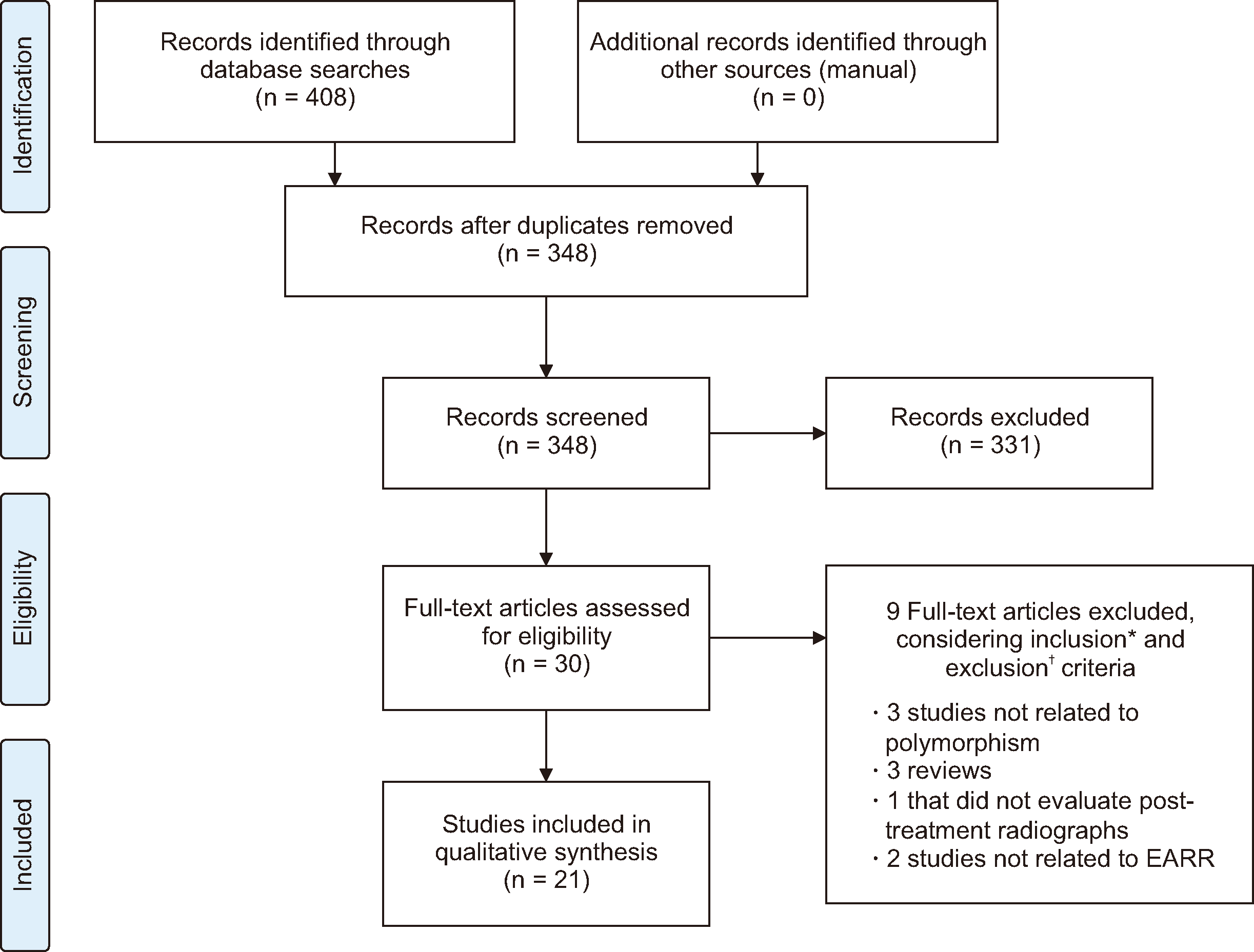
Our electronic search yielded 408 abstracts and titles (Figure 1). We inputted these references into the Reference Manager Software® (Reference Manager, Thomson Reuters, version 12.0.3; Toronto, ON, Canada). After removing duplicates, 348 abstracts and titles were independently reviewed and analyzed by two calibrated reviewers (ILAL, JDC). The calibration process involved determination of the inclusion and exclusion criteria. The reviewers thoroughly discussed these criteria, which were applied to 20% of the retrieved studies. This exercise was repeated until adequate agreement was obtained based on Cohen’s kappa coefficient (kappa: 0.74).
Of the 348 abstracts reviewed, 30 were selected for full-text analysis. To include all related articles, we contacted one author to obtain the full texts of articles that were not accessible online. After the full-text analysis, 21 studies were included in this systematic review (Figure 1).

Data extraction

Data were independently extracted by the same two reviewers (ILAL, JDC). Inter-examiner conflicts were resolved by consensus. The extraction was performed according to the manufacturer’s instructions.
The forms used for data extraction included the year of publication, location, study design, population, sample size, participants’ age, teeth evaluated, EARR quantification criteria, main results, conclusions, and types of gene polymorphisms (Table 1). Most of the described gene polymorphisms were of the single nucleotide polymorphism (SNP) type, identified by the same “rs” (reference SNP) number. However, one polymorphism was of the variable number tandem repeat (VNTR) type.8 Data on genotype distribution and allelic frequency were also included in this systematic review (Table 2).
As different types of polymorphisms have been described, a qualitative analysis was conducted. However, the data were not sufficiently comparable for meta-analysis.

RESULTS

Characteristics of studies

Twenty-one studies were included in this systematic review: 15 case-control studies and six cross-sectional studies (Figure 1). In general, studies recruited cases from university services,1,8,9,11,14-17,21-23,28,29 with seven studies also including patients from private practice,1,9,19-21,24,30 and four studies recruited patients exclusively from private practice.11,18,25,27 The sample sizes ranged from 5414,15 to 48029 individuals. The age of participants ranged from 8.4 to 55.4 years.27
The outcomes were based on tooth dimensions (root and crown lengths) measured directly on radiographs obtained before and after treatment. Lateral cephalometric,1,9,14,16,18,22,25,26,28,30 panoramic,1,9,14,16,18-22,25,26,28-30 periapical,11,24 and occlusal radiographs were used, in addition to cone beam computed tomography (CBCT).23 Control groups comprised the following: (1) non-exposed individuals; (2) patients treated with fixed orthodontic appliances; and (3) patients with a history of EARR of < 2 mm. Three categories of control groups were seen in this review: (1) orthodontically treated patients with an EARR of < 2 mm;11-18,21-26,28 (2) orthodontically treated patients from the same family (parents or siblings) with an EARR of < 2 mm1,9 and (3) orthodontically treated patients with resorption lower than the apical third.22 Furthermore, most studies included in this systematic review performed PCR-RFLP analysis for polymorphisms.1,9,11-18,22-25,28,30
Different teeth were evaluated among the articles retrieved: maxillary central incisors,1,9,11,14,15,18-21,23-30 lateral maxillary incisors,11,14,18-21,26-29 maxillary canine,19,20 maxillary premolars,14,16,22 mandibular central incisors and mandibular molars,1,9,15,18,25 and mandibular first molars.18,25,30

Data synthesis

Qualitative analysis of polymorphisms and EARR
Not all studies described the same variables. There was considerable clinical and methodological heterogeneity as studies have described different types of polymorphisms and SNPs. One study specifically examined tooth-based samples rather than patient-based samples; in this study, IL1B (rs1143634) was not associated with EARR (P > 0.05).15 However, another study found a significant association between IL1B (rs1143634) and EARR (95% confidence interval [CI]: 1.9–21.2) after orthodontic treatment.1 Three other studies examined the association between EARR, gene polymorphisms, and OTM in endodontically treated teeth.14,16,22 They demonstrated that it was not IL1A (rs1800587) but variations in IL1B SNPs that might contribute to the occurrence of EARR.14,16 Furthermore, variants of allele 1 of IL1RN (rs419598) are associated with an increased risk of post-orthodontic EARR in endodontically treated teeth.22 In genetics, an SNP is a variation at a single position in the DNA that can influence disease and drug response. A ‘CC’ genotype implies that both alleles at a gene are cytosine, while a ‘T’ genotype has at least one thymine allele, and a ‘TT’ genotype implies that both alleles are thymine. Homozygosity refers to the presence of identical alleles (either CC or TT) in a specific gene. A haplotype is a set of inherited genes that can provide insight into an individual’s genetic predispositions. Microsatellite polymorphisms involve variations in the short DNA sequences used for genetic testing. Linkage disequilibrium describes how certain alleles at different loci are associated more frequently than expected by chance, aiding genetic mapping and disease studies. Only homozygous individuals for the T allele of IL1RN were more prone to orthodontic-induced EARR.21 IL1RN and P2RXT analyses have indicated that the presence of SNPs in these genes can predispose individuals to EARR.18 IL1RN VNTR polymorphism 86-bp in two introns was also related to EARR, specifically in girls.8
The STAG2 gene, located on the X chromosome, was found to be associated with an increased risk of EARR, particularly restricted to the male sex.9 Another study demonstrated that IRAK1 polymorphism is a protective variant of EARR.20 For genes encoding OPG, RANK, IL1, and IL1RN, the effect changed from protective to deleterious depending on the duration of treatment and the age of the patient.19
Regarding RANKL polymorphisms, no significant association was found with EARR. Among RANK polymorphisms, only rs12455775 was associated with EARR. Regarding OPG polymorphisms, an association was found between EARR and rs3102724, rs2875845, rs1032128, and rs3102728.24
TNSALP, TNFα, and TNFRSF11A polymorphisms were also investigated. It was concluded that TNFRSF11A (rs1805034) is associated with EARR (P = 0.02), but TNSALP and TNFα are not.9 Additionally, SPP1 gene polymorphisms (rs9138 and rs11730582) were related to genetic susceptibility of EARR: rs9138 CC vs. CA/AA (odds ratio: 4.10; 95% CI: 1.03–16.35); rs9138 AA vs. CA/CC (odds ratio: 0.20; 95% CI: 0.05–0.81); rs11730582 CC vs. CT/TT (odds ratio: 11.68; 95% CI: 1.12–121.06); and rs9138 CT vs. CC/TT (odds ratio: 0.035; 95% CI: 0.062–0.90).26 One study observed that the presence of a specific P2RX7 (rs208294) was significantly associated with EARR (P = 0.0028) (Table 1).27,28
According to Ciurla et al.25 the effect of SNPs in CASP1, TNFRSF11B, and IL 6 genes on EARR was not confirmed however, Guo et al.23 showed that IL 6 increased the risk of EARR.
Marañón-Vásquez et al.30 showed that individuals with the AA genotype of VDR rs2228570 had a higher risk of EARR than those with the AG and GG genotypes. In addition, the incidence of EARR in heterozygous rs2228570 was lower than that in homozygotes. In addition, participants with the CCG haplotype (rs1544410-rs7975232-rs731236) in the VDR showed a lower EARR than those who did not carry this haplotype. Regarding CYP27B1 rs4646536, participants with at least one G allele showed a lower risk of EARR than those with homozygote AA. Although these results were not significant after multiple test adjustments, potential associations may still be suggested.30

DISCUSSION

Herein, we conducted a systematic review on the association of EARR, OTM, and gene polymorphisms.

Assessment of bias in included studies

The present review presents no bias related to the language or year of publication. The search strategy identified only articles published in English1,9,11,14-30 between 2003 and 2023. Although there is a possibility of bias due to the small sample sizes of some reports, this is a relatively new field of investigation, and the retrieved studies presented high methodological quality.

Assessment of methodological quality

All case-control studies presented an adequate definition of EARR through radiographic evaluation.8,11,14-17,22,26,27 The cases analyzed were from the same family or clinic, and the study participants were controlled for age.8,11,14-17,22,26,27 The control group comprised orthodontically treated patients with an EARR of < 2 mm,11-17,22 orthodontically treated patients from the same family (parents or siblings) with an EARR of < 2 mm, and orthodontically treated patients with resorption limited to the apical third.27 Individuals presenting with EARR of ≥ 2 mm11,14-17 or resorption equal to or more than the apical third27 were considered as cases. In addition, the same method of ascertainment (PCR-RFLP analysis) was employed for both cases and controls.
All cross-sectional studies in this systematic review included clinically diagnosed cases of EARR.1,9 Secure records with no indication of EARR at the start of the study were presented and radiographs of the patients after the end of treatment were used to evaluate the presence of EARR. Therefore, the follow-up was considered adequate. The details of blind processes have rarely been reported.

Strength of evidence

In 1975, EARR was described as familial,31 and in 1997, a hypothesis of genetic influence on EARR was proposed using a sib-pair model with an estimated heritability of approximately 70%.32 The current understanding is that EARR has a multifactorial etiology influenced by a combination of environmental and host factors. Genetic predisposition to an exaggerated inflammatory response may contribute to EARR in some patients during OTM. Accordingly, many studies have attempted to demonstrate the association between gene polymorphisms involved in inflammatory responses and EARR. In this regard, the IL1 gene is the most frequently described, as it may be an early critical mediator in inflammatory processes.33 IL1 plays a critical role in the inflammatory response related to orthodontic treatment; it recruits large immune cells and affects transmigration to the tooth movement area. Moreover, IL1 directly or indirectly amplifies osteoclast differentiation,34 affecting root resorption. Elevated IL1β levels in gingival crevicular fluid and gingival tissues during OTM further support its role in tooth resorption.35
Some studies have investigated IL1 polymorphisms in the context of EARR; however, conflicting results have been reported, mainly associated with ethnic populations. These discrepancies may arise due to the geographic restrictions of each study.15 Tomoyasu et al.15 demonstrated that IL1B (rs1143634) was not associated with a predisposition to EARR. Accordingly, a meta-analysis revealed that IL1B polymorphisms were not found to be associated with the susceptibility to EARR.36 In contrast, other studies found an association between IL1 polymorphisms and EARR following OTM.1,17 Studies report that the presence of haplotype +3,954 and –889 with “T” allele increases the production of IL1B and IL1A, respectively,8,15,17 suggesting an association with EARR.1,11,14 A link between EARR and IL1B SNPs has also been described.25 On the contrary, other studies found that the presence of “CC” genotype increase the risk of EARR.1,11,27 This result agrees with a previous study showing that the absence of IL1B gene causes root resorption in mouse molar.13
IL1A (rs1800587) and IL1B (rs1143634) polymorphisms were analyzed in four case-control studies.8,14,18,21 Linhartova et al.8 found no association between IL1 (rs1800587 and rs1143634) and EARR, while Iglesias-Linares et al.14 reported a relationship between IL1B (rs1143634) and EARR. Although both studies used similar methods, the number and age of the participants could explain these differences. The study by Linhartova et al.8 included 106 participants (mean age: 15.0 ± 4.1 years in EARR group and 15.2 ± 5.3 years in the control group), whereas Iglesias-Linares et al.14 included 54 patients (mean age: 22.9 ± 6.3 years). Silva et al.19 showed that for IL1, the effect changed from protective to deleterious depending on the duration of treatment and the age of the patient.
Perrier et al.,37 demonstrated that allele 2 (two repeats) of IL1RN (VNTR 86bp) polymorphism downregulates interleukin-1 receptor antagonist (IL1Ra) production in saliva, increasing IL1 expression. One study that evaluated the role of IL1RN polymorphism VNTR 86bp in EARR8 showed that the allele 2 variant was specifically related to EARR and OTM in girls.8 Individuals that are homozygous for the T allele of IL1RN rs419598 were more prone to EARR during orthodontic treatment.21 There is further evidence that “TT” genotype of IL1RN rs419598 increases the risk of EARR in endodontically treated teeth.22 In addition, IL1RN rs419598 and VNTR 86bp haplotypes are in linkage disequilibrium, affecting IL1Ra and IL1B production.22 Another study18 concluded that the presence of IL1RN SNP (rs419598) may predispose individuals to EARR.
Al-Qawasmi et al.,1 Bastos Lages et al.,11 and Iglesias-Linares et al.14,16,22 suggested a possible link between IL1A (rs1800587), IL1B (rs1143634), and EARR. Moreover, Ciurla et al.25 showed that IL1B increased the odds of developing EARR four-fold. Although Al-Qawasmi et al.1 and Bastos Lages et al.11 used similar methods, the ethnic diversity of the samples may have influenced the results. They found that the “CC” genotype increases the risk of EARR, although the presence of “T” allele 4-fold increased the level of IL1B.9 In contrast, Iglesias-Linares et al.14,16,22 evaluated endodontically treated teeth, which may have been a confounding factor.
TNFSF11A, involved in RANK encoding, plays a crucial role in osteoclastogenesis, a major mechanism involved in mineral matrix destruction. An important association between TNFRSF11A rs1805034 microsatellite polymorphism (D18S64) and EARR has been discussed based on linkage disequilibrium between them.9 Although TNSALP is important for cementum formation and mineralization and TNFα is critical in bone remodeling, no evidence of association or linkage disequilibrium was found between them and EARR.9
SPP1, which regulates the release of pro- and anti-inflammatory mediators (mainly IL1B) by human monocytes, has also been studied. Iglesias-Linares et al.26 found that “C” allele of SPP1 gene polymorphisms “rs9138 and rs11730582” increase the risk of EARR, and they are associated with elevated osteonectin expression.
The purinergic receptor (P2X) is a member of the cationic channel family, in which receptor P2RX7 plays an important role in the immune response, mainly in bone metabolism, inflammation (upstream of IL1B maturation and release), cell proliferation, and the central nervous systems.38 Association between EARR and P2RX7 gene polymorphisms “rs208294 and rs1718119” have been reported; the “T” allele was associated with a protective role while “G” allele increased the risk for EARR.27 Borilova Linhartova et al.28 also found that variability in P2RX7 may be an important factor contributing to the etiopathogenesis of EARR. Furthermore, an association of caspase 1—involved with IL1B maturation and release—with EARR was investigated; the “T” allele of rs530537 increases the risk of EARR.
Altogether, existing evidence suggests that IL1 is associated with EARR, however, pinpointing the specific IL1 allele (IL1A, IL1B, or IL1RN) implicated in EARR remains challenging. Several factors contribute to this uncertainty. First, the retrieved studies exhibited significant heterogeneity in gene polymorphism analyses. Second, variations in radiographic techniques—ranging from periapical and occlusal radiographs to CBCT—yield varying measurement accuracies of EARR. This could represent bias in the diagnosis of EARR. Additionally, the lack of important data related to the sample, such as sex and age, are often absent.
Moreover, isolated studies have explored other candidate genes. These include TNFRSF11A, TNFRSF11B, SPP1, IL6, STAG2, VDR, IRAK1, and IL17. However, further research is needed to confirm the possible association of these genes with EARR and OTM.

CONCLUSIONS

This systematic review demonstrated that the majority of studies pointed to a possible association between EARR, OTM, and IL1 gene polymorphisms. Yet, the specific IL1 allele driving this association remains elusive. Additionally, P2RX7 may be an important factor contributing to the etiopathogenesis of EARR. TNFRSF11A, SPP1, IL1RN, IL6, TNFRSF11B, STAG2, VDR, IRAK1, IL17, CASP1/ICE and CASP5 have been identified in isolated studies. Further research is recommended to unravel the genetic susceptibility underlying EARR associated with orthodontic treatments. Furthermore, identifying specific genes associated with EARR, given its multifactorial nature, will enhance orthodontic management of patients at a high risk of EARR.

Notes

AUTHOR CONTRIBUTIONS

Conceptualization: ILAL, JDC. Data curation: ILAL, JDC. Formal analysis: ILAL, JDC. Investigation: All authors. Methodology: All authors. Project administration: ILAL. Resources: ILAL, JDC, FRM, DAM. Software: ILAL, JDC, FRM, DAM. Supervision: ILAL. Validation: ILAL, JDC, FRM, DAM. Visualization: ILAL, JDC, FRM, DAM. Writing–original draft: ILAL, JDC. Writing–review & editing: ILAL, ALCÁA, YDAP, BEBM.

CONFLICTS OF INTEREST

No potential conflict of interest relevant to this article was reported.

FUNDING

None to declare.

REFERENCES

1. Al-Qawasmi RA, Hartsfield JK Jr, Everett ET, Flury L, Liu L, Foroud TM, et al. 2003; Genetic predisposition to external apical root resorption. Am J Orthod Dentofacial Orthop. 123:242–52. https://doi.org/10.1067/mod.2003.42. DOI: 10.1067/mod.2003.42. PMID: 12637896.
2. Brezniak N, Wasserstein A. 2002; Orthodontically induced inflammatory root resorption. Part II: the clinical aspects. Angle Orthod. 72:180–4. https://pubmed.ncbi.nlm.nih.gov/11999942/.
3. Harris EF, Robinson QC, Woods MA. 1993; An analysis of causes of apical root resorption in patients not treated orthodontically. Quintessence Int. 24:417–28. https://pubmed.ncbi.nlm.nih.gov/8234648/.
4. Harris EF, Butler ML. 1992; Patterns of incisor root resorption before and after orthodontic correction in cases with anterior open bites. Am J Orthod Dentofacial Orthop. 101:112–9. https://doi.org/10.1016/0889-5406(92)70002-R. DOI: 10.1016/0889-5406(92)70002-R.
5. Hartsfield JK Jr, Everett ET, Al-Qawasmi RA. 2004; Genetic factors in external apical root resorption and orthodontic treatment. Crit Rev Oral Biol Med. 15:115–22. https://doi.org/10.1177/154411130401500205. DOI: 10.1177/154411130401500205.
6. Brezniak N, Wasserstein A. 1993; Root resorption after orthodontic treatment: part 2. Literature review. Am J Orthod Dentofacial Orthop. 103:138–46. https://doi.org/10.1016/S0889-5406(05)81763-9. DOI: 10.1016/S0889-5406(05)81763-9.
7. Killiany DM. 1999; Root resorption caused by orthodontic treatment: an evidence-based review of literature. Semin Orthod. 5:128–33. https://doi.org/10.1016/s1073-8746(99)80032-2. DOI: 10.1016/S1073-8746(99)80032-2.
8. Linhartova P, Cernochova P, Izakovicova Holla L. 2013; IL1 gene polymorphisms in relation to external apical root resorption concurrent with orthodontia. Oral Dis. 19:262–70. https://doi.org/10.1111/j.1601-0825.2012.01973.x. DOI: 10.1111/j.1601-0825.2012.01973.x.
9. Al-Qawasmi RA, Hartsfield JK Jr, Everett ET, Flury L, Liu L, Foroud TM, et al. 2003; Genetic predisposition to external apical root resorption in orthodontic patients: linkage of chromosome-18 marker. J Dent Res. 82:356–60. https://doi.org/10.1177/154405910308200506. DOI: 10.1177/154405910308200506.
10. Ngan DC, Kharbanda OP, Byloff FK, Darendeliler MA. 2004; The genetic contribution to orthodontic root resorption: a retrospective twin study. Aust Orthod J. 20:1–9. https://doi.org/10.2478/aoj-2004-0001. DOI: 10.2478/aoj-2004-0001. PMID: 302ddfe014f546da87a2eedd90b9f5dc.
11. Bastos Lages EM, Drummond AF, Pretti H, Costa FO, Lages EJ, Gontijo AI, et al. 2009; Association of functional gene polymorphism IL-1beta in patients with external apical root resorption. Am J Orthod Dentofacial Orthop. 136:542–6. https://doi.org/10.1016/j.ajodo.2007.10.051. DOI: 10.1016/j.ajodo.2007.10.051.
12. Gülden N, Eggermann T, Zerres K, Beer M, Meinelt A, Diedrich P. 2009; Interleukin-1 polymorphisms in relation to external apical root resorption (EARR). J Orofac Orthop. 70:20–38. https://doi.org/10.1007/s00056-009-8808-6. DOI: 10.1007/s00056-009-8808-6.
13. Hartsfield JK Jr. 2009; Pathways in external apical root resorption associated with orthodontia. Orthod Craniofac Res. 12:236–42. https://doi.org/10.1111/j.1601-6343.2009.01458.x. DOI: 10.1111/j.1601-6343.2009.01458.x. PMID: 19627526. PMCID: PMC5161460.
14. Iglesias-Linares A, Yañez-Vico R, Ballesta-Mudarra S, Ortiz-Ariza E, Ortega-Rivera H, Mendoza-Mendoza A, et al. 2012; Postorthodontic external root resorption is associated with IL1 receptor antagonist gene variations. Oral Dis. 18:198–205. https://doi.org/10.1111/j.1601-0825.2011.01865.x. DOI: 10.1111/j.1601-0825.2011.01865.x.
15. Tomoyasu Y, Yamaguchi T, Tajima A, Inoue I, Maki K. 2009; External apical root resorption and the interleukin-1B gene polymorphism in the Japanese population. Orthod Waves. 68:152–7. https://doi.org/10.1016/j.odw.2009.05.002. DOI: 10.1016/j.odw.2009.05.002.
16. Iglesias-Linares A, Yañez-Vico RM, Ortiz-Ariza E, Ballesta S, Mendoza-Mendoza A, Perea E, et al. 2012; Postorthodontic external root resorption in root-filled teeth is influenced by interleukin-1β polymorphism. J Endod. 38:283–7. https://doi.org/10.1016/j.joen.2011.12.022. DOI: 10.1016/j.joen.2011.12.022.
17. Iglesias-Linares A, Yañez-Vico RM, Ballesta S, Ortiz-Ariza E, Mendoza-Mendoza A, Perea E, et al. 2012; Interleukin 1 gene cluster SNPs (rs1800587, rs1143634) influences post-orthodontic root resorption in endodontic and their contralateral vital control teeth differently. Int Endod J. 45:1018–26. https://doi.org/10.1111/j.1365-2591.2012.02065.x. DOI: 10.1111/j.1365-2591.2012.02065.x. PMID: 22621214.
18. Ciurla A, Marruganti C, Doldo T, Szymańska J. 2021; Association between polymorphisms in the IL-1β, TNFRSF11B, CASP1, and IL-6 genes and orthodontic-induced external apical root resorption. J Clin Med. 10:4166. https://doi.org/10.3390/jcm10184166. DOI: 10.3390/jcm10184166. PMID: 34575287. PMCID: PMC8469359.
19. Silva HC, Lavado N, Canova F, Lopez MG, Regateiro FJ, Pereira SA. 2022; Influence of clinical factors on the protective or deleterious impact of genetic variants in orthodontically induced external root resorption: an observational study. BMC Oral Health. 22:270. https://doi.org/10.1186/s12903-022-02306-y. DOI: 10.1186/s12903-022-02306-y. PMID: 341a6ad901f54d82aebda8e7a517af1a.
20. Pereira S, Nogueira L, Canova F, Lopez M, Silva HC. 2016; IRAK1 variant is protective for orthodontic-induced external apical root resorption. Oral Dis. 22:658–64. https://doi.org/10.1111/odi.12514. DOI: 10.1111/odi.12514.
21. Iglesias-Linares A, Sonnenberg B, Solano B, Yañez-Vico RM, Solano E, Lindauer SJ, et al. 2017; Orthodontically induced external apical root resorption in patients treated with fixed appliances vs removable aligners. Angle Orthod. 87:3–10. https://doi.org/10.2319/02016-101.1. DOI: 10.2319/02016-101.1.
22. Iglesias-Linares A, Yañez-Vico RM, Ballesta-Mudarra S, Ortiz-Ariza E, Mendoza-Mendoza A, Perea-Pérez E, et al. 2013; Interleukin 1 receptor antagonist (IL1RN) genetic variations condition post-orthodontic external root resorption in endodontically-treated teeth. Histol Histopathol. 28:767–73. https://doi.org/10.14670/HH-28.767.
23. Guo Y, He S, Gu T, Liu Y, Chen S. 2016; Genetic and clinical risk factors of root resorption associated with orthodontic treatment. Am J Orthod Dentofacial Orthop. 150:283–9. https://doi.org/10.1016/j.ajodo.2015.12.028. DOI: 10.1016/j.ajodo.2015.12.028.
24. Simas Netta Fontana MLS, Pereira FA, Tanaka OM, Trevilatto PC. Borges de Castilhos B. Machado de Souza C. 2019; Association of clinical variables and polymorphisms in RANKL, RANK, and OPG genes with external apical root resorption. Am J Orthod Dentofacial Orthop. 155:529–42. https://doi.org/10.1016/j.ajodo.2018.05.016. DOI: 10.1016/j.ajodo.2018.05.016. PMID: 30935608.
25. Ciurla A, Szymańska J, Płachno BJ, Bogucka-Kocka A. 2021; Polymorphisms of encoding genes IL1RN and P2RX7 in apical root resorption in patients after orthodontic treatment. Int J Mol Sci. 22:777. https://doi.org/10.3390/ijms22020777. DOI: 10.3390/ijms22020777. PMID: 33466672. PMCID: PMC7828778. PMID: 0ff96776f6c8480da7ade324ad85defe.
26. Iglesias-Linares A, Yañez-Vico RM, Moreno-Fernández AM, Mendoza-Mendoza A, Orce-Romero A, Solano-Reina E. 2014; Osteopontin gene SNPs (rs9138, rs11730582) mediate susceptibility to external root resorption in orthodontic patients. Oral Dis. 20:307–12. https://doi.org/10.1111/odi.12114. DOI: 10.1111/odi.12114.
27. Sharab LY, Morford LA, Dempsey J, Falcão-Alencar G, Mason A, Jacobson E, et al. 2015; Genetic and treatment-related risk factors associated with external apical root resorption (EARR) concurrent with orthodontia. Orthod Craniofac Res. 18(Suppl 1):71–82. https://doi.org/10.1111/ocr.12078. DOI: 10.1111/ocr.12078. PMID: 25865535. PMCID: PMC4810020.
28. Borilova Linhartova P, Cernochova P, Kastovsky J, Vrankova Z, Sirotkova M, Izakovicova Holla L. 2017; Genetic determinants and postorthodontic external apical root resorption in Czech children. Oral Dis. 23:29–35. https://doi.org/10.1111/odi.12564. DOI: 10.1111/odi.12564.
29. Iber-Díaz P, Senen-Carramolino R, Iglesias-Linares A, Fernández-Navarro P, Flores-Mir C, Yañez-Vico RM. 2020; GWAS of post-orthodontic aggressive external apical root resorption identified multiple putative loci at X-Y chromosomes. J Pers Med. 10:169. https://doi.org/10.3390/jpm10040169. DOI: 10.3390/jpm10040169. PMID: 33066413. PMCID: PMC7712155.
30. Marañón-Vásquez G, Küchler EC, Hermann S, Paddenberg E, Schröder A, Baratto-Filho F, et al. 2023; Association between genetic variants in key vitamin-D-pathway genes and external apical root resorption linked to orthodontic treatment. Eur J Oral Sci. 131:e12916. https://doi.org/10.1111/eos.12916. DOI: 10.1111/eos.12916. PMID: 36683003.
31. Newman WG. 1975; Possible etiologic factors in external root resorption. Am J Orthod. 67:522–39. https://doi.org/10.1016/0002-9416(75)90298-5. DOI: 10.1016/0002-9416(75)90298-5.
32. Harris EF, Kineret SE, Tolley EA. 1997; A heritable component for external apical root resorption in patients treated orthodontically. Am J Orthod Dentofacial Orthop. 111:301–9. https://pubmed.ncbi.nlm.nih.gov/9082853/. DOI: 10.1016/S0889-5406(97)70189-6.
33. Dinarello CA. 2002; The IL-1 family and inflammatory diseases. Clin Exp Rheumatol. 20(5 Suppl 27):S1–13. https://pubmed.ncbi.nlm.nih.gov/14989423/. PMID: 14989423.
34. Wei S, Kitaura H, Zhou P, Ross FP, Teitelbaum SL. 2005; IL-1 mediates TNF-induced osteoclastogenesis. J Clin Invest. 115:282–90. https://doi.org/10.1172/JCI23394. DOI: 10.1172/JCI23394. PMID: 15668736. PMCID: PMC544608.
35. Grieve WG 3rd, Johnson GK, Moore RN, Reinhardt RA, DuBois LM. 1994; Prostaglandin E (PGE) and interleukin-1 beta (IL-1 beta) levels in gingival crevicular fluid during human orthodontic tooth movement. Am J Orthod Dentofacial Orthop. 105:369–74. https://pubmed.ncbi.nlm.nih.gov/8154462/. DOI: 10.1016/S0889-5406(94)70131-8.
36. Wu FL, Wang LY, Huang YQ, Guo WB, Liu CD, Li SG. 2013; Interleukin-1β +3954 polymorphisms and risk of external apical root resorption in orthodontic treatment: a meta-analysis. Genet Mol Res. 12:4678–86. https://doi.org/10.4238/2013.October.18.6. DOI: 10.4238/2013.October.18.6. PMID: 24222244.
37. Perrier S, Coussediere C, Dubost JJ, Albuisson E, Sauvezie B. 1998; IL-1 receptor antagonist (IL-1RA) gene polymorphism in Sjögren's syndrome and rheumatoid arthritis. Clin Immunol Immunopathol. 87:309–13. https://doi.org/10.1006/clin.1998.4520. DOI: 10.1006/clin.1998.4520. PMID: 9646842.
38. Ursu D, Ebert P, Langron E, Ruble C, Munsie L, Zou W, et al. 2014; Gain and loss of function of P2X7 receptors: mechanisms, pharmacology and relevance to diabetic neuropathic pain. Mol Pain. 10:37. https://doi.org/10.1186/1744-8069-10-37. DOI: 10.1186/1744-8069-10-37. PMID: 24934217. PMCID: PMC4072620.

Figure 1
PRISMA flow diagram showing the process of article selection.
EARR, external apical root resorption.
*Inclusion criteria: epidemiological studies (cross-sectional, case-control, cohort and clinical trials) that evaluated the association of genetic polymorphisms and EARR in patients treated with fixed appliances; studies that used polymerase chain reaction-restriction fragment length polymorphism analysis to evaluate genetic polymorphisms; and, studies that reported pre- and post-orthodontic treatment.
Exclusion criteria: reviews, articles or other systematic reviews; case reports; case series; descriptive studies; opinion articles; abstracts; letters to the editor; laboratory and/or animal studies; subjects with cleft lip, palate or both; studies on individuals with other craniofacial deformities/syndromes; studies on unrelated topics that had no association with genetics or those that presented data of EARR not caused by orthodontic treatment.
kjod-54-5-284-f1.tif
Table 1
Characteristics of the case-control and cross-sectional studies included in this systematic review
Study/country Design Population Sample size analyzed (case) Sample size analyzed (control) Participant age (yr) Tooth evaluation Measurement criteria Gene polymorphism Result Conclusion
Al-Qawasmi et al.1 (2003)/USA Cross-sectional University clinic and private orthodontic practice 118 - 12.1 ± 1.89 MxCI with the longest root, MndCI with the longest root, and the mesial and distal roots of both the MndFM Ceph and PAN
IL1A (rs1800587)
IL1B (rs1143634)
IL1β allele 1 had a 5.6-fold (95% CI: 1.9–21.2) increased risk of EARR greater than 2 mm compared with those who are not homozygous for the IL-1B allele 1 (P = 0.004) IL1B polymorphisms are associated with EARR
Al-Qawasmi et al.9 (2003)/USA Cross-sectional University clinic and private orthodontic practice 124 - 12.3 ± 1.82 MxCI with the longest root, MndCI with the longest root, and the mesial and distal roots of both the MndFM Ceph and PAN
TNSALP (rs not informed)
TNFα (rs1800629)
TNFRSF11A (rs1805034)
TNFRSF11A (LOD = 2.5; P = 0.02)
No evidence of linkage was found with EARR and the TNFα and TNSALP genes
TNFRSF11A is associated with EARR
Bastos Lages et al.11 (2009)/Brazil Case-control Private orthodontic practice 23 38 18.9 ± 5.2 MxCI, MxLI Periapical radio-graphs IL1B (rs1143634) 2/2 vs. 1/2 + 1/1, OR = 4.00; 95% CI:1.23–12.9; P = 0.0349 The IL1B polymorphism associated with EARR
Tomoyasu et al.15 (2009)/Japan Case-control University clinic 54 -
Male 19
Female 21
MxCI, MndCI, FM Ceph and PAN IL1B (rs1143634)
IL1B
Maxillary central incisor (P = 0.47) Mandibular central incisor (P = 0.48) Mandibular first molar, metal root (P = 0.08) Mandibular first molar, distal root (P = 0.22)
No association
Iglesias-Linares et al.17 (2012)/Spain Case-control University clinic 73 73 23.78 ± 5.91 Maxillary root-filled PM and the contralateral tooth with a vital pulp Ceph and PAN
IL1B (rs1143634)
IL1A (rs1800587)
CC vs. CT/TT 5.143 1.38–19.10 (P = 1.00)
TT vs. CT/CC 2.032 1.99–14.773 (P = 0.031)
CT vs. CC/TT 10.66 0.72–158.50 (P = 0.625)
IL1B polymorphisms are associated with a twofold increased predisposition to have EARR secondary to orthodontic treatment in endodontically treated teeth
Linhartovaet al. 8 (2013)/Czech Republic Case-control University clinic 32 74
15.0 ± 4.1
and
15.2 ± 5.3
MxCI, MxLI Ceph and PAN
IL1A (rs1800587)
IL1B (rs1143634)
IL1RN (86pb VNTR)
IL1RN
Variants in girls – short allele P = 0.020, OR = 2.50; 95% CI: 1.13–5.53
No significant role of IL1A and IL1B variants in EARR
IL1RN may be associated with EARR, especially in girls
Iglesias-Linares et al.14 (2012)/Spain Case-control University clinic 25 29
22.25 ± 5.30
and
23.89 ± 5.03
MxCI, MxLI with the longest root Ceph and PAN
IL1A (rs1800587)
IL1B (rs1143634)
IL1RN (rs419598)
IL1B
CC vs. CT⁄ TT OR = 3.477 (1.12–10.72); P = 0.027
IL1A
CC vs. CT⁄ TT OR = 2.51 (0.8–7.57); P = 0.097
IL1R
TT vs. CC⁄TC OR = 6.750 (2.04–22.27); P = 0.001
Variations in the IL1RN and not only in the IL1B gene are determinants of a predisposition to post-orthodontic EARR
Iglesias-Linares et al.16 (2012)/Spain Case-control University clinic 39 54
24.54 ± 5.85
and
23.89 ± 5.72
Upper second root-filled PM Ceph and PAN
IL1A (rs1800587)
IL1B (rs1143634)
IL1B
CC vs. CT/TT OR: 2.54 (1.05–6.12); P = 0.035
TT vs. CT/CCOR: 11.59 (1.36–98.61); P = 0.006
CT vs. CC/TT OR: 0.12 (0.03–0.39); P = 0.001
IL1B gene polymorphisms are associated with EARR
Iglesias-Linares et al.22(2013)/Spain Case-control University clinic 39 54
24.54 ± 5.85
and
23.89 ± 5.72
Upper second root-filled PM Ceph and PAN IL1RN (rs419598)
IL1RN
CC vs. CT/TT OR: 10.857 (3.97–29.6); P = 0.001
CT vs. TT/CC OR: 0.18 (0.06–0.50); P = 0.001
CC vs. CT/CC OR: 0.10 (0.13–0.83); P = 0.011
IL1RN polymorphisms are associated with an increased risk of suffering post-orthodontic EARR in root-filled teeth
Iglesias-Linares et al.26 (2014)/Spain Case-control University clinic 37 50
24.70 ± 5.95
and
23.80 ± 5.33
MxCI, MxLI with the longest root Ceph and PAN SPP1(rs9138, rs11730582)
rs9138
CC vs. CA/AA OR: 4.10 1.03–16.35 (P = 0.045)
AA vs. CA/CC OR: 0.200.05–0.81 (P = 0.025)
CA vs. CC/AA OR: 0.8 0.20–3.53 (P = 0.823)
rs11730582
CC vs. CT/TTOR: 11.681.12–121.06 (P = 0.039)
TT vs. CT/CCOR: 0.450.07–2.80 (P = 0.39)
CT vs. CC/TTOR: 0.0350.062–0.90 (P = 0.035)
Specific allele (not informed) of the SPP1 is associated with genetic susceptibility to EARR
Sharab et al.27 (2015)/USA Case-control Private orthodontic practice 67 67
15.78 ± 1.13
and
15.79 ± 1.14
MxCI, MxLI Occlusal
P2RX7 (rs208294, rs1718119, rs2230912)
CASP1/ICE (rs580253,rs530537)
CASP5 (rs554344)
IL1B (rs1143634)
IL1A (rs1800587)
IL1Ra (rs419598)
P2RX7, rs208294
CC: 28 (41.8%)
CT: 33 (49.3%)
TT: 6 (9.0%)
P = 0.0028
P2RX7, rs1718119
GG: 32 (47.8%)
GA: 25 (37.3%)
AA: 10 (14.9%)
P2RX7, rs2230912
AA: 53 (79.1%)
AG: 13 (19.4%)
GG: 1 (1.5%)
CASP1/ICE, rs530537
TT: 24 (35.8%)
TC: 30 (44.8%)
CC: 13 (19.4%)
CASP1/ICE, rs580253
GG: 52 (77.6%)
GA: 14 (20.9%)
AA: 1 (1.5%)
CASP5, rs554344
GG: 52 (77.6%)
GC: 14 (20.9%)
CC: 1 (1.5%)
IL1B, rs1143634
GG: 37 (55.2%)
GA: 26 (38.8%)
AA: 4 (6.0%)
IL1Ra, rs419598
TT: 41 (61.2%)
TC: 23 (34.3%)
CC: 3 (4.5%)
P = 0.0533
IL1A, rs1800587
GG: 28 (41.8%)
GA: 28 (41.8%)
AA: 11 (16.4%)
P2RX7 rs208294 was associated with EARR
Borilova Linhartova et al.28 (2017)/Czech Republic Case-control University clinic 30 69 15.0 ± 4.7 MxCI, MxLI rooth and crown lengths Ceph and PAN IL-17(rs2275913); SPP1 (rs11730582, rs9138); P2RX7(rs208294, Tyr155His, rs1718119); 11B (rs3102735, rs2073618) No significant differences were observed in allele or genotype frequencies of all seven studied SNPs. Specific haplotype of P2RX7 (rs208294 and rs1718119) modified the risk of EARR development (P < 0.05) The variability in the P2RX7 gene may be important factor contributing to the etiopatho-genesis of post-orthodontic EARR
Ciurla et al.25 (2021)/Poland Case-control Private orthodontic practice 40 61 22.9 ± 6.3 MxCI, MxLI, MndCI, MndLI, FM Ceph and PAN IL1RN (rs419598) and P2RX7 (rs208294) P2RX7 (rs208294) and IL1RN (rs419598) modified the risk of EARR development (P < 0.05) The analysis of the P2RX7 and IL1RN gene polymorphisms showed that the presence of SNPs of these genes may predispose individuals to EARR
Guo et al.23 (2016)/China Case-control University clinic 174 - 14.07 ± 3.10 MxCI CBCT IL1RN (rs419598); IL-6 SNP (rs1800796) The IL-6 SNP rs1800796 GC was associated with EARR, and root resorption IL-6 SNP rs1800796 GC is a risk factor for EARR
Ciurla et al.18 (2021)/Poland Case-control Private orthodontic practice 101 - 21.32 ± 7.28 MxCI, MxLI, MndCI, MndLI, FM Ceph and PAN IL-1β, TN- FRSF11B, CASP1, and IL-6 A significant association was found between EARR presence and the SNP for the IL-1β gene but not for the TNFRSF11B, CASP1, and the IL-6 genes. IL-1β gene increases the odds of developing EARR by around four times A significant association between EARR occurrence and the SNP for the IL-1β gene. Conversely, the effect of SNPs for CASP1, TNFRSF11B, and IL-6 genes on EARR presence was not confirmed by the present study
Borges de Castilhos et al.24 (2019)/Brazil Case-control University clinic and private orthodontic practice 178 160 14.9 (8–21) MxCI Periapical radio-graphs RANKL, RANK, OPG For polymor-phisms of RANKL, no significant association was found with EARR. For RANK polymorphisms, only rs12455775 was associated with EARR Regarding OPG polymorphisms, an association of rs3102724, rs2875845, rs1032128, and rs3102728 with EARR was found Regarding the analysis of polymorphisms in the genes RANKL, RANK, and OPG, several SNPs in RANK and OPG were associated with EARR, but only the association of the allele A of the rs3102724 in OPG remained after multivariate analysis
Silva et al.19 (2022)/Portugal Cross-sectional University clinic and private orthodontic practice 195 -
< 14 (n = 63); 14–18
(n = 85);
18 > (n = 47)
MxCI, MxLI, MxCanine PAN rs1143634 (in IL1B gene) and rs3102735 (TNFRSF11B gene, encoding OPG) and (rs315952 from IL1RN), rs1805034 from TNFRSF11A, encoding RANK, and rs1718119 from P2RX7 For genes encoding OPG, RANK and the IL1 and IL1RN, the effect of analyzed variants changed from protective to deleterious depending on the duration of treatment and the age of the patient This work shows that in OIEARR the impact of genetic susceptibility factors is dynamic changing according to clinical variables
Iber-Díaz et al.29 (2020)/Spain Cohort University clinic 101 361
21.52 ±
11.65
and
22.83 ± 11.66
MxCI, MxLI PAN Genes located in the X chromosome, specifically, STAG2 (rs151184635) and RP1-30E17.2 (rs55839915)
Two novel putative genes located in the X chromosome, specifically, STAG2 gene, rs151184635 and RP1-30E17.2 gene, rs55839915, were associated with aggressive EARR
These variants were found to be associated with an increased risk of aEARR, particularly restricted to male. Marginal associations were found at previously studied variants such as SPP1: rs11730582, P2RX7: rs1718119, and TNFRSF11A: rs8086340, found solely in females
Multiple putative genetic variants located at chromosomes X and Y are potentially implicated in an extreme phenotype of aggressive EARR
Marañón-Vásquez et al.30 (2023)/Germany Cross-sectional University clinic and private orthodontic practice 143 - 13.5 ± 4.5 MxCI, FM Ceph and PAN VDR rs731236 – TaqI (A/G); VDR rs7975232 – ApaI (C/A); VDR rs1544410 – BsmI (T/C); VDR rs2228570 – FokI (A/G); GC rs4588 (T/G); CYP27B1 rs4646536 (G/A); CYP24A1 rs927650 (T/C) Individuals carrying the AA genotype in VDR rs2228570 had a 21% higher EARRmol than those having AG and GG genotypes. EARRmol in heterozygous rs2228570, was 12% lower than for homozygotes. Participants with the CCG haplotype (rs1544410-rs7975232-rs731236) in VDR had an EARRmol 16% lower than those who did not carry this haplotype. Regarding CYP27B1 rs4646536, EARRinc in participants who had at least one G allele was 42% lower than for homozygotes AA Although these results did not remain significant after multiple testing adjustment, potential associ- ations may still be suggested
Pereira et al.20 (2016)/Portugal Cross-sectional University clinic and private orthodontic practice 195 - 17.24 ± 6.8 MxCI, MxLI, MxCanine PAN IL1B (rs1143634), IL1RN (rs315952) IRAK1 (rs1059703) Homozygosity/ hemizygosity for variant C from IRAK1 gene (P = 0.018) proved to be a protective factor IRAK1 polymorphism is proposed as a protective variant for EARR
Iglesias-Linares et al.21 (2017)/Spain Case-control University clinic and private orthodontic practice 174 198
28.48 ±
13.6
and
26.29 ± 13.66
MxCI, MxLI root PAN IL1B (rs1143634), IL1RN (rs419598), SPP1 (rs9138/rs11730582) Only subjects homozygous for the T allele of IL1RN (rs419598) were more prone to OIEARR during orthodontic treatment Only subjects homozygous for the T allele of IL1RN (rs419598) were more prone to OIEARR during orthodontic treatment

Values are presented as number only, mean ± standard deviation, or mean (range).

MxCI, maxillary central incisor; MxLI, maxillary lateral incisor; MndCI, mandibular central incisor; MndFM, mandibular first molar; FM, first molar; PM, premolars; Ceph, lateral cephalogram; PAN, panoramic radiograph; EARRmol, external apical root resorption of the lower first molars; EARRinc, external apical root resorption of the upper central incisors; OR, odds ratio; CI, confidence interval; EARR, external apical root resorption; LOD, logarithm of odds; OIEARR, orthodontically induced external apical root resorption; VNTR, variable number tandem repeat; MndLI, mandibular central incisor; SNP, single-nucleotide polymorphism; CBCT, cone beam computed tomography; MxCanine, maxillary canine adenine.

Table 2
Gene profiles evaluated in the retrieved articles
Gene SNP Previous designation Allele Position Function
IL1A rs1800587 –889 C > G Chr: 2q14; NG_008850.1:g.5012C > G UTR-5; NA
IL1B rs1143634 +3,954 C > T Chr: 2q14; NG_008851.1:g.8967C > T Synonymous [Phe105Phe]
IL1RN rs419598 +2,018 T > C Chr: 2q14.2; NG_021240.1:g.16738T > C Synonymous [Ala23Ala]
VNTR 86bp +2,018 C > T Chr: 2q14.2 NA
rs315952 NA T > C Chr: 2q13; NG_021240.1:g.19835T > C Synonymous [Ser130Ser]
TNFa rs1800629 –308 G > A Chr: 6p21.3; NG_007462.1:g.4682G > A nearGene-5; NA
TNFRSF11A rs1805034 NA C > T Chr: 18q21.2; NG_008098.1:g.39694C > T Missense [Ala192Val]
P2RX7 rs208294 +489 T > C Chr: 12q24; NG_011471.2:g.34576T > C Missense [Tyr155His]
rs1718119 +1,068 T > C Chr: 12q24; NG_011471.2:g.49426G > T Missense [Ala348Thr]
rs2230912 NA A > G Chr: 12q24; NG_011471.2:g.56519A > G Missense [Gln357Arg]
SPP1 rs9138 NA A > C Chr: 4q22.1; NG_030362.1:g.12541A > C UTR´3; NA
rs11730582 –443 T > C Chr: 4q22.1; NG_030362.1:g.4620T > C nearGene-5; NA
Casp1/ICE rs580253 NA C > T Chr: 11q23; NG_029124.1:g.10370C > T Synonymous [Leu163Leu]
Casp1 rs530537 NA A > G Chr: 11q23; NG_029124.1:g.12345A > G Intron 7; NA
Casp5 rs554344 NA C > G Chr: 11q22.2-q22.3; NG_029124.1:g.
15661C > G
nearGene-5; NA
IL-17 rs2275913 –197 A > G
TNFRSF11B rs3102735 –163 C > T
rs2073618 +1,181 C > G [Lys3Asn]
SPP1 rs11730582 –443 T > C
rs9138 +1,239 A > C
IL-6 rs1800796 NA C > G
RANK rs12455775 NA G > T Chr: 18q22.1
OPG rs3102724 NA A > G Chr: 8q24
rs2875845 NA G > A
rs1032128 NA A > G
rs3102728 NA C > T
STAG2 rs151184635 Stromal antigen 2 [Source:HGNC Symbol;Acc:11355]
rs55839915 RP1-30E17.2; Clone-based (Vega) gene
VDR rs731236 A > G Chr: 12q13.11; (GRCh37) 48,238,757 Synonymous variant
rs7975232 C > A Chr: 12q13.11; (GRCh37) 48,238,837 Intron variant
rs1544410 T > C Chr: 12q13.11; (GRCh37) 48,239,835 Intron variant
rs2228570 A > G Chr: 12q13.11; (GRCh37) 48,272,895 Missense variant
CYP27B1 rs4646536 G > A Chr: 12q14.1; (GRCh37) 58,157,988 Intron variant
GC rs4588 T > G Chr: 4q13.3; (GRCh37) 72,618,323 Missense variant
CYP24A1 rs927650 T > C Chr: 20q13.2; (GRCh37) 52,772,741 Intron variant
IRAK1 rs1059703 T > C Chr: Xq28; NG_008387.1:g.11514C > T Missense [Ser532Leu]

IL1RN specific polymorphism, variable number tandem repeat of an 86bp in the second intron (VNTR 86bp) on chromosome 2q was also included in this analysis.

SNP, single-nucleotide polymorphism; rs, reference SNP; NA, not available; NG, Reference Sequence Gene Mapping.

TOOLS
Similar articles