Abstract
A reverse sural flap is a surgical procedure to repair soft tissue defects, usually in the ankle region. This procedure involves moving a tissue flap from the calf to cover a defect in the ankle. The flap is turned 180° so that the tissue around the wound is supplied with blood by the vessels at the base of the flap, typically preserving the sural nerve and artery. This method is particularly valuable when thick and robust tissue is required to cover defects resulting from traumatic injuries, chronic wounds, or post-skin tumor removal when the local tissue is insufficient for direct closure. In this case, a patient who had undergone surgery for a chronic ulcerative lesion on the Achilles tendon three years prior to presentation at the authors’ hospital was treated using a half-width reverse sural flap. Modifications to the sural flap design may be crucial considering the surgical history, blood supply, and defect size around the lower leg. In particular, previous surgeries for lower leg fractures or ligament damage may limit blood supply and require flap design modifications.
Reconstruction of heel wounds remains a difficult problem for the surgeon, particularly when deep essential structures such as bones, joints, neuromuscular structures, or tendons are exposed.
The characteristics of the heel, absence of an intervening muscle between the skeleton and skin, and limited movement of the overlying skin make covering soft tissues challenging.1,2)
Maintaining an equilibrium between aesthetics and stability is crucial in addressing the weightbearing and functional movement requirements of the heel. The scarcity of local soft tissue coverage frequently requires the use of distant or microvascular flaps. Additionally, reconstructing the Achilles tendon region poses challenges owing to its slim and non-expandable structure, insufficient blood supply in the wound area, and tendency for notable swelling.2,3)
In this case the previous surgery was performed for Achilles tendon rupture and we needed to modify the reverse sural flap design because of concerns about limitations on the blood supply required for flap survival. This patient, who had undergone surgery 3 years previously due to Achilles tendon rupture, had a 10 cm longitudinal scar crossing the central part of the calf. If a traditional reverse sural flap is used for surgery, there may be insufficient blood supply to adequately attach the flap. Therefore, it is crucial to design the flap such that it does not extend beyond the scar, while adequately covering the defect with a sufficient size. Using handheld Doppler, we confirmed the perforators and elevated a small half-width reverse sural flap with a lateral shift.
In this case, a patient with a 3 cm×6 cm soft tissue defect in the right Achilles tendon was successfully treated with a half-width reverse sural flap because of the patient’s surgical history and condition. This case report was approved by the Institutional Review Board of Kangwon National University Hospital (IRB no. 2024-03-008).
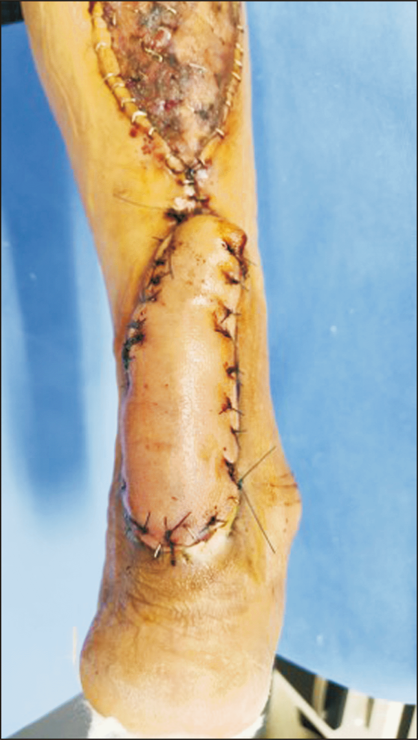
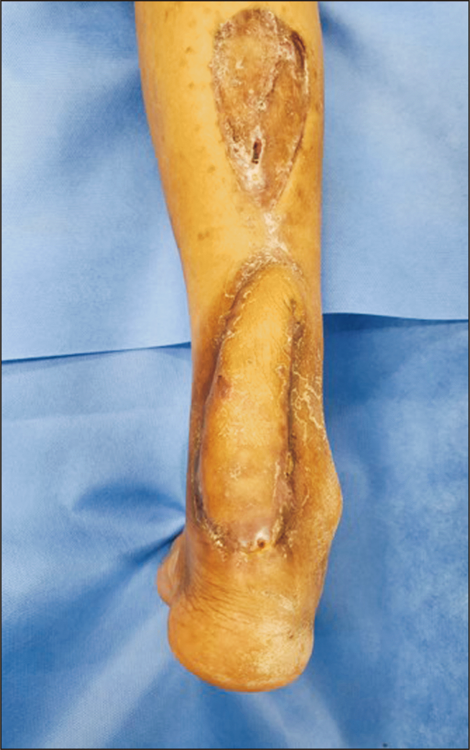
A 52-year-old male patient with alcoholic liver cirrhosis and diabetes, who had undergone surgery 3 years prior to presentation for a ruptured Achilles tendon, was admitted to the hospital with a three-month-old soft tissue defect. Preoperative laboratory results revealed an HbA1c level of 7%, total bilirubin of 2.41 mg/dL, AST level of 37 U/L, ALP level of 176 U/L, and GGT level of 77 U/L. The patient used a splint and crutches for ambulation prior to the surgery. This patient underwent incision and drainage for a right Achilles tendon abscess in the orthopedic surgery department two months prior to presentation. Despite the application of vacuum-assisted closure to the wound in the month before surgery, no improvement was evident. We managed the preoperative infection with intravenous ciprofloxacin 400 mg bid (HK Inno.N) and aseptic dressings. He was transferred from orthopedic surgery department to plastic and reconstructive surgery department for the reconstruction of the tissue defect of the right Achilles tendon (Fig. 1A). He had a 10 cm longitudinal scar crossing the central part of his calf (Fig. 1B). Therefore, a 3 cm×6 cm sized elliptical shaped flap was designed on the lateral portion of his right lower leg, situated 15 cm above the upper margin of the lesion. The pivot point was marked 8 cm above the lateral malleolus and the incision line was designed along the previous scar. Unhealthy tissue around the lesion was removed using a no. 15 surgical blade and scissors, and the skin was incised along the incision line from distal region to proximal region using a no. 15 surgical blade. Electrocautery was performed to reach the gastrocnemius fasciata. The subfascial plane was dissected proximally to distally, and the short saphenous vein and lateral sural nerve were tied. The subfascial plane was dissected along the pivot point of the fasciocutaneous flap to ensure that it reached and transposed over the lesion. To reduce tension, the subcutaneous layer was further dissected laterally at the wound margin and the flap was rotated counterclockwise and secured to the lesion using a stapler (Fig. 1C). After fixing the fasciocutaneous flap with nylon 2-0 and a stapler, an 8 cm×15 cm thick skin graft was obtained from the posterior aspect of the right thigh using an electrodermatome (0.2 mm thickness; Zimmer Biomet Holdings, Inc.) and sutured with a stapler (Fig. 2). A splint was applied to the right anterior tibia, and surgery was concluded after dressing.
The flap was designed to ensure that it did not extend beyond the scar in the middle of the calf, while maintaining a sufficient size to cover the defect. Using portable Doppler, the perforators were confirmed, and a flap with a half-width design different from that of the traditional flap was elevated.
Three months after surgery, the patient was able to walk well, and there were no complications such as pain or swelling (Fig. 3).
The reconstruction of heel soft tissue defects remains a difficult problem because of the unique challenges associated with this area. The complex anatomy, weightbearing function, limited local tissue availability, and need for long-term functional and aesthetic success make it a persistent challenge in the field of reconstructive surgery. The surgical plan can differ by dividing the location of the tissue defect in the heel into a weightbearing area (anterior heel) and a nonweightbearing area (posterior heel). Owing to their similarity to the skin of the heel, medial plantar artery flaps are preferred for reconstructing defects in the anterior weightbearing portion of the heel. Achilles tendon injuries and calcaneal fractures are frequently associated with defects of the posterior heel. Skin grafting is recommended when the wound is superficial. The majority of studies show that if the defect is deep and exposes the calcaneus or Achilles tendon, reverse sural artery and lateral calcaneal artery flaps are the most frequently performed procedures.1-4)
Surgical options include exposure of the Achilles tendon to a nonweightbearing area, such as local flaps, medial plantar artery flaps, reverse sural artery flaps, cross-leg flaps, and free flaps. Based on the reconstructive ladder concept, skin graft surgery can be considered prior to flap surgery for defect coverage; however, reconstruction of tendon-exposed defects is not possible with a skin graft because tendons are fibrous connective structures with a relatively low blood supply.1-4)
The majority of studies on deep defects with exposure of the calcaneus or Achilles tendon, reverse sural artery, and lateral calcaneal artery flap have been conducted.4,5) In the sural region of the posterior lower leg, two rows of perforators are distributed. These perforators originate from the peroneal artery, with three located in the posterolateral aspect. Additionally, the three perforators originated from the posterior tibial artery and were located in the posteromedial aspect. It is noteworthy that lateral peroneal artery perforators exhibit a more robust development than those of the medial posterior tibial artery.3) In addition, flap surgery involves the transfer of both tissue and blood arteries, and it is important to consider the vascular state of the flap.6) Therefore, conventional flap surgery may be risky for patients with comorbidities who have poor vascular status at operative sites. Therefore, abnormal calf tissue and central scars were avoided by designing a reverse sural flap with perforators in the posterolateral aspect.
The goal of heel reconstruction is to create a durable, natural-looking, and functional cover that fits into regular shoes. Fasciocutaneous flaps offer benefits such as skin color that closely resembles that of the lost tissue and a reduction in the overall surgical time. These flaps are particularly advantageous because of their versatility and reliability, given their consistent vascular anatomical patterns. Cross-leg flaps are outdated because of significant patient limitations and restricted positioning. Free flaps, which primarily rely on the gracilis, are effective in addressing substantial soft tissue defects in the heel and lower third of the leg. However, microsurgery involves extended procedures and are costly for both patients and hospitals, especially in terms of postoperative care.7,8)
The reverse sural flap is frequently used in reconstruction of the Achilles tendon area; however, complications such as congestion may arise. Congestion and flap loss are acknowledged when utilizing a subcutaneous tunnel.9) This appears to be associated with various factors, such as constriction of the pedicle caused by the skin's limited elasticity over the tunnel roof in the islanded variant, valvular incompetence, edema in the pedicle region, compartment syndrome, or compression of the pedicle due to hematoma.10)
We were concerned about blood flow in the previously operated Achilles tendon area. Therefore, we did not create a subcutaneous tunnel but instead covered the skin defect with a split-thickness skin graft. We reduced the pedicle width from the standard 4~5 cm to 2.5 cm to better accommodate the vascular condition of the recipient site and reduce the pressure on the folded area at the pivot point. Although reducing the pedicle width could potentially increase the risk of venous congestion and partial flap loss, our patient did not experience these complications. This favorable outcome was attributed to the use of a half-width reverse sural flap for reconstruction of the previous surgical site. The patient’s postoperative course was uneventful and weightbearing ambulation commenced one month after surgery.11)
This study had some limitations as it was based on a single case. Owing to the lack of published evidence on the use of the half-width reverse sural flap in the Achilles tendon area, it was not possible to directly compare the results of this study with those of other patient populations. Further analysis of the half-width reverse sural flap according to etiology is necessary. This analysis aimed to determine whether modifications of the reverse sural flap are more or less responsive in cases with soft tissue defects in the Achilles tendon area and surrounding scars. Such findings could potentially assist surgeons in successfully treating soft tissue defects in the Achilles tendon area.
The reverse sural flap needs to be personalized depending on factors such as the defect size, location, and history of surgery. In this case, we used half-width reverse sural flaps because of the presence of a previous incision line for Achilles tendon rupture surgery performed 3 years ago. Despite the compromised blood supply, we created a half-width flap after Doppler examination of the perforator artery. In conclusion, reverse sural flap, even in cases with a history of previous surgery, can be adequately performed by modifying the flap design as long as the perfusion of the perforators is maintained. Therefore, in the lower leg with a history of surgery, the use of a half-width reverse sural flap is a valuable and suitable first-choice option.
REFERENCES
1. Dhamangaonkar AC, Patankar HS. 2014; Reverse sural fasciocutaneous flap with a cutaneous pedicle to cover distal lower limb soft tissue defects: experience of 109 clinical cases. J Orthop Traumatol. 15:225–9. doi: 10.1007/s10195-014-0304-0. DOI: 10.1007/s10195-014-0304-0.
2. Chang SM, Li XH, Gu YD. 2015; Distally based perforator sural flaps for foot and ankle reconstruction. World J Orthop. 6:322–30. doi: 10.5312/wjo.v6.i3.322. DOI: 10.5312/wjo.v6.i3.322.
3. Altinkaya A, Yazar S, Bengur FB. 2021; Reconstruction of soft tissue defects around the Achilles region with distally based extended peroneal artery perforator flap. Injury. 52:1985–92. doi: 10.1016/j.injury.2021.04.015. DOI: 10.1016/j.injury.2021.04.015.
4. Krishna D, Chaturvedi G, Khan MM, Cheruvu VPR, Laitonjam M, Minz R. 2021; Reconstruction of heel soft tissue defects: an algorithm based on our experience. World J Plast Surg. 10:63–72. doi: 10.29252/wjps.10.3.63. DOI: 10.52547/wjps.10.3.63.
5. Hashmi DPM, Musaddiq A, Ali DM, Hashmi A, Zahid DM, Nawaz DZ. 2021; Long-term clinical and functional outcomes of distally based sural artery flap: a retrospective case series. JPRAS Open. 30:61–73. doi: 10.1016/j.jpra.2021.01.013. DOI: 10.1016/j.jpra.2021.01.013.
6. Dastagir K, Gamrekelashvili J, Dastagir N, Limbourg A, Kijas D, Kapanadze T, et al. 2023; A new fasciocutaneous flap model identifies a critical role for endothelial Notch signaling in wound healing and flap survival. Sci Rep. 13:12542. doi: 10.1038/s41598-023-39722-1. DOI: 10.1038/s41598-023-39722-1.
7. Novriansyah R, Prabowo I, Laras S. 2021; Non-microsurgical bipedicled reverse sural fasciocutaneous flap with preservation of medial and lateral sural cutaneous nerve: current surgical management of skin defect after traumatic Achilles tendon rupture - a case report. Int J Surg Case Rep. 78:259–64. doi: 10.1016/j.ijscr.2020.12.027. DOI: 10.1016/j.ijscr.2020.12.027.
8. Talukdar A, Yadav J, Purkayastha J, Pegu N, Singh PR, Kodali RK, et al. 2019; Reverse sural flap - a feasible option for oncological defects of the lower extremity, ankle, and foot: our experience from Northeast India. South Asian J Cancer. 8:255–7. doi: 10.4103/sajc.sajc_11_19. DOI: 10.4103/sajc.sajc_11_19.
9. de Rezende MR, Saito M, Paulos RG, Ribak S, Abarca Herrera AK, Cho ÁB, et al. 2018; Reduction of morbidity with a reverse-flow sural flap: a two-stage technique. J Foot Ankle Surg. 57:821–5. doi: 10.1053/j.jfas.2017.11.020. DOI: 10.1053/j.jfas.2017.11.020.
10. Mohamed AY, Ibrahim YB, Taşkoparan H, Çi Çek Eİ, May H. 2022; Reverse sural flap for anteromedial ankle and dorsal foot soft-tissue defect following an injury: a case report. Ann Med Surg (Lond). 84:104935. doi: 10.1016/j.amsu.2022.104935. DOI: 10.1016/j.amsu.2022.104935.
11. Johnson L, Liette MD, Green C, Rodriguez P, Masadeh S. 2020; The reverse sural artery flap: a reliable and versatile flap for wound coverage of the distal lower extremity and hindfoot. Clin Podiatr Med Surg. 37:699–726. doi: 10.1016/j.cpm.2020.05.004. DOI: 10.1016/j.cpm.2020.05.004.
Figure 1
Clinical photograph. (A) A 6 cm × 4 cm tissue defect in the right Achilles tendon. (B) A 10 cm longitudinal scar crossing the central part of the calf. (C) A 3 cm × 6 cm elliptical fasciocutaneous flap was elevated from the lateral aspect of the right lower leg. The pivot point was marked at 8 cm above the right lateral malleolus.


 PDF
PDF Citation
 Print
Print


 XML Download