Dear Editor,
Thyridium endophyticum, formerly known as Phialemoniopsis endophytica, is an uncommon mold species frequently isolated from the environment or plants [1-3]. To our knowledge, only one clinical case of T. endophyticum infection, manifesting as an asymptomatic subcutaneous nodule, has been reported [4]. We present a case of painful tenosynovitis attributed to T. endophyticum in a patient with preexisting medical conditions who underwent synovectomy and repair surgery in the right wrist. This possibly is the first documentation of T. endophyticum as a human pathogen capable of causing severe tenosynovitis. The Institutional Review Board of Chonnam National University Bitgoeul Hospital (CNUBH), Gwangju, Korea, approved this study and waived the requirement for informed consent (CNUBH-EXP- 2024-003).
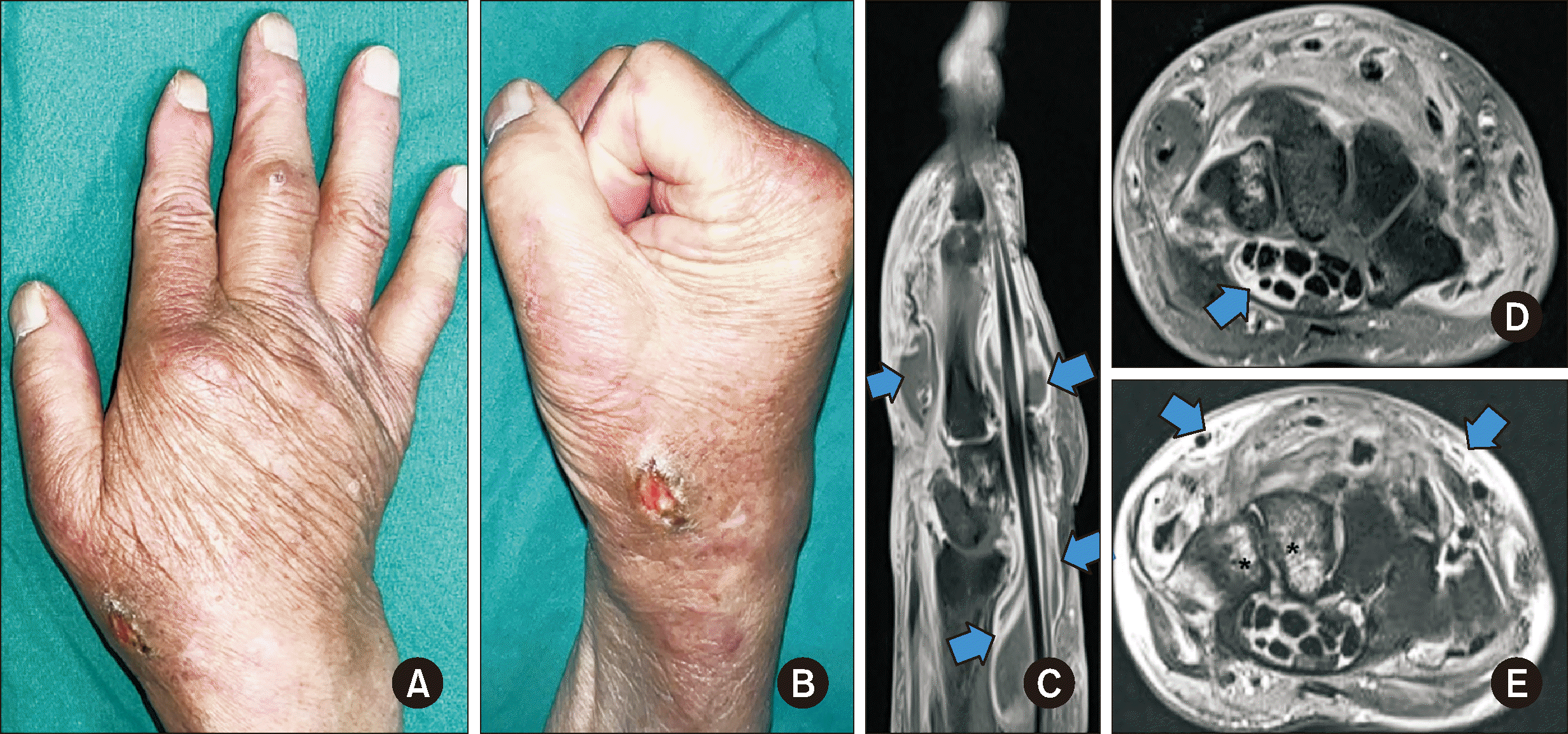
A 72-yr-old man presented to the CNUBH with right wrist pain in January 2023. Four months prior, he had undergone surgery for a total tear in his right extensor pollicis brevis tendon at a local hospital. The patient’s medical history included diabetes, dilated cardiomyopathy, atrial fibrillation, and seronegative rheumatoid arthritis, for which he received 10 mg of methotrexate weekly, and 20 mg of leflunomide and 5 mg of prednisolone daily. Physical examination revealed general swelling of the distal forearm and hand, although tenderness was not pronounced. A small ulcerative lesion was observed on the skin near the radial head (Fig. 1A and 1B). Magnetic resonance imaging of the right hand revealed multiple fluid collections, circular enhancement of the flexor and extensor tendon sheaths, and edema in the superficial soft tissues, skin, and carpal bones (Fig. 1C–1E). Laboratory tests revealed mild leukocytosis (10.96×10⁹/L, reference interval [RI]: 4.8–10.8×10⁹/L), an elevated C-reactive protein level (2.0 mg/dL, RI: 0–0.3 mg/dL), and an erythrocyte sedimentation rate of 43 mm/hr (RI: 2–9 mm/hr). Two months later, the patient underwent a dorsal tenosynovectomy and tendon transfer surgery. During the surgery, severe tenosynovitis, multiple tendon ruptures, granulation tissues, and a greenish discharge were noticed.
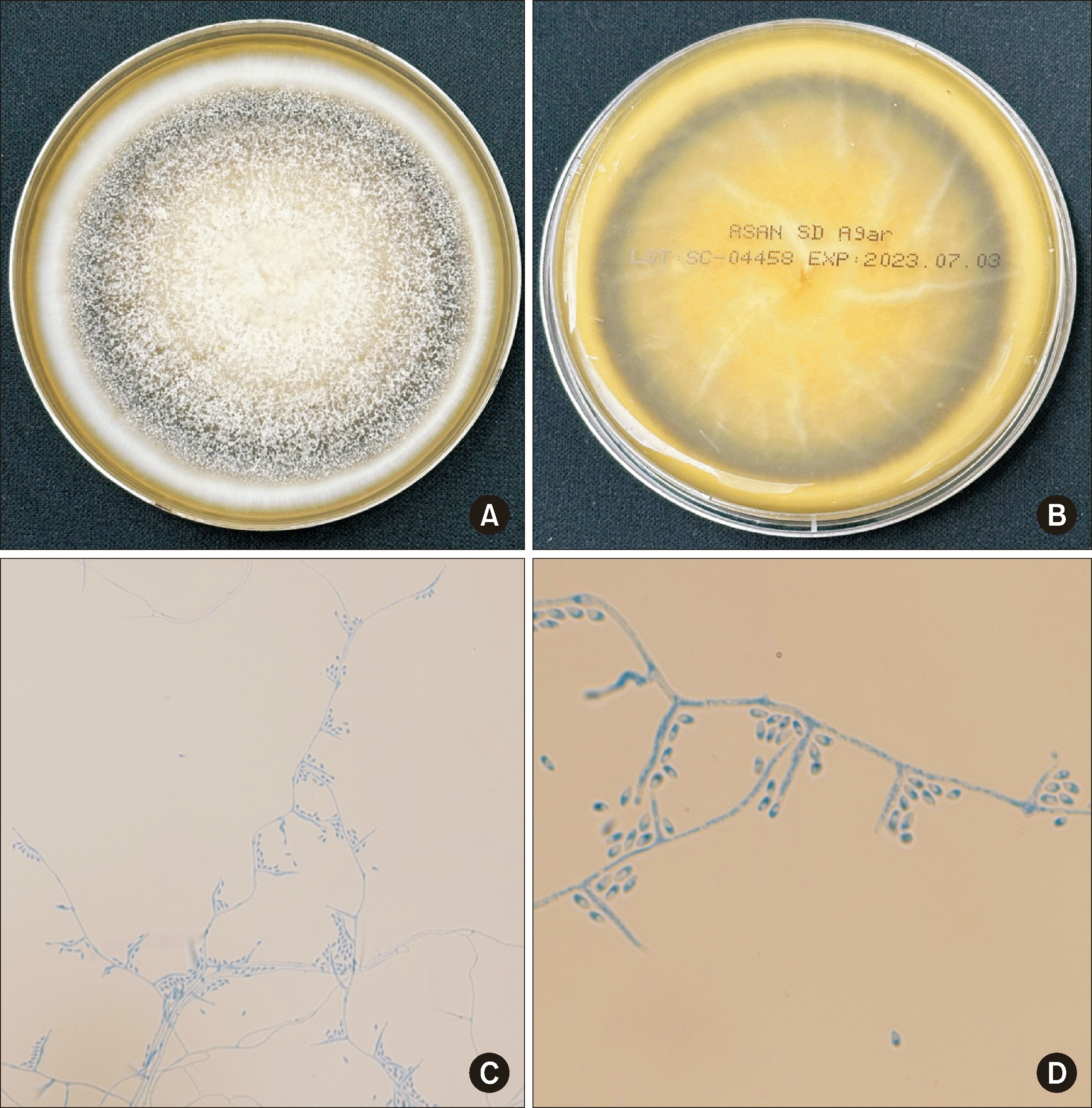
In pus and tissue cultures, mold-like fungi appeared in four out of seven sets. The cultured specimen formed a spreading colony with a white or creamy surface on Sabouraud dextrose agar at 30°C. After four weeks, it developed a dark-brown area with annular structures and radial folds (Fig. 2A and 2B). Slide culture revealed broad, branched hyaline hyphae and oval microconidia (Fig. 2C and 2D). The microorganism was identified as T. endophyticum based on the sequencing of the internal transcribed spacer region (100% [535/535 bp] identity with MT271971.1), D1/D2 regions of the 28S rRNA gene (100% [571/571 bp] identity with KT799559.1), actin gene (99.8% [609/610 bp] identity with KT799552.1), and β-tubulin gene (99.6% [496/498 bp] identity with KT799561.1). Antifungal susceptibility testing using the broth dilution method according to CLSI M38-A3 revealed the following minimal inhibitory concentrations (MICs): amphotericin B, 0.25 μg/mL; itraconazole, 2 μg/mL; posaconazole, 0.5 μg/mL; voriconazole, 1 μg/mL; anidulafungin, 4 μg/mL; caspofungin, >8 μg/mL; and micafungin, >8 μg/mL. Consequently, the patient was administered liposomal amphotericin B 340 mg/day for four days, followed by oral voriconazole 400 mg/day. The patient completed a six-month course of voriconazole, and at the three-month post-treatment follow-up, no signs of recurring infection were observed.
Thyridium (Phialemoniopsis) comprises 33 species, which rarely cause human infection [2]. Most reported human infections by Thyridium species were cutaneous or subcutaneous and typically occurred after trauma or skin injury [4-10]. Among Phialemoniopsis species, Phialemoniopsis curvatum (formerly Phialemonium curvatum) induces invasive infections, such as endocarditis and fungemia, in immunocompromised patients [5, 6]. We present the first case of tenosynovitis caused by T. endophyticum in a patient with multiple underlying diseases. Considering that T. endophyticum is commonly found in the environment and on plants, the wound from previous surgery may have been exposed to T. endophyticum during the patient’s agricultural activities. T. endophyticum possibly caused an invasive infection, given his compromised immune status [4].
Thyridium species, similar to other dematiaceous molds, are not easily identifiable using conventional clinical laboratory methods; therefore, sequence-based identification is essential for accurate species-level identification [2-4]. Thyridium has been recently revised taxonomically and phylogenetically using several gene regions [2]. We reliably identified the isolate as T. endophyticum by sequencing four genes.
Several cases of subcutaneous infection caused by Phialemoniopsis species were successfully treated with oral voriconazole, with or without prior intravenous amphotericin B [4, 7]. Our T. endophyticum isolate showed MIC values of 0.25 μg/mL for amphotericin B and 1 μg/mL for voriconazole. Our patient was successfully treated with intravenous amphotericin B followed by oral voriconazole therapy, suggesting its viability in treating T. endophyticum infections.
Notes
AUTHOR CONTRIBUTIONS
Conceptualization: Kang SJ; Methodology: Cho SH, Kwon YJ, and Byun SA; Investigation: Cho SH, Kwon YJ, Shin JH, Choi HW, Heo SH, Lim JH, Kim MS, Lee Y, and Kang SJ; Visualization: Cho SH, Heo SH, Lim JH, Kim MS, and Kang SJ; Project administration: Kwon YJ, Shin JH, and Kang SJ; Supervision: Shin JH and Kang SJ; Writing – original draft: Cho SH, Kwon YJ, Shin JH, and Kang SJ; Writing – review & editing: Kwon YJ, Shin JH, and Kang SJ.
References
1. Perdomo H, García D, Gené J, Cano J, Sutton DA, Summerbell R, et al. 2013; Phialemoniopsis, a new genus of Sordariomycetes, and new species of Phialemonium and Lecythophora. Mycologia. 105:398–421. DOI: 10.3852/12-137. PMID: 23099515.
2. Sugita R, Tanaka K. 2022; Thyridium revised: synonymisation of Phialemoniopsis under Thyridium and establishment of a new order, Thyridiales. MycoKeys. 86:147–76. DOI: 10.3897/mycokeys.86.78989. PMID: 35145340. PMCID: PMC8825628. PMID: 37ccf211b34b41118d266bd3c64a504a.
3. Su L, Deng H, Niu YC. 2016; Phialemoniopsis endophytica sp. nov., a new species of endophytic fungi from Luffa cylindrica in Henan, China. Mycol Progress. 15:48. DOI: 10.1007/s11557-016-1189-5.
4. Ito A, Yamada N, Kimura R, Tanaka N, Kurai J, Anzawa K, et al. 2017; Concurrent double fungal infections of the skin caused by Phialemoniopsis endophytica and Exophiala jeanselmei in a patient with microscopic polyangiitis. Acta Derm Venereol. 97:1142–4. DOI: 10.2340/00015555-2734. PMID: 28654130. PMID: 2d2dd51b1fdd49b283aa42100c913999.
5. Rivero M, Hidalgo A, Alastruey-Izquierdo A, Cía M, Torroba L, Rodríguez-Tudela JL. 2009; Infections due to Phialemonium species: case report and review. Med Mycol. 47:766–74. DOI: 10.3109/13693780902822800. PMID: 19888810.
6. Tang M, Li J, Xia F, Min C, Liu Z, Hu Y, et al. 2022; Acute post-cataract endophthalmitis due to Phialemoniopsis curvata: a rare case report. Infect Drug Resist. 15:1651–7. DOI: 10.2147/IDR.S359481. PMID: 35422644. PMCID: PMC9004724.
7. Desoubeaux G, García D, Bailly E, Augereau O, Bacle G, De Muret A, et al. 2014; Subcutaneous phaeohyphomycosis due to Phialemoniopsis ocularis successfully treated by voriconazole. Med Mycol Case Rep. 5:4–8. DOI: 10.1016/j.mmcr.2014.04.001. PMID: 24936402. PMCID: PMC4052356. PMID: b3a14ce58d434dc2b496645f87338152.
8. Alvarez Martinez D, Alberto C, Riat A, Schuhler C, Valladares P, Ninet B, et al. 2021; Phialemoniopsis limonesiae sp. nov. causing cutaneous phaeohyphomycosis in an immunosuppressed woman. Emerg Microbes Infect. 10:400–6. DOI: 10.1080/22221751.2021.1892458. PMID: 33634736. PMCID: PMC7946049. PMID: 3c93faee05954e6b85b40ea30f64afdd.
9. Tsang CC, Chan JF, Ip PP, Ngan AH, Chen JH, Lau SK, et al. 2014; Subcutaneous phaeohyphomycotic nodule due to Phialemoniopsis hongkongensis sp. nov. J Clin Microbiol. 52:3280–9. DOI: 10.1128/JCM.01592-14. PMID: 24966363. PMCID: PMC4313151.
10. Patolia H, Bansal E. 2021; Two rare cases of fungal bursitis due to Phialemoniopsis pluriloculosa. IDCases. 24:e01095. DOI: 10.1016/j.idcr.2021.e01095. PMID: 33898253. PMCID: PMC8055602.
Fig. 1
Clinical and magnetic resonance imaging (MRI) findings. (A and B) General swelling of the hand and wrist and a small, ulcerated lesion under the thumb were observed. (C–E) MRI of the right hand revealing multiple fluid collections and circular enhancement of the flexor and extensor tendon sheaths (arrows) on gadolinium-enhanced sagittal (C) and axial (D) T1-weighted images, consistent with tenosynovitis. (E) Axial T2-weighted image with fat saturation revealing bone marrow edema of the carpal bones (asterisks) and edema of the dorsal superficial soft tissue and skin (arrows).

Fig. 2
Macroscopic and microscopic findings of the Thyridium endophyticum isolate. (A and B) Colony of T. endophyticum. Front (A) and back side (B) of a Sabouraud dextrose agar plate after four weeks of culture at 30°C. (C and D) Microscopic examination of a slide culture revealing hyaline hyphae with microconidia (lactophenol cotton blue stain, 400× [C] and 1,000× [D]).


 PDF
PDF Citation
 Print
Print


 XML Download