Journal List > Ann Lab Med > v.44(6) > 1516088443

Huang, Li, Li, Cao, Zhong, Liu, Lin, Lin, Fang, Zeng, Su, Li, Liang, Zhu, Lin, Huang, Yang, and Jiang: Exploring Appropriate Reference Intervals and Clinical Decision Limits for Glucose-6-Phosphate Dehydrogenase Activity in Individuals From Guangzhou, China

Abstract

Background

Quantitative detection of glucose-6-phosphate dehydrogenase (G6PD) is commonly done to screen for G6PD deficiency. However, current reference intervals (RIs) of G6PD are unsuitable for evaluating G6PD-activity levels with local populations or associating G6PD variants with hemolysis risk to aid clinical decision-making. We explored appropriate RIs and clinical decision limits (CDLs) for G6PD activity in individuals from Guangzhou, China.

Methods

We enrolled 5,852 unrelated individuals between 2020 and 2022 and screened their samples in quantitative assays for G6PD activity. We conducted further investigations, including G6PD genotyping, thalassemia genotyping, follow-up analysis, and statistical analysis, for different groups.

Results

In Guangzhou, the RIs for the G6PD activities were 11.20–20.04 U/g Hb in male and 12.29–23.16 U/g Hb in female. The adjusted male median and normal male median (NMM) values were 15.47 U/g Hb and 15.51 U/g Hb, respectively. A threshold of 45% of the NMM could be used as a CDL to estimate the probability of G6PD variants. Our results revealed high hemolysis-risk CDLs (male <10% of the NMM, female <30% of the NMM), medium hemolysis-risk CDLs (male 10%–45% of the NMM, female 30%–79% of the NMM), and low hemolysis-risk CDLs (male ≥45% of the NMM, female ≥79% of the NMM).

Conclusions

Collectively, our findings contribute to a more accurate evaluation of G6PD-activity levels within the local population and provide valuable insights for clinical decision-making. Specifically, identifying threshold values for G6PD variants and hemolysis risk enables improved prediction and management of G6PD deficiency, ultimately enhancing patient care and treatment outcomes.

INTRODUCTION

Reference intervals (RIs) and clinical decision limits (CDLs) constitute vital information from laboratories that support the interpretation of laboratory results [1]. They are critical for health assessments, disease diagnosis, treatment monitoring, and prognostic judgments. An RI is commonly defined as 95% of the range of a certain indicator in a healthy population [2]. Owing to variants in population and measurement methods, RIs can differ across different regions and laboratories [3]. CDLs refer to specific thresholds, where values above or below the threshold are associated with a significantly higher risk of adverse clinical outcomes or are used to help diagnose the presence of a specific disease. CDLs are established based on comparisons with gold-standard diagnostic results or clinical outcomes in patients. When laboratory results exceed the CDL threshold, they can support clinical decision-making, such as diagnosis or treatment. CDLs vary for different purposes.
Glucose-6-phosphate dehydrogenase (G6PD) deficiency is the most common human enzymopathy, affecting over 500 million people worldwide [4, 5]. Quantitative detection of G6PD is a commonly used screening method for assessing G6PD deficiency. Improving the interpretation of quantitative G6PD-detection results requires a predetermined definition of normal (100%) G6PD activity. In 2013, Domingo, et al. [6] introduced a standardized method for calculating normal G6PD activity, involving two steps: (1) calculating the initial median (M0) value of the male population and (2) recalculating the median for the male population with values of more than 10% of the M0, designated as the adjusted male median (AMM). In 2018, the WHO acknowledged the aforementioned method for calculating normal G6PD activity [7]. In 2022, the WHO proposed a new definition and calculation method for normal G6PD activity: (1) male individuals with abnormal G6PD expression are excluded by genetic testing (typically targeting prevalent G6PD variants in a specific geographic region, such as the 18 common G6PD variants in China), and (2) the median G6PD activity in the remaining male population is calculated and referred to as the normal male median (NMM) [5].
Currently, for quantitative G6PD detection, laboratories typically utilize the G6PD-activity RI provided by test manufacturers (1,300–3,600 U/L). RI values are not specific to any region and fail to combine AMM, NMM, and CDL data to facilitate clinical decision-making. We established reliable and region-specific RIs for G6PD activity to improve the assessment of G6PD activity levels in a local population. We calculated the AMM and NMM values for G6PD activity to define normal G6PD activity in individuals from Guangzhou, China and better interpret the quantitative G6PD results. We also used appropriate G6PD-activity CDLs to provide a reference for the probability of G6PD variants and hemolysis-risk assessment based on G6PD activity.

MATERIALS AND METHODS

Participants and data collection

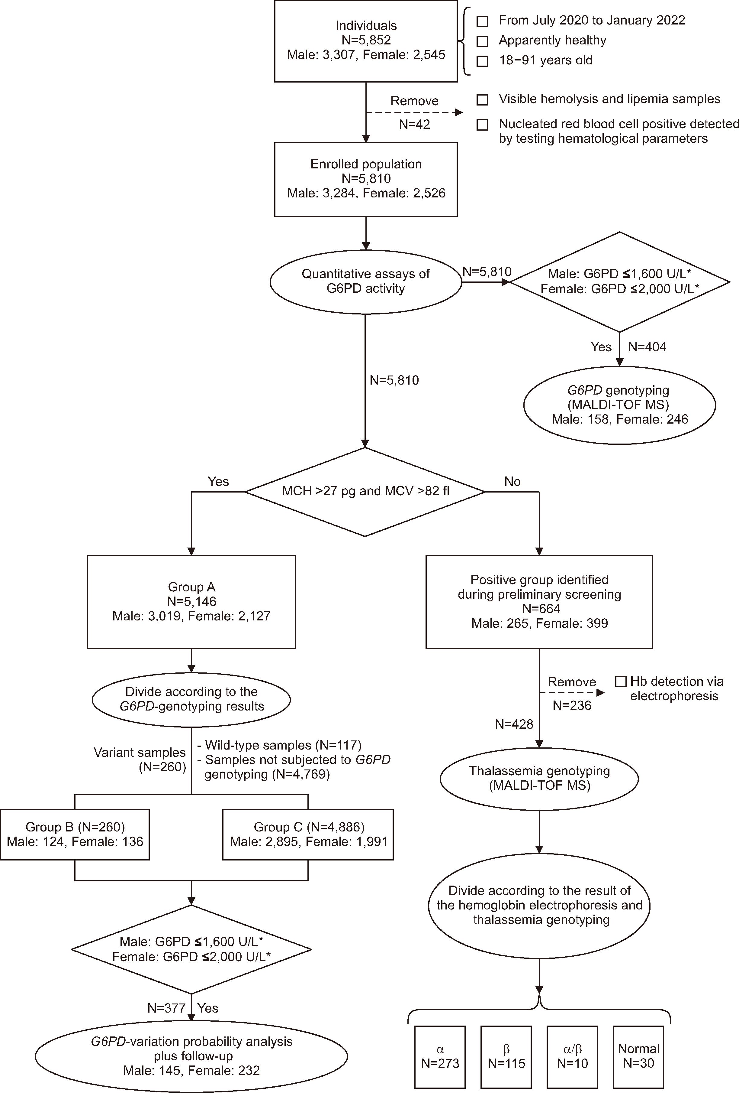
From July 2020 to January 2022, we enrolled 5,852 individuals, including 3,307 male (aged 18–91 yrs) and 2,545 female (aged 18–89 yrs), in a single-center study. All individuals were healthy and visited Zhujiang Hospital, Southern Medical University, Guangzhou, China, for a physical examination. The physical examination included testing for hematological parameters such as the mean corpuscular Hb (MCH) mean corpuscular volume (MCV), as well as Hb levels (assessed via electrophoresis) and biochemical parameters (liver and kidney function, myocardial enzymes, blood glucose, and blood lipid levels). The remaining blood samples were collected for quantitative assays of G6PD activity and G6PD and thalassemia genotype determinations. Individuals whose blood samples showed evident hemolysis and lipemia or nucleated red blood cells were excluded from this study. The study protocols were approved by the Ethics Committee of Zhujiang Hospital of Southern Medical University, Guangzhou, China (approval number 2018-JYYXB-002). All procedures performed in this study involving human participants were in accordance with the guidelines of the 1964 Declaration of Helsinki and its later amendments or comparable ethical standards. Informed consent was obtained from all individual participants included in the study. The study protocol is presented as a flowchart in Fig. 1, and a more detailed description of the protocol is provided in Supplemental Data Text S1.

Quantitative assays of G6PD activity

G6PD activity was detected using a G6PD Detection Assay Kit (Antu Co., Ltd., Beijing, China), which measures changes in the absorbance of the reduced form of nicotinamide adenine dinucleotide phosphate. Absorbance readings at 340 nm were taken at 37°C to calculate the G6PD-activity values (U/L). G6PD activity was normalized to the amount of Hb (U/g Hb), as recommended by the International Council for Standardization in Hematology (ICSH) and WHO. All tests were conducted according to the manufacturer’s instructions. The reliability of the test results was monitored by calibration and using the controls provided by Antu Co., Ltd. in each test run.

G6PD genotyping

Genomic DNA was extracted from peripheral venous blood using a DNA Extraction System and Kit (Tianlong, Co., Ltd., Xian, China), according to the manufacturer’s instructions. G6PD variants were identified using a method based on multiplex polymerase chain reaction technology and matrix-assisted laser desorption ionization time-of-flight mass spectrometry (MALDI-TOF-MS; Darui Biotechnology Co., Ltd., Guangzhou, China), according to the manufacturer’s instructions. This kit enables qualitative detection of 18 G6PD variants in human peripheral blood genomic DNA that are common in China, namely c.95A>G, c.202G>A, c.473G>A, c.383T>C, c.392G>T, c.487G>A, c.493A>G, c.592C>T, c.835A>G, c.871G>A, c.1004C>A, c.1024C>T, c.1339G>A, c.1360C>T, c.1376G>T, c.1387C>T, c.1388G>A, and c.1466C>T.

Thalassemia genotyping

Genomic DNA was extracted from peripheral venous blood using a DNA Extraction System and Kit (Tianlong, Co., Ltd.), according to the manufacturer’s instructions. Variants were identified using a method based on the target-allele-specific probe single-base extension and traditional single-base extension method to detect 28 α-/β-thalassemia variants by single-tube MALDI-TOF-MS. The principle of the method, reagents and instrumentation used, and steps followed were published previously [8].

Follow-up analysis

A follow-up analysis was conducted on a subset of individuals with low G6PD activity (male: G6PD ≤1,600 U/L; female: G6PD ≤2,000 U/L). The follow-up included telephone calls and case review analysis. The follow-up questions included: (1) Did you have any of these symptoms before: dark-colored urine or blood in the urine, paleness, jaundice, shortness of breath, dizziness, weakness, and back and/or abdominal pain [7]? If yes, were you hospitalized as a result? (2) Have you ever taken any of the following drugs: analgesics, antipyretics, or antibacterials [9]? If yes, did you experience any adverse reactions? (3) Have you ever consumed fresh broad beans and experienced any adverse reactions? (4) Do you have a history of adverse reactions to other drugs or foods? (5) Does anyone in your family suffer from G6PD deficiency? If yes, do they have any of the conditions mentioned in the previous four questions?

Statistical analysis

Data were analyzed in accordance with the EP28-A3c guidelines issued by the CLSI [2]. Excel 2016 (Microsoft Corporation, Redmond, WA, USA), SPSS 26.0 (IBM Corporation, Armonk, NY, USA), GraphPad Prism 9.2.0 (GraphPad Software, San Diego, CA, USA), and Origin 2021 (OriginLab Corporation, Northampton, MA, USA) were used for statistical analyses and data processing. The Kolmogorov–Smirnov test was used to determine the normal distribution of G6PD activity. We used Tukey’s method to eliminate discrete values. RIs were calculated using nonparametric methods. Differences between groups were tested using the Mann–Whitney U-test. Statistical significance was set at P<0.05.

RESULTS

Distribution of G6PD activity

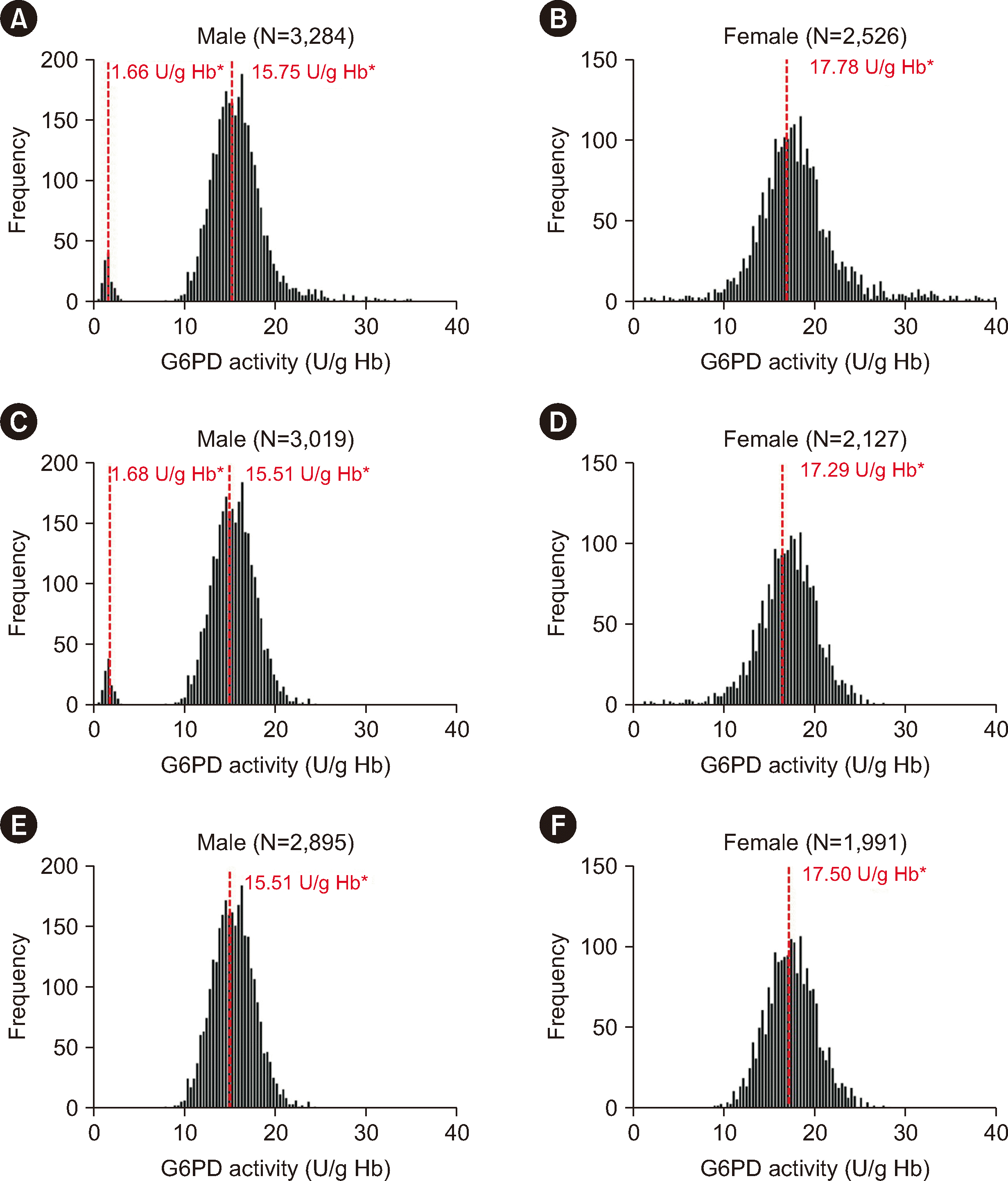
The distributions of G6PD activities in three male and three female groups are shown in Fig. 2, and a detailed description of the data shown in Fig. 2 is presented in Supplemental Data Text S2.

Effects of thalassemia on G6PD activity

Supplemental Data Fig. S1 shows that the measured values of G6PD activity in thalassemia-positive individuals were higher than those in thalassemia-negative individuals, as determined during preliminary screening, irrespective of sex or type of thalassemia (P<0.001).

AMM and NMM determinations

Normal G6PD activity is generally represented by the AMM and NMM, which are based on different criteria and calculation methods. We calculated the M0, AMM, and NMM values separately for two groups: Group 1 (all male in the enrolled population, N=3,284) and Group 2 (all male in Group A, N=3,019) (Supplemental Data Table S1). The M0 and AMM values of each group were calculated as follows: First, the M0 values for male in each group were calculated. The M0 of Group 1 was 15.60 U/g Hb, and that of Group 2 was 15.01 U/g Hb. Group 1 included 53 individuals with a G6PD activity of less than 1.56 U/g Hb, and Group 2 had 38 individuals with a G6PD activity of less than 1.501 U/g Hb. After removing these individuals from both groups, the median was recalculated to obtain the AMM for each group. The NMM value of each group was calculated as follows. Group 1 (N=3,284) had 138 individuals with a G6PD variant, and Group 2 (N=3,019) had 124 individuals with a G6PD variant. After removing these individuals from both groups, the median was recalculated to obtain the NMM for each group. Statistical analysis showed significant differences between AMM1 and AMM2 (P<0.001) and between NMM1 and NMM2 (P<0.001). However, the difference between the AMM and NMM values of both groups was not significant (NS).

Establishment of RIs

We established the RIs for G6PD activity in two groups: Group A (thalassemia-negative group based on preliminary screening, N=5,146) and Group C (thalassemia-negative group with a normal G6PD genotype based on preliminary screening, N=4,886). We checked for outliers and calculated 95% RIs (Table 1). We identified significant differences in G6PD activities between male and female in Groups A and C (P<0.001) and between female in both groups (P=0.036). However, no statistically significant difference in G6PD activity was found between the two groups (NS) with male.

Relationship between G6PD-activity levels and G6PD-variant probabilities

The relationship between G6PD-activity levels and G6PD-variant probabilities is shown in Table 2. When the G6PD activity was less than or equal to 45% NMM in male and female, the positive rate for a G6PD variant was 100%. When G6PD activity was greater than 45%, zero male were positive for genetic variants in G6PD, whereas female had a variant-positivity rate ranging from 30% to 91.3%.

Follow-up analysis

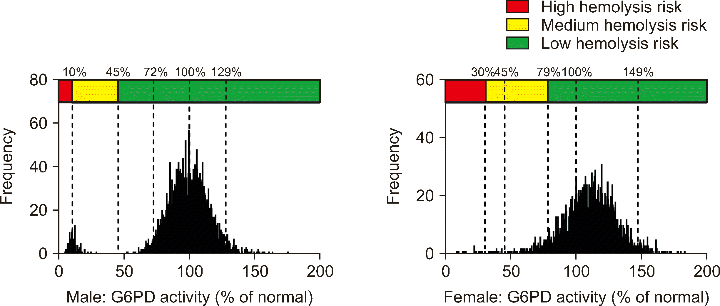
Follow-up analysis was successful with 235 individuals: of these 235 individuals, 7 had hemolysis, and 3 did not, but their relatives did (Supplemental Data Table S2, Table 3). All individuals with hemolysis avoided exposure to triggers after the diagnosis of G6PD deficiency and did not develop hemolysis again during the study. The distribution of G6PD activities in the follow-up population is shown in Supplemental Data Fig. S2. Based on follow-up-analysis data combined with previous reports and guidelines [5, 10], we used CDLs of 10% and 45% NMM for male and 30% and 79% NMM for female to assess the risk of acute hemolysis. The RIs and CDLs for G6PD activity in the study region are shown in Fig. 3.

DISCUSSION

G6PD deficiency is a common inherited hematological disorder in southern China [11]. Several reports have shown G6PD-activity distributions for different populations [6, 12]. Consistent with previous reports, in this study, the G6PD activity in the enrolled population showed a bimodal distribution in male and a unimodal distribution in female, both before and after removing samples through positive preliminary screening for thalassemia. The G6PD activities of male and female in the high range had a narrower distribution, and the overall distribution shifted to the left, suggesting that thalassemia may influence the measured G6PD activity. After excluding individuals with G6PD variants, the G6PD activities of male and female showed a unimodal distribution in accordance with a normal distribution (NS).
Thalassemia is another common inherited hematological disorder in southern China [13]. Notably, G6PD activity is higher in individuals with thalassemia [14-16]. Concordantly, our results showed that G6PD activity was higher in both male and female in the thalassemia group than in the non-thalassemia group. These findings may reflect the compensatory production of new red blood cells in individuals with thalassemia, resulting in a false increase in the measured G6PD-activity values. Quantitative detection of G6PD in individuals with thalassemia cannot truly reflect the G6PD activity level. To establish regional RIs for G6PD activity, individuals with thalassemia must be excluded.
The AMM and NMM values are based on different criteria for defining normal (100%) G6PD activity. Unlike previous studies, we compared the calculated AMM and NMM values between the two groups. The differences between AMM1 and AMM2 (NS) and between NMM1 and NMM2 (P<0.001) were statistically significant, indicating that thalassemia can increase the AMM and NMM values. No statistical difference was found between AMM and NMM values among both populations (P>0.05), indicating that the AMM and NMM results were very similar. Theoretically, the NMM is more accurate than the AMM, although it involves G6PD genotyping. Conducting G6PD genotyping for all samples is expensive, complex, and time-consuming, and it requires considerable human and material resources. We propose that AMM can be used to estimate NMM and that AMM2 (15.47 U/g Hb) and NMM2 (15.51 U/g Hb) can be used to define normal G6PD activity (100%) in adults in this region.
Excluding thalassemia-positive samples identified during screening, we established the RIs for G6PD activities in Groups A and C. G6PD activities between male and female differed significantly (P<0.001). No statistical difference in G6PD activity was found between the two groups (NS) among male. However, a statistical difference was observed between both groups among female (P=0.036). We believe that this difference was likely owing to the removal of female heterozygotes from Group C through G6PD genetic testing, which made the RIs more accurate. Therefore, we selected Group C as the reference population to establish the RIs for G6PD activity for male (11.20–20.04 U/g Hb, 72%–129% of normal G6PD activity) and female (12.29–23.16 U/g Hb, 79%–149% of normal G6PD activity) in the local adult population.
Currently, no region-specific RIs for G6PD activity have been established in China. Test manufacturers provide the RIs for G6PD activity in most regions. We established RIs for G6PD activities that differed from the RIs provided by manufacturers (1,300–3,600 U/L, 54%–149% of normal G6PD activity), with the lower limit of our RI being higher. Specifically, we standardized the RI using Hb levels and accounted for different factors, such as thalassemia and gender differences. Standardizing G6PD activity using Hb levels is recommended by both the WHO and ICSH [7, 9]. However, the manufacturers did not provide standardized G6PD-activity RIs. In a prospective study of G6PD deficiency in 74,114 healthy adults from 21 provinces and cities in China, Ying, et al. [11] found that the mean G6PD-activity values in normal male and female were 15.49±2.67 U/g Hb and 18.01±3.37 U/g Hb, respectively. Consistently, in this study, the mean G6PD-activity values of male and female in Group C were 15.51±2.24 U/g Hb and 17.51±2.69 U/g Hb, respectively.
Increased G6PD-activity values were associated with decreased G6PD-variant probabilities in both male and female; the decreasing trend in male was more evident, whereas that in female was more gradual. Heterozygous female showed a wide range of G6PD levels, consistent with previously published studies and guidelines [7, 17-19]. All male and female homozygotes had G6PD activities of less than 45% of the NMM, which is consistent with the 2022 WHO guidelines for G6PD deficiency [5]. In 2022, the WHO established a new classification scheme for G6PD variants using thresholds of 20%, 45%, 60%, and 150% NMM and indicated that no variants have been identified with median G6PD-activity values in male hemizygous and/or female homozygous individuals that fell between 45% and 60%. We found that a threshold of 45% of the NMM could serve as a CDL to estimate the probability of a G6PD variant and indicate the necessity for G6PD testing in individuals. When the G6PD activity was greater than 45% NMM, the probability of a G6PD variant was 0% for male and between 30% and 91.3% for female. Further G6PD genetic testing is necessary for female but not for male in this region. The limitations of using 45% NMM to estimate the probability of a G6PD variant should be noted. The results of studies conducted in China and other countries revealed some rare variants that are classified as class IV variants (class C, 60%–150% of the NMM), such as c.660C>G (G6PD São Paulo) [20, 21], c.152C>T, c.290A>T, and c.1285A>G (G6PD Yucatan) [22]. Estimating the G6PD-variant probability using 45% NMM only applies to common G6PD variants. We detected 18 common variants in G6PD in human peripheral blood from Chinese individuals, as outlined in the materials and methods section.
Foods and drugs that trigger hemolysis in G6PD-deficient individuals include fava beans, antimalarial drugs, analgesics, antipyretics, and antibacterial agents [9]. Although antimalarial drugs are rarely used in this region, edible fava beans and other oxidative drugs and some traditional Chinese medicines (including honeysuckle and bezoar) are often used. G6PD is expressed abundantly in the human body, and many people do not develop hemolytic symptoms even when the enzymatic activity of G6PD is below the normal RI. This indicates that the risk for hemolysis in individuals is not well assessed using the G6PD activity as an RI. We correlated G6PD-activity levels with the risk of acute hemolysis through follow-up analysis (Supplemental Data Table S2, Table 3), which provided a reference for establishing CDLs based on G6PD-activity levels to evaluate the risk for hemolysis in individuals with G6PD deficiency in the region in the future.
Our follow-up analysis revealed that most individuals did not know their status and had no symptoms or complications. Two compound heterozygous female and four homozygous female had no symptoms. Only seven individuals (cases 1–7) showed hemolysis symptoms; all seven individuals tested positive for a G6PD variant, and G6PD activity below the RI established in this study, and hemolysis did not re-occur after it was resolved. They paid special attention to their diet and medications after hemolysis occurred. These findings suggest that G6PD deficiency can greatly reduce the risk of hemolysis by preventing exposure to oxidative stress factors after diagnosis and that standardized life guidance is of great importance for individuals with G6PD deficiency. Cases 8–10 had no hemolytic symptoms, but their relatives showed symptoms of hemolysis, suggesting that relatives of individuals with G6PD deficiency should also pay attention to preventing G6PD deficiency. In cases where hemolysis symptoms occurred during the follow-up period, the triggers included fava beans, sulfonamides, analgesics, and antipyretics, and the symptoms included jaundice, blood in the urine, paleness, and rash. These findings suggest that more attention should be paid to these triggers and symptoms to prevent, diagnose, and manage G6PD deficiency in this region.
CDLs were previously established to better assess the risk of acute hemolysis in individuals with G6PD deficiency. In 2016, the WHO predicted the risk of acute hemolysis with primaquine treatment based on G6PD-activity levels (10%, 30%, and 80% of the AMM) in male and female [10]. In 2019, a cut-off value of 70% was used to evaluate the risk for hemolysis in individuals who took tafenoquine [23]. In 2020, Commons, et al. [24] recommended a threshold of 70% normal G6PD activity to evaluate tafenoquine use in terms of hemolysis risk. These CDLs are associated with antimalarial drug use. Antimalarial drugs were not a trigger in Guangzhou because malaria is not endemic to the area. No CDLs have been established for this region that can be used to assess the risk of hemolysis in individuals with G6PD deficiency.
We classified G6PD-activity levels into high, medium, and low hemolysis risk groups. This classification utilized CDLs of 10% and 45% of the NMM for male and 30% and 79% of the NMM for female. The basis for the specific classifications is shown in Supplemental Data Text S3. People with high and medium hemolysis risk should establish G6PD profiles and indicate their G6PD-deficiency status. People at high risk for hemolysis should be prohibited from taking fava beans, sulfonamides, analgesics, and antipyretics, as well as oxidative drugs. People at medium risk for hemolysis should be cautious when taking these drugs, taking them only after assessment and guidance from professional doctors and undergoing close observation. When acute hemolysis occurs, patients should immediately stop taking suspicious food and drugs. Consumption of fava beans and the aforementioned drugs is generally considered safe for people with a low risk of hemolysis.
In summary, based on the complex genetic background of the high prevalence of G6PD deficiency and thalassemia in Guangzhou, our findings contribute to a more accurate evaluation of G6PD activity levels within the local population and provide valuable insights for clinical decision-making. Specifically, the identification of threshold values for G6PD variants and hemolysis risk enables improved prediction and management of the associated conditions, ultimately enhancing patient care and treatment outcomes. In the future, establishing hemolysis risk threshold values for each commonly consumed drug in this region will enhance clinical drug decision-making guidance.
ACKNOWLEDGEMENTS

None.

Notes

AUTHOR CONTRIBUTIONS

Jiang L designed the study; Huang Z, Li Z, Li Y, Cao Y, Zhong S, Liu J, Lin ZQ, Zeng J, Su Z, Li H, Liang J, Zhu B, Lin ZP, and Huang Y contributed to data acquisition and enzyme activity detection. Lin L, Fang Y, and Yang X contributed to genotype identification. Huang Z and Li Z performed data analysis and wrote the manuscript. All the authors have read and approved the final version of the manuscript.

CONFLICTS OF INTEREST

None declared.

RESEARCH FUNDING

None declared.

Appendix

SUPPLEMENTARY MATERIALS

Supplementary materials can be found via https://doi.org/10.3343/alm.2023.0477

References

1. Ozarda Y, Sikaris K, Streichert T, Macri J. IFCC Committee on Reference intervals and Decision Limits (C-RIDL). 2018; Distinguishing reference intervals and clinical decision limits - a review by the IFCC Committee on Reference Intervals and Decision Limits. Crit Rev Clin Lab Sci. 55:420–31. DOI: 10.1080/10408363.2018.1482256. PMID: 30047297.
2. CLSI. 2010. Defining, establishing, and verifying reference intervals in the clinical laboratory; Approved Guideline. 3rd ed. Clinical and Laboratory Standards Institute;Wayne, PA: CLSI EPC28-A3c.
3. Coşkun A, Sandberg S, Unsal I, Cavusoglu C, Serteser M, Kilercik M, et al. 2021; Personalized reference intervals in laboratory medicine: a new model based on within-subject biological variation. Clin Chem. 67:374–84. DOI: 10.1093/clinchem/hvaa233. PMID: 33188412.
4. Luzzatto L, Ally M, Notaro R. 2020; Glucose-6-phosphate dehydrogenase deficiency. Blood. 136:1225–40. DOI: 10.1182/blood.2019000944. PMID: 32702756.
5. World Health Organization. Technical consultation to review the classification of glucose-6-phosphate dehydrogenase (G6PD). https://www.who.int/publications/m/item/WHO-UCN-GMP-MPAG-2022.01. Updated on Mar 2023.
6. Domingo GJ, Satyagraha AW, Anvikar A, Baird K, Bancone G, Bansil P, et al. 2013; G6PD testing in support of treatment and elimination of malaria: recommendations for evaluation of G6PD tests. Malar J. 12:391. DOI: 10.1186/1475-2875-12-391. PMID: 24188096. PMCID: PMC3830439.
7. World Health Organization. Guide to G6PD deficiency rapid diagnostic testing to support P. vivax radical cure, 2018. https://www.who.int/publications/i/item/9789241514286. Updated on June 2018.
8. Chen Q, Yang X, Huang W, Li Z, Xu M, Li Y, et al. 2023; Target-allele-specific probe single-base extension (TASP-SBE): a novel MALDI-TOF-MS strategy for multi-variants analysis and its application in simultaneous detection of α-/β-thalassemia mutations. Hum Genet. 142:445–56. DOI: 10.1007/s00439-023-02520-w. PMID: 36658365.
9. Roper D, Layton M, Rees D, Lambert C, Vulliamy T, De la Salle B, et al. 2020; Laboratory diagnosis of G6PD deficiency. A British Society for Haematology guideline. Br J Haematol. 189:24–38. DOI: 10.1111/bjh.16366. PMID: 31991476.
10. World Health Organization. Testing for G6PD deficiency for safe use of primaquine in radical cure of P. vivax and P. ovale. https://www.who.int/publications/i/item/WHO-HTM-GMP-2016.9. Updated on Sep 2016.
11. He Y, Zhang Y, Chen X, Wang Q, Ling L, Xu Y. 2020; Glucose-6-phosphate dehydrogenase deficiency in the Han Chinese population: molecular characterization and genotype-phenotype association throughout an activity distribution. Sci Rep. 10:17106. DOI: 10.1038/s41598-020-74200-y. PMID: 33051526. PMCID: PMC7555859.
12. Oo NN, Bancone G, Maw LZ, Chowwiwat N, Bansil P, Domingo GJ, et al. 2016; Validation of G6PD point-of-care tests among healthy volunteers in Yangon, Myanmar. PLoS One. 11:e0152304. DOI: 10.1371/journal.pone.0152304. PMID: 27035821. PMCID: PMC4818080. PMID: 82b35d5e7b9a40e691f67427c1401dc2.
13. Lai K, Huang G, Su L, He Y. 2017; The prevalence of thalassemia in mainland China: evidence from epidemiological surveys. Sci Rep. 7:920. DOI: 10.1038/s41598-017-00967-2. PMID: 28424478. PMCID: PMC5430438. PMID: 5fe99403708b4a458e51ee0db8191f6a.
14. Ye W, Liang Z, Xu Y. 2020; Correlation analysis of G6PD activity level and thalassemia. China Pract Med. 15:95–7.
15. Huang S, Zhu W, Chen Q. 2012; Relationship between G6PD activity and thalassemia. Chin J Gen Pract. 10:1140–1.
16. Tang L, Feng R. 2021; Correlation analysis between carrying different thalassemia genes and G6PD activity. J Chin Prescrip Drug. 19:159–60.
17. Pfeffer DA, Ley B, Howes RE, Adu P, Alam MS, Bansil P, et al. 2020; Correction: quantification of glucose-6-phosphate dehydrogenase activity by spectrophotometry: a systematic review and meta-analysis. PLoS Med. 17:e1003311. DOI: 10.1371/journal.pmed.1003311. PMID: 32706838. PMCID: PMC7380886. PMID: 257666b4af3142bb8fd3d91a5ba44f72.
18. Domingo GJ, Advani N, Satyagraha AW, Sibley CH, Rowley E, Kalnoky M, et al. 2019; Addressing the gender-knowledge gap in glucose-6-phosphate dehydrogenase deficiency: challenges and opportunities. Int Health. 11:7–14. DOI: 10.1093/inthealth/ihy060. PMID: 30184203. PMCID: PMC6314154.
19. World Health Organization. How to use a G6PD rapid diagnostic test (for detecting glucose-6-phosphate dehydrogenase deficiency): a guide for training at health facility level. https://www.who.int/publications/i/item/WHO-CDS-GMP-2018.15. Updated on Aug 2018.
20. Oliveira RAG, Oshiro M, Hirata MH, Hirata RDC, Ribeiro GS, Medeiros TMD, et al. 2009; A novel point mutation in a class IV glucose-6-phosphate dehydrogenase variant (G6PD Sao Paulo) and polymorphic G6PD variants in Sao Paulo State, Brazil. Genet Mol Biol. 32:251–4. DOI: 10.1590/S1415-47572009005000033. PMID: 21637675. PMCID: PMC3036924.
21. Lee J, Park J, Choi H, Kim J, Kwon A, Jang W, et al. 2017; Genetic profiles of Korean patients with glucose-6-phosphate dehydrogenase deficiency. Ann Lab Med. 37:108–16. DOI: 10.3343/alm.2017.37.2.108. PMID: 28028996. PMCID: PMC5203987.
22. Liu Z, Yu C, Li Q, Cai R, Qu Y, Wang W, et al. 2020; Chinese newborn screening for the incidence of G6PD deficiency and variant of G6PD gene from 2013 to 2017. Hum Mutat. 41:212–21. DOI: 10.1002/humu.23911. PMID: 31489982.
23. Lacerda MVG, Llanos-Cuentas A, Krudsood S, Lon C, Saunders DL, Mohammed R, et al. 2019; Single-dose tafenoquine to prevent relapse of Plasmodium vivax malaria. N Engl J Med. 380:215–28. DOI: 10.1056/NEJMoa1710775. PMID: 30650322. PMCID: PMC6657226.
24. Commons RJ, McCarthy JS, Price RN. 2020; Tafenoquine for the radical cure and prevention of malaria: the importance of testing for G6PD deficiency. Med J Aust. 212:152–153.e1. DOI: 10.5694/mja2.50474. PMID: 32036613. PMCID: PMC7064913.
25. Li Z, Huang Z, Liu Y, Cao Y, Li Y, Fang Y, et al. 2023; Genotypic and phenotypic characterization of glucose-6-phosphate dehydrogenase (G6PD) deficiency in Guangzhou, China. Hum Genomics. 17:26. DOI: 10.1186/s40246-023-00473-9. PMID: 36949502. PMCID: PMC10035184. PMID: 7b247618716b450f8dd08c2481028acf.

Fig. 1

Study protocol. During our preliminary screening, we identified a thalassemia-negative group, a thalassemia-negative group with an abnormal G6PD genotype, and a thalassemia-negative group with a normal G6PD genotype (Groups A–C, respectively).

Abbreviations: G6PD, glucose-6-phosphate dehydrogenase; MALDI-TOF MS, matrix-assisted laser desorption/ionization time-of-flight mass spectrometry; MCH, mean corpuscular Hb; MCV, mean corpuscular volume; α, group with α-thalassemia; β, group with β-thalassemia; α/β, group with α/β complex thalassemia; Normal, group with thalassemia and a normal G6PD genotype.
*The screening criteria were based on one of our previous studies [25].
alm-44-6-487-f1.tif
Fig. 2

Distributions of glucose-6-phosphate dehydrogenase (G6PD) activities. (A and B) G6PD-activity distributions in the study population. (C and D) G6PD-activity distributions in Group A (thalassemia-negative group identified during preliminary screening). (E and F) G6PD-activity distributions in Group C (thalassemia-negative group with a normal G6PD genotype identified during preliminary screening). The distributions shown in panels A–D did not follow a normal distribution, whereas those in panels E and F exhibited a normal distribution.

*The asterisks indicate the median of each peak.
alm-44-6-487-f2.tif
Fig. 3

RIs and CDLs for G6PD activity in individuals from Guangzhou, China. For male, the CDLs were 10% and 45%, the RI was 72%–129%, and NMM2 was used as the 100% value. For female, the CDLs were 30%, 45%, and 79%; the RI was 79%–149%; and NMM2 was used as the 100% value. The G6PD activity (% of normal) is calculated as a percentage by dividing the G6PD (U/g Hb) result by the NMM2 value (15.51 U/g Hb).

Abbreviations: RI, reference interval; CDL, clinical decision limit; G6PD, glucose-6-phosphate dehydrogenase; NMM2, normal male median in group 2.
alm-44-6-487-f3.tif
Table 1

Establishment of reference intervals for G6PD

Variables Group A* Group C*
Male Female Male Female
N1 3,019 2,127 2,895 1,991
Distribution Bimodal, abnormal distribution (P<0.001) Unimodal, abnormal distribution (P<0.001) Unimodal, normal distribution (NS) Unimodal, normal distribution (NS)
N2 155 64 37 27
N3 2,864 2,063 2,858 1,964
Distribution Unimodal, normal distribution (NS) Unimodal, normal distribution (NS) Unimodal, normal distribution (NS) Unimodal, normal distribution (NS)
G6PD activity, U/g Hb
Mean 15.51 17.27 15.51 17.51
SD 2.26 2.96 2.24 2.69
Minimum 8.91 9.05 9.51 10.35
Maximum 21.94 25.15 21.80 24.99
P2.5 11.16 11.06 11.20 12.29
P50 15.50 17.34 15.50 17.48
P97.5 20.05 23.23 20.04 23.16
Reference interval 11.16–20.05 11.06–23.23 11.20–20.04 12.29–23.16
G6PD activity, % of normal 72–129 71–150 72–129 79–149

*Group A is the thalassemia-negative group identified during preliminary screening, and Group C is the thalassemia-negative group with a normal G6PD genotype identified during preliminary screening.

The distributions after removing discrete values are shown.

The G6PD activity (% of normal) is calculated as a percentage by dividing the G6PD (U/g Hb) result by the NMM2 value (15.51 U/g Hb).

Abbreviations: N1, total number of people; N2, number of discrete values; N3, total number of people after removing discrete values; NMM2, normal male median in group 2; P2.5, 2.5th percentile; P50, 50th percentile; P97.5, 97.5th percentile; U/g Hb, units per g of Hb; G6PD, glucose-6-phosphate dehydrogenase; NS, not significant.

Table 2

Relationship between the G6PD activity level and G6PD variant probability

G6PD activity, % of normal* Cases with G6PD variants/total cases (positive rate, %)
Male Female
<10 43/43 (100) 3/3 (100)
10–<20 77/77 (100) 6/6 (100)
20–<30 4/4 (100) 9/9 (100)
30–<45 0/0 (0) 16/16 (100)
45–<60 0/6 (0) 21/23 (91.3)
60–<70 0/12 (0) 29/39 (74.4)
70–<80 0/2 (0) 25/49 (51.0)
80–<90 0/1 (0) 18/57 (31.6)
90–<110 ND 9/30 (30.0)

*The G6PD activity (% of normal) is calculated as a percentage by dividing the G6PD (U/g Hb) result by the NMM2 value (15.51 U/g Hb).

Abbreviations: G6PD, glucose-6-phosphate dehydrogenase; ND, not tested for G6PD variants; NMM2, normal male median in group 2.

Table 3

Ten cases where either the individual or their family has a history of hemolysis

No. case* Age, yrs Sex G6PD activity,
U/g Hb
G6PD activity, % of normal G6PD genotype Occurrences of hemolysis Onset age of hemolysis occurred Trigger(s) Clinical manifestation Hospitalization Note
1 56 Male 0.86 5.5 c.1376G>T hemizygote 1 School age Sulfonamides, analgesic-antipyretic Blood in the urine Yes -
2 23 Male 1.09 7.0 c.1376G>T hemizygote 1 School age Fava beans Jaundice Yes -
3 33 Male 1.51 9.7 c.1388G>A hemizygote 1 School age Fava beans -§ Yes -
4 29 Male 2.29 14.7 c.95A>G hemizygote 1 Neonatal period - Neonatal jaundice No|| -
5 37 Female 4.26 27.5 c.1376G>T heterozygote 1 School age Fava beans -§ Yes -
6 56 Female 9.02 58.2 c.871G>A heterozygote 1 Preschool age Sulfonamides -§ No Her father has G6PD deficiency
7 37 Female 10.09 65.0 c.1376G>T heterozygote 1 Neonatal period - Neonatal jaundice No|| -
8 41 Female 6.22 40.1 c.1388G>A heterozygote 0 - - - - Her daughter: neonatal jaundice
9 41 Female 8.43 54.3 c.95A>G heterozygote 0 - - - - Her nephew: paleness after taking an analgesic-antipyretic
10 37 Female 10.18 65.6 c.1388G>A heterozygote 0 - - - - Her son: neonatal jaundice

*Patients 1–7 had hemolysis, whereas patients 8–10 showed no symptoms, but their relatives had hemolysis.

The G6PD activity (% of normal) is calculated as a percentage by dividing the G6PD (U/g Hb) result by the NMM2 value (15.51 U/g Hb).

Hospitalization was due to sudden acute hemolysis.

§Patients 3, 5, and 6 forgot their symptoms.

||Patients 4 and 7 underwent no additional hospitalization and had no symptoms of hemolysis after the last follow-up.

Abbreviations: G6PD, glucose-6-phosphate dehydrogenase; NMM2, normal male median in group 2.

TOOLS
Similar articles