Abstract
The upper lip is a functionally and aesthetically important area of the face. Therefore, reconstruction of an upper lip defect needs sufficient consideration to ensure functional and aesthetic recovery. Several methods, such as wedge resection, rotation flaps, advancement flaps, and myomucosal advancement flaps, have been used to reconstruct vermilion defects. However, it is challenging to reconstruct a vermilion defect because of the possibility of residual asymmetry or scars and restrictions to normal lip movement after the reconstruction. We present the case of a 51-year-old female that had an upper lip vermilion defect caused by a dog bite. The lip defect was reconstructed using a mucosal V-Y advancement flap. This mucosal flap was based on the orbicularis oris muscle with a branch of the superior labial artery to ensure sufficient blood supply. Therefore, flap survival was excellent, and there was no constriction of the flaps. Moreover, the color and contour were matched to the adjacent lip tissue, and re-establishment of the white roll and adequate lip volume were achieved. This mucosal V-Y advancement flap technique represents a reliable method to repair mucosal defects without vascular compromise of the flap.
The upper lip, consisting of cutaneous and vermilion portions, is an aesthetically and functionally important region of the face. It is a protruding part of the face that can easily get injured, resulting in various defects1. However, if appropriate treatment is not provided, upper lip defects can cause severe functional and aesthetic problems related to facial expression, appearance, pronunciation, and chewing2. Hence, appropriate reconstruction is necessary for functional and aesthetic recovery of the upper lip. Various types of reconstruction, such as primary closure, advancement flap, z-flap, and local or free flap, are used, depending on the size and depth of the defect3-5. Among them, several methods, including wedge resection, rotation flaps, advancement flaps, and myomucosal advancement flaps, have been used to reconstruct vermilion defects6-8. These treatments usually showed good functional and aesthetic recovery without severe complications. However, reconstruction using adjacent skin or lip tissue may leave asymmetry in the upper lip, and the remaining scar tissue may cause restrictions to normal lip movement. To prevent these disadvantages, the V-Y advancement flap, which uses inner lip mucosa, will reduce asymmetry and scar tissue formation.
This paper reports a case of using a mucosal V-Y advancement flap to reconstruct an upper lip vermilion defect after a dog bite.
A 51-year-old female patient visited Kyungpook National University Hospital emergency medicine (EM) department to treat an upper lip defect caused by a dog bite. Upon clinical examination, a left upper lip vermilion defect was identified from below the left cupid bow apex to 5 mm medial to the left commissure, having 15-mm vertical and 21-mm horizontal dimensions, with an irregular vermilion border. The vermilion defect was 1/3 of the total upper lip vermilion. Depth of the defect was mucosal thickness of vermilion partially involved with orbicularis oris muscle at the center of the defect.(Fig. 1. A)
According to our EM protocol, tetanus immunoglobulin (Hyper-TET, GC Biopharma) and diphtheria-tetanus booster vaccine (diTeBooster, AccessPharm) are administered after an animal bite. However, the patient’s pet dog was already immunized, and the patient had received a tetanus immunization 4 months prior. Therefore, the patient did not receive an additional tetanus immunization at EM. Antibiotics were administered to prevent secondary infection caused by dog bites. After continually checking, no signs of infection were observed for 8 days before lip reconstruction under general anesthesia.
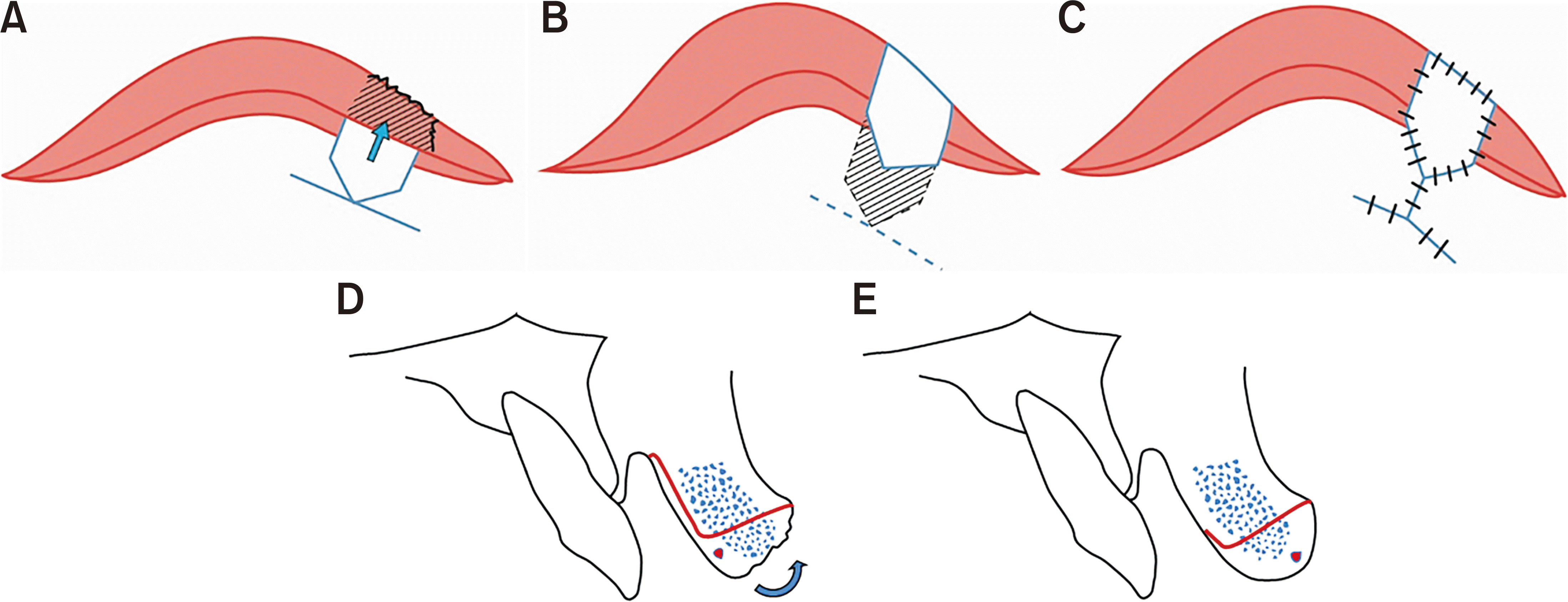
Since the defect extended over the vermilion border around 1 mm to the skin and was limited to the white roll, reconstruction of the defect was planned to use a mucosal V-Y advancement flap. Before preparing the mucosal flap, it is necessary to debride the wound and excise the devitalized tissue surrounding the vermilion defect. The incision was performed to the superficial layer of the orbicularis oris muscle and dissected from the white lip. Since the blood supply to the mucosal flap is based on the orbicularis oris muscle, dissection was performed carefully. The superior labial artery was included in the mucosal part of the flap. After the initial incision and dissection, horizontal, full thickness transection of the orbicular oris muscle was performed below the shield shaped mucosal flap, which is attached to the orbicularis muscle. Then the flap was vertically rotated and advanced superiorly into the vermilion defect. The vermilion defect margin served as the base of the triangular flap, and the tip of the flap became the fornix. Since the orbicularis oris muscle has sufficient movement, this mucosal flap can easily cover the defect without causing tension during lip motions. The vascular supply of the flap was based on the labial artery and orbicularis oris muscle. Finally, the vermilion defect was completely covered by the mucosal flap. The advanced flap was accurately sutured to the superior margin of the defect with 6-0 non-absorbable sutures, and the new vermilion border was established. The vertical closure of the inner lip mucosa was started from above the vestibular sulcus using the 5‐0 absorbable suture. An additional horizontal vestibular incision was made medially to release the tension of the vertical suture.(Fig. 1) The concept of the surgical step is demonstrated in Fig. 2.
Postoperatively, the surgical area healed uneventfully without any signs of inflammation or flap necrosis, hematoma, or wound dehiscence. Since the volume of the flap was rather than thick compared to the opposite side, the flap volume was reduced by a small vertical wedge excision of vermilion mucosa at 2 months after the surgery. In the follow-up check, the flap showed good healing, and harmony with adjacent structures was confirmed. Moreover, the color and texture of the mucosal flap were not visibly asymmetric or distorted and maintained normal lip movement at 6 months and 1 year postoperatively.(Fig. 3)
This study was approved by the Institutional Review Board of Kyungpook National University Dental Hospital (No. KNUDH-2024-04-01-00).
Various types of defects can be seen on the upper lip from trauma and benign or malignant tumors. Several reconstruction methods are used depending on the size and depth of the defect3,9. The upper lip is important in determining facial profile, so functional and aesthetic recovery should be carefully considered during defect reconstruction. An upper lip defect is adjacent to anatomical structures, such as the philtrum, nose, cheek, skin, and mucosa, meaning reconstruction is performed considering these structures10. Since the skin laxity of the upper lip is relatively limited compared to that of the lower lip, and the central structure of the face is located near the upper lip, it is challenging to reconstruct a lip defect. Various techniques have been suggested to reconstruct an upper lip vermilion defect, with one of these being horizontal advancement of the mucosal flap. This flap was fabricated by undermining beneath the upper lip mucosa and was simple to design. However, the flap volume is limited, and it is difficult to achieve lip fullness because of flap contraction after surgery4.
If the defect is large and it is difficult to make a local flap, a cross-lip flap from the lower lip or a tongue flap from the anterior tongue can be utilized. However, both techniques require temporary immobilization of the lip or tongue for blood supply of the flap and also a second surgery to cut the original pedicle 2 weeks after the initial surgery. For these reasons, the labial mucosa was regarded as a better choice for a vermilion defect. These V-Y advancements of the labial mucosa show good color matching, but scars and lip asymmetry can occur after reconstruction. Furthermore, lip volume may be decreased and lip movement restricted due to muscle contraction11. Even though the above-mentioned potential complications have been suggested, the V-Y advancement flap reported from the previous report showed quite acceptable results2.
In this case report, an upper lip defect caused by a dog bite was successfully treated with a mucosal V-Y advancement flap. This flap could also be termed a myomucosal flap, which contains the orbicularis oris muscle with a branch of the superior labial artery. The superior labial artery is located transversally and supplies the mucosa and orbicularis oris muscle. The vermilion defect was successfully reconstructed by advancing the myomucosal flap via superior mobilization and repositioning the mucosal flap to cover the defects. Because the mucosal flap has a good blood supply, the flap can be adapted to the vermilion border without risk of flap necrosis.
This flap is a good option for vermilion defect reconstruction because harvesting is not complicated. Since the flap uses the inner mucosal area, there is no visible scarring or major differences in the lip movement after sufficient healing. In addition, the color between the mucosa and upper lip is similar, so the color matching after surgery was satisfactory. Since the volume and length of the lip were symmetrical after the mucosal V-Y advancement flap, it is also more effective for aesthetic restoration than a local flap using the adjacent lip and structure. It is important to note that if there is tension after suture for V-Y advancement, there is the possibility of a residual whistling deformity6.
However, since the mucosal V-Y advancement flap does not include cutaneous tissue, it can only be used for defects limited to the vermilion. Thus, a double V-Y advancement flap or another local flap should be considered if the vermilion defect extends to the cutaneous portion12. This flap uses only inner mucosa, so its application may be limited if the vermilion defect is large and includes skin area. In addition, other reconstruction methods should be considered for defects that include the philtral ridge13. Since this is a single case report, additional studies with a number of cases need to be investigated.
In conclusion, this mucosal V-Y advancement flap technique is reliable and easy to perform to repair mucosal defects and can be expected to yield excellent color matching to the original lip without vascular compromise of the flap.
Notes
Authors’ Contributions
G.J.S. conceptualized the study and participated in the treatment and study design. H.W.Y and D.K. collected the patient’s data. G.J.S. drafted the manuscript. H.W.Y and D.K. reviewed the manuscript. T.G.K. organized the study and critically revised and finalized the manuscript.
Ethics Approval and Consent to Participate
This study was approved by the Institutional Review Board of the Kyungpook National University Dental Hospital (No. KNUDH-2024-04-01-00).
References
1. Daraei P, Calligas JP, Katz E, Etra JW, Sethna AB. 2014; Reconstruction of upper lip avulsion after dog bite: case report and review of literature. Am J Otolaryngol. 35:219–25. https://doi.org/10.1016/j.amjoto.2013.11.008. DOI: 10.1016/j.amjoto.2013.11.008. PMID: 24332929.

2. Jin X, Teng L, Zhang C, Xu J, Lu J, Zhang B, et al. 2011; Reconstruction of partial-thickness vermilion defects with a mucosal V-Y advancement flap based on the orbicularis oris muscle. J Plast Reconstr Aesthet Surg. 64:472–6. https://doi.org/10.1016/j.bjps.2010.07.017. DOI: 10.1016/j.bjps.2010.07.017. PMID: 20709612.
3. Malard O, Corre P, Jégoux F, Durand N, Dréno B, Beauvillain C, et al. 2010; Surgical repair of labial defect. Eur Ann Otorhinolaryngol Head Neck Dis. 127:49–62. https://doi.org/10.1016/j.anorl.2010.04.001. DOI: 10.1016/j.anorl.2010.04.001. PMID: 20822758.

4. Madorsky S, Meltzer O. 2020; Myomucosal lip island flap for reconstruction of small to medium lower lip defects. Facial Plast Surg Aesthet Med. 22:200–6. https://doi.org/10.1089/fpsam.2020.0068. DOI: 10.1089/fpsam.2020.0068. PMID: 32255366. PMCID: PMC7312741.

5. Pepper JP, Baker SR. 2013; Local flaps: cheek and lip reconstruction. JAMA Facial Plast Surg. 15:374–82. https://doi.org/10.1001/jamafacial.2013.1608. DOI: 10.1001/jamafacial.2013.1608. PMID: 24051684.

6. Srivastava S. 1989; Reconstruction of traumatic loss of vermilion and mucocutaneous junction of the lips. Br J Plast Surg. 42:526–9. https://doi.org/10.1016/0007-1226(89)90038-6. DOI: 10.1016/0007-1226(89)90038-6. PMID: 2804516.

7. Kolhe PS, Leonard AG. 1988; Reconstruction of the vermilion after "lip-shave". Br J Plast Surg. 41:68–73. https://doi.org/10.1016/0007-1226(88)90147-6. DOI: 10.1016/0007-1226(88)90147-6. PMID: 3345410.

8. Lane JE, Kent DE. 2007; Repair of vermilion Mohs defect with unilateral axial myocutaneous advancement flap. Dermatol Surg. 33:1502–4. discussion 1504. https://doi.org/10.1111/j.1524-4725.2007.33324.x. DOI: 10.1111/j.1524-4725.2007.33324.x. PMID: 18076619.

9. Martin TJ, Zhang Y, Rhee JS. 2008; Options for upper lip reconstruction: a survey-based analysis. Dermatol Surg. 34:1652–8. https://doi.org/10.1111/j.1524-4725.2008.34342.x. DOI: 10.1111/j.1524-4725.2008.34342.x. PMID: 19018829.

10. Boson AL, Boukovalas S, Hays JP, Hammel JA, Cole EL, Wagner RF Jr. 2021; Upper lip anatomy, mechanics of local flaps, and considerations for reconstruction. Cutis. 107:144–8. https://doi.org/10.12788/cutis.0205. DOI: 10.12788/cutis.0205. PMID: 33956606.

11. Nicholas MN, Liu A, Chan AR, Jia J, Fuller K, Eisen DB. 2021; Postoperative outcomes of local skin flaps used in oncologic reconstructive surgery of the upper cutaneous lip: a systematic review. Dermatol Surg. 47:1047–51. https://doi.org/10.1097/DSS.0000000000003063. DOI: 10.1097/DSS.0000000000003063. PMID: 33927091.

12. Duan M, Yue C, Dai Y, Wu Y, Peng J. 2024; Technical solution for reconstruction of upper lip defect by bilateral V-Y advanced flaps. J Am Acad Dermatol. 90:e67–8. https://doi.org/10.1016/j.jaad.2022.09.047. DOI: 10.1016/j.jaad.2022.09.047. PMID: 36206935.
13. Drosou A, Trieu D, Goldberg LH. 2017; Reconstruction of a postoperative mohs defect of the upper cutaneous and vermilion lip involving cupid's bow. Dermatol Surg. 43 Suppl 1:S96–8. https://doi.org/10.1097/DSS.0000000000001034. DOI: 10.1097/DSS.0000000000001034. PMID: 28079636.

Fig. 1
A. The lip wound was thoroughly debrided before reconstruction. B. Design of the mucosal V-Y advancement flap in the inner mucosal area. C. Completion of the mucosal V-Y advancement flap dissection based on the orbicularis oris muscle. An additional horizontal incision was made in the orbicularis oris muscle to facilitate flap mobilization. D. The shield-shaped flap was moved superiorly to approximate the superior flap margin to the vermilion border. E. The V-Y suture was placed between the inner mucosa and flap. F. After reconstruction of the upper lip defect, good color matching and volume were observed.

Fig. 2
Schematic illustration of the mucosal V-Y advancement flap. A-C. The mucosal incision was made at the inner lip mucosa. The mobilized mucosal flap was approximated to the vermilion-skin border. The inner vestibule was sutured in a Y-shaped manner. D, E. Cross section of the flap. The labial artery was included in the mucosal flap, and dissection proceeded.

Fig. 3
A. Clinical findings of the lip immediately after the surgery. B. One-month postoperative photograph. Excessive lip volume from the mobilized flap was revised after excision of the redundant lip tissue. C. Six-month postoperative view. D. One year after the reconstruction, both the volume and width of the flap were symmetrical and matched the original color.


 PDF
PDF Citation
 Print
Print


 XML Download