

 PDF
PDF Citation
 Print
Print


I. Introduction
Mandibular condyle hyperplasia bone disease, which occurs in the maxillofacial region, is known to cause severe facial asymmetry and malocclusion due to excessive growth of the mandibular condyle1. While it has been reported to be common in females2, the cause of onset has not been fully elucidated3. It is difficult to improve the facial appearance of affected patients; thus, orthognathic surgery is often combined with other treatments1. In such cases, there is no clear standard regarding whether the condylectomy and orthognathic surgical procedures should be performed at the same time, or whether orthognathic surgery should be separated into two stages, including concerns regarding time required for recovery.
For orthognathic treatment, an approach including starting with conventional orthodontic treatment became popular in the 1970s4, though patients treated in that manner were forced to endure facial deformity and tooth function deterioration during the preoperative orthodontic treatment period that lasted for 12 to 24 months4,5. To solve this problem, surgery-first and surgery-early approaches, in which orthognathic surgery is performed with or without a shortened period of preoperative orthodontic treatment, have recently been developed6. These approaches have gained support from both clinicians and patients because of the significantly shorter total treatment time and superior postoperative stability7. Presented here are findings of a surgery-early approach for orthodontic treatment, in which condylectomy and orthognathic surgery were performed at the same time for a patient with facial asymmetry and mandibular condyle hyperplasia.
Go to : 
II. Case Report
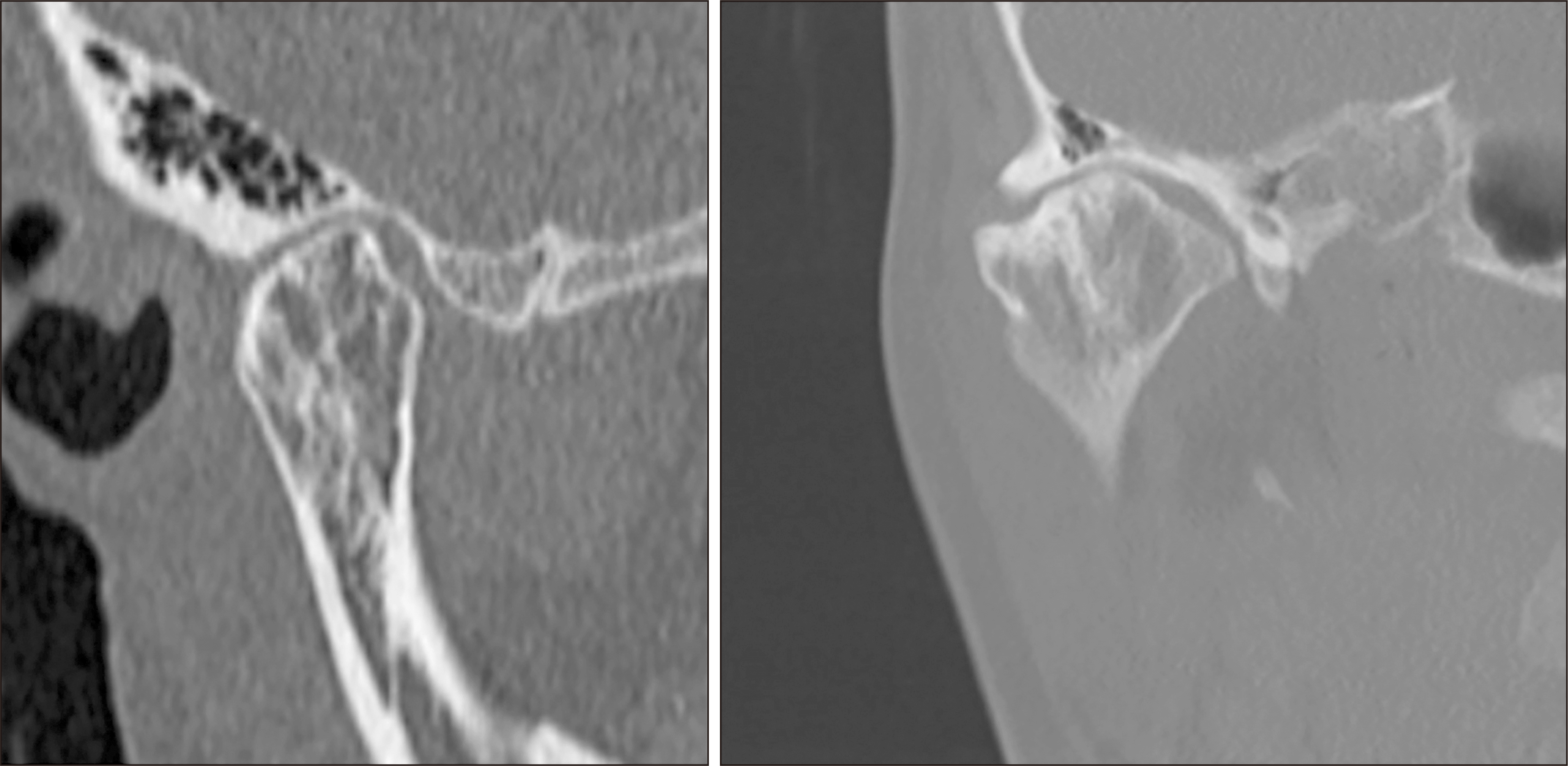
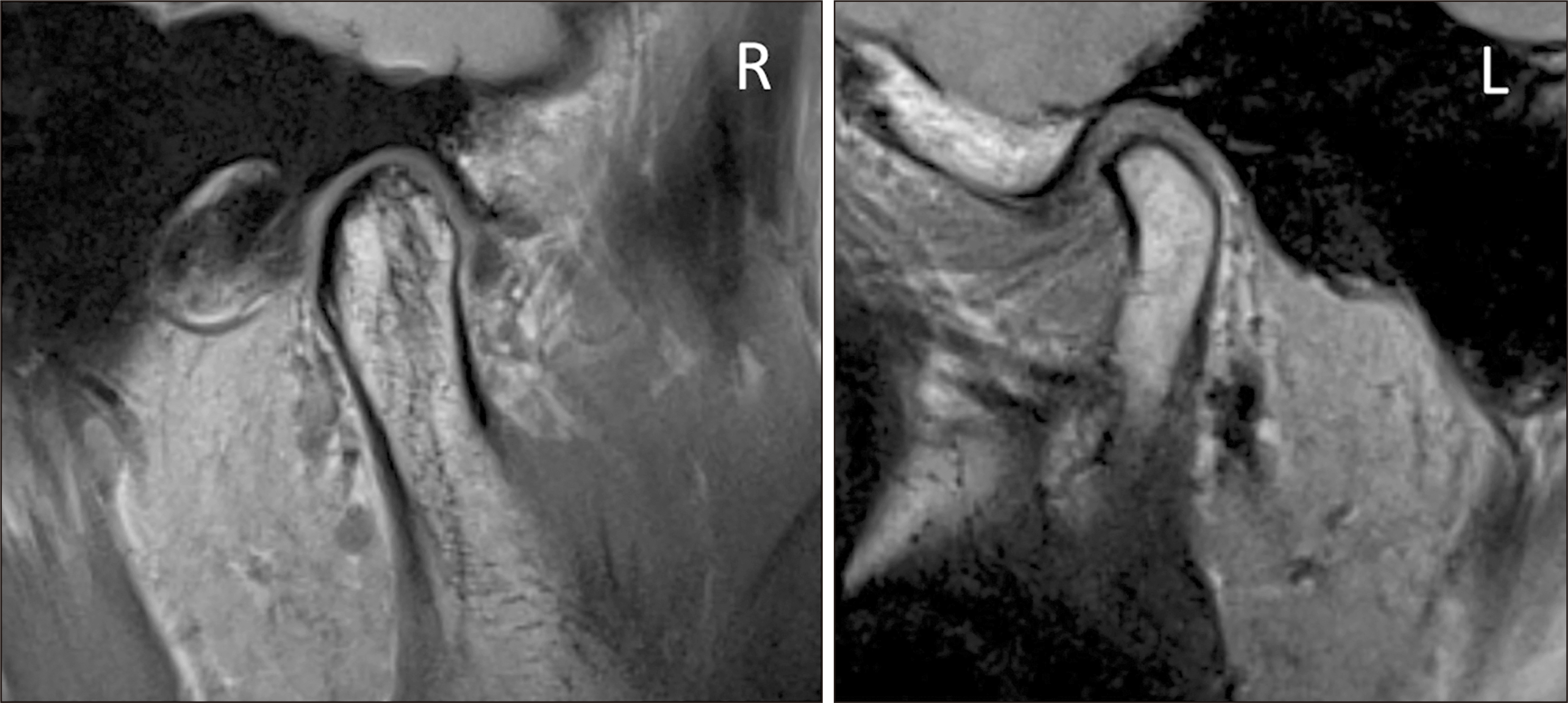
In March 2018, a female aged 28 years at the time of the initial examination was presented to our department with chief complaints of left deviation of the mandible and right-side temporomandibular joint (TMJ) noise. A facial examination indicated skeletal Class III, concave type, and severe left deviation of the molars.(Fig. 1) Intraoral findings included a left-sided anterior to molar counterbite, marked leftward tilt of the occlusal plane, and 8.5-mm leftward deviation of the midline of the mandibular dentition. Cephalometric findings showed anterior nasal spine of –3.0° and Wits appraisal of –10.0 mm, while template cephalometric analysis results clearly indicated a skeletal class III mandible.(Fig. 2) Furthermore, panoramic X-ray imaging showed excessive height of the right mandibular branch, computed tomography (CT) findings showed irregular hyperplasia of the right mandibular head (Fig. 3), and bone scintigraphy showed increased accumulation of 99mTc in the right mandibular head. Anterior displacement of the bilateral TMJ discs was also indicated by magnetic resonance imaging (MRI).(Fig. 4)

 | Fig. 2A, B. Cephalometric radiographs. C. Panoramic radiograph image. D. Cephalometric template analysis. (CDS: craniofacial drawing standards, Ave: average)
|
The patient provided informed consent after receiving an explanation of the advantages and disadvantages of treatment. Preoperative orthodontic treatment was performed for 3 months to deal with problems associated with a surgery-early approach. Specifically, since it was difficult to attach brackets to the maxillary tooth, leveling and alignment of the mandibular tooth were performed first. Then, just prior to the planned surgery, brackets were attached to the maxillary tooth using a bite plane along with passive rectangular wires.(Fig. 5)

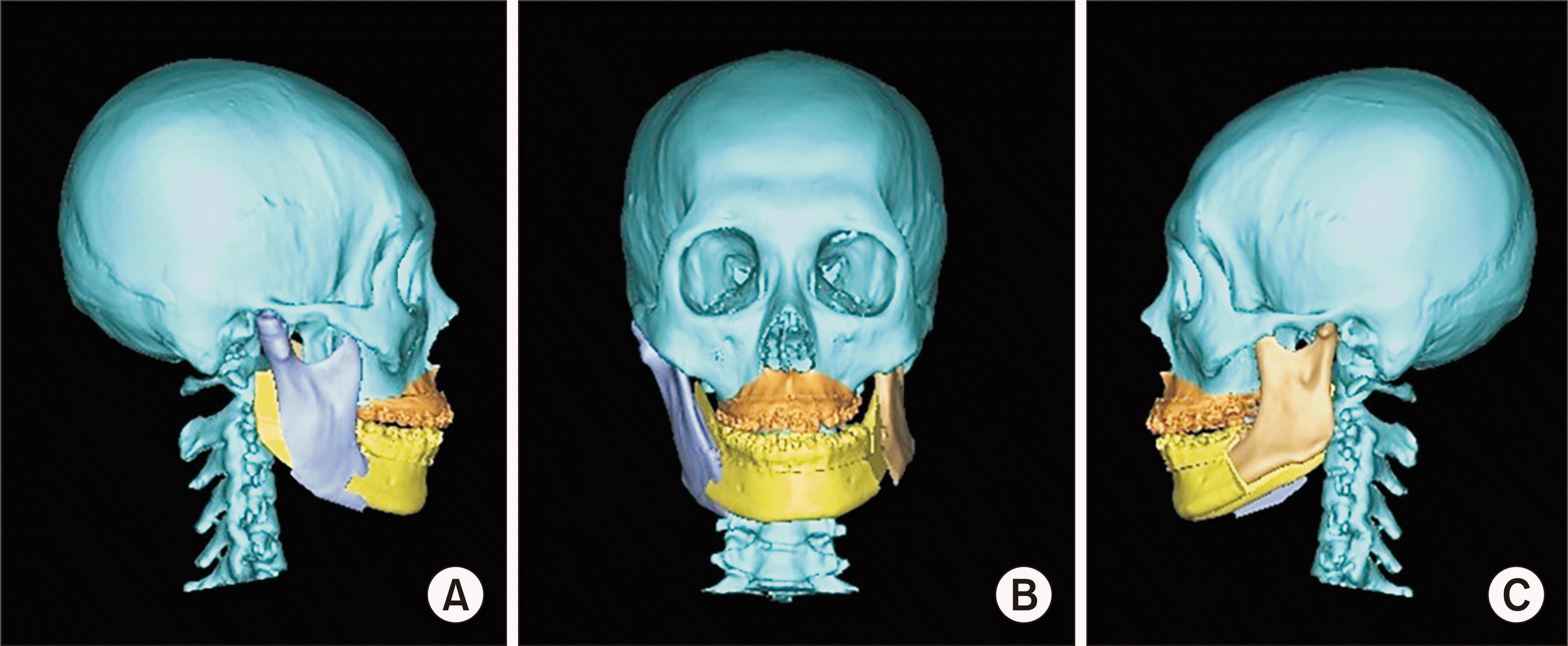
Following 3 months of preoperative orthodontic treatment as noted above, which provided good results, a right mandibular condylectomy under general anesthesia was performed in October 2018 based on a diagnosis of facial asymmetry due to hyperplasia of the right mandibular condyle. The procedure included a Le Fort I osteotomy, right mandibular sagittal split ramus osteotomy, and left mandibular inverted L ramus osteotomy.(Fig. 6) The mandibular condyle resection area was evaluated using a three-dimensional (3D) jawbone model prior to surgery, then the procedure was performed at a height of 19 mm from the mandibular parietal. For orthognathic surgery, pre-surgery evaluations were performed using 3D simulation imaging.(Fig. 7) Examination of the conventional mandibular branch sagittal segmentation showed an excessive amount of downward movement of the mandibular angle on the left side; thus, a left-sided inverted L ramus osteotomy was selected. Using the simulation model, the position of the artificial condylar head was confirmed so as not to contact any part of the mandibular fossa in the intercuspal position. In addition, during the operation, when the mandible was moved in opening and lateral movements, the artificial condylar head was positioned such that it did not exert excessive pressure on the bone of the mandibular fossa.
 | Fig. 6Intraoperative photographs. A. The joint capsule was exposed by a preauricular incision to perform condylectomy. B. Resected mandibular head lesion. C. The osteotomy design was step-type and subspinal, and the procedure was performed using a reciprocating saw. The surgeon then moved the osteotomes toward the lateral nasal wall and pterygomaxillary junction to obtain the final maxillary down-fracture. The maxilla was then stabilized utilizing a titanium miniplate. A medial osteotomy cut was made at the level of the lingula and parallel with the occlusal plane using a fissure burr with consecutive cuts performed medial to the external oblique ridge. A buccal cut was performed vertically from the distal portion of the second molar to the inferior border of the mandible. Fragments were separated using a thin osteotome and forceps. After confirmation of a complete mandibular split, an acrylic occlusal splint was used to position the distal segment. The fragments were then stabilized utilizing a titanium miniplate.
|
The postoperative orthodontic treatment course is shown in Fig. 8. Placement of a multi-bracket appliance combined with mini-screw usage was performed to improve occlusion. The duration of postoperative orthodontic treatment was 6.8 months, while total treatment time was 14.6 months. At the end of treatment, the severe facial asymmetry seen at the initial visit was significantly improved, and there was a balanced profile for the lateral aspect of the mandibular prognathism. Intraoral examination findings showed that the mandibular midline, which had been significantly deviated, was aligned with the facial and maxillary midlines, with normal occlusion both aesthetically and functionally improved. These good results were obtained within a relatively short period, and the patient experienced great satisfaction with smiling more naturally and enjoying conversation with others. At 1 year after surgery, no retrograde changes in skeletal structure or occlusion were observed, and stable maxillofacial morphology and occlusal function were maintained.(Fig. 9) A TMJ evaluation showed bilateral noises as observed at the initial visit, though the patient remained in good condition with no other symptoms. Three years after surgery, CT findings revealed cortical bone formation on the resected surface of the right mandibular condyle, and MRI findings showed that the right side of the TMJ disc was in a normal position, while the left side was anteriorly displaced, the same as before surgery.(Fig. 10)
 | Fig. 8A. Leveling of upper dentition begun, 24 days after orthognathic surgery. B. Leveling of upper dentition continued, 1.7 months after orthognathic surgery. C. Class II elastics were used for correction of Class II denture, 2.7 months after orthognathic surgery. D. Class II elastics continued, 3.8 months after orthognathic surgery. E. Closing space of upper dentition, 5.1 months after orthognathic surgery. F. Detailing and finishing, 6.8 months after orthognathic surgery.
|


Go to :
III. Discussion
Although the cause of mandibular condyle hyperplasia is unclear8, individuals aged 11 to 30 years are often affected9. Furthermore, female predominance has been previously reported in several studies10,11, while Beltran et al.12 also noted that female hormones may be involved. Findings in the present case were consistent with those factors, though the relationship to female hormones is unknown because no related tests were performed.
Clinical photography and 2D or 3D imaging are generally used for diagnosis of mandibular condyle hyperplasia, as those have been reported effective to identify abnormal growth in the oral cavity. However, single photon emission computed tomography has been found to have low levels of specificity and sensitivity for condylar hyperplasia, with concordance rates ranging from 30% to 60%13,14. Obwegeser and Makek15 classified unilateral mandibular condylar hyperplasia by type of malformation, and when that leads to severe maxillofacial asymmetry and malocclusion, condylectomy can be performed, which halts progression16. On the other hand, another report recommended orthognathic surgery instead of a mandibular condylectomy procedure for cases of facial asymmetry shown by bone scintigraphy to have low activity in the condyle17. In the present case, CT, MRI, and bone scintigraphy were used for diagnostic imaging, and a high level of activity was observed in the right mandibular condyle. In addition, severe facial asymmetry and significant leftward deviation of the midline of the mandibular dentition were observed; thus, it was determined that not only condylectomy, but also orthognathic surgery, should be performed.
Controversy exists regarding whether to perform condylectomy and orthognathic surgery simultaneously or in two stages for patients with unilateral mandibular condylar hyperplasia with facial asymmetry. Those who argue that the procedure should be performed at the same time note potential improvement in patient quality of life due to the shorter operative and treatment times, including orthodontic treatment, as well as the ability to deter development of TMJ symptoms associated with occlusal head interference that occurs after condylectomy18. On the other hand, a report stating that the procedures should be performed in two stages noted that early mouth opening training is necessary to avoid contracture after a mandibular condylectomy, while intermaxillary fixation is necessary to stabilize the jaw position after orthognathic surgery, and also pointed out conflicting needs related to postoperative changes in the position of the mandibular condyle leading to degenerative changes in the TMJ19. Unfortunately, there is no detailed report regarding differences in treatment periods between simultaneous and two-stage surgery, though it is expected that the time will be shortened with a simultaneous procedure, as noted above. Interestingly, Nolte et al.20 also reported cases in which good results were obtained with only orthodontics and without orthognathic surgery after condylectomy, including when two stages were initially considered, suggesting that even if two-stage treatment is selected at the planning stage, it may end up as a single operation with a treatment period that is not excessive.
For the present patient, simultaneous performance of a mandibulectomy and orthognathic surgery was chosen, which resulted in a relatively short total treatment period of 14.6 months, with no problems encountered during the postoperative correction process. However, based on this experience with only one case, the authors cannot say that a simultaneous condylectomy and surgery-early approach will be effective in most cases of mandibular head hyperplasia, and recognize that evaluations in future studies will be necessary.
Go to :
 XML Download