Abstract
Background
By analyzing small bowel capsule endoscopy (SBCE) performed in two large hospitals in Kazakhstan, we aimed to explore the characteristics of patients representative of Central Asia and the technical characteristics of SBCE.
Methods
SBCE cases were retrospectively analyzed. A descriptive analysis was conducted on the patients’ demographic data, diagnosis, and clinical symptoms. The results of SBCE, such as the lesions found, transit time and retention rate in the stomach and small bowel, and bowel visualization quality, were analyzed. Complications related to SBCE were investigated.
Results
SBCE was performed in 123 patients. Abdominal pain (81.3%) and chronic diarrhea (66.7%) were the most common symptoms, followed by weight loss (25.2%) and gastrointestinal bleeding (15.4%). The most common disease was Crohn’s disease (52.0%). Definite lesions, such as ulcers, polyps, and bleeding, were identified in 55.3% of patients. SBCE was successfully completed in all cases except for 11 (9.1%). The average small bowel transit time was 4 hours and 28 minutes. Excellent visualization (>75% of mucosa) was reported in 82.5% of patients. No patients experienced complications.
Capsule endoscopy (CE) was commercialized in the 2000s and is now widely used to diagnose diseases of the small intestine. It is primarily used to determine the cause of gastrointestinal (GI) blood loss or hemorrhage (e.g., ulcers, tumors, or vascular malformations in the small intestine) and chronic iron deficiency anemia. It can also be used to determine the extent of small intestinal involvement or the distribution of celiac disease and to diagnose lymphangiectasia.
Kazakhstan, located in Central Asia, is a large area in the middle of the Eurasian continent and is known for its diverse ethnic distribution of Asians and Europeans. Various diseases are noted with ethnic diversity, including celiac disease, which is rare in Asia. Recently, Kaibullayeva et al. [1] reported the prevalence of inflammatory bowel disease (IBD) in Kazakhstan for the first time in Central Asia and reported that the age- and sex-adjusted IBD prevalence was 113.9 per 100,000 people.
Despite the active research on disease epidemiology, the government's control over health care costs limits the ability to perform a variety of tests in Kazakhstan and other Central Asian countries. The introduction of CE was carried out by one of our authors in 2011 on the basis of the city clinic No. 2 in Astana. However, the use of CE was mainly in private clinics since public clinics had insufficient funding. In particular, it is uncommon in Kazakhstan due to its high cost. Therefore, no reports of diseases are found mainly in the small intestine through CE in Central Asia as well as in Kazakhstan.
In 2022, the CE system was introduced to two medical institutions in two regions of Kazakhstan and has been actively used to diagnose various diseases. In particular, a recently developed bidirectional CE was used, and skilled experts applied artificial intelligence (AI) as a reading aid technology.
The purpose of this study was to retrospectively analyze the experience of CE in Kazakhstan and report the indications for CE, types of diseases and lesions found through CE, and various information related to CE in Kazakhstani patients.
Ethical statements: This study was approved by the Institutional Review Board of the Pusan National University Yangsan Hospital (IRB No. 55-2023-014). The requirement for informed consent was waived because of the retrospective nature of the study.
Between May 2022 and February 2023, 123 CEs were conducted at the National Research Oncology Center in Astana and the Asfendiyarov Kazakh National Medical University Hospital in Almaty, Kazakhstan. These two institutions are the largest medical institutions in two cities, the capital and the first economic city, and patients were either local patients or referred from other regions. Experienced gastroenterologists and endoscopists (KB, AG, LY, and JK) from both medical institutions selected patients who needed a diagnosis of small intestinal disease according to the guidelines [2].
This project was implemented as follows: all CEs were performed on an outpatient basis. After receiving sufficient explanations about the examination and analysis, all patients signed a consent form and underwent the examination. The patient’s bowel was prepared using polyethylene glycol or diet control. The patient visited an outpatient clinic, had a CE receiver installed, swallowed the CE on their own, returned home, and submitted the receiver the next day. After obtaining images from the patient, the images were encrypted, transmitted, and analyzed remotely. Division of the first and last points of the small intestine, visible region evaluation, and lesion detection were performed with an AI algorithm, and the analyzed images were evaluated and confirmed by a gastroenterologist with more than 13 years of CE reading experience. The images were also read separately by endoscopists from two medical institutions in Kazakhstan, and the two readings were combined to make an expert diagnosis of the patient. CE was performed using MiroCam MC2000 (IntroMedic Co. Ltd.), which is a bidirectional image capture system allowing for AI analysis to detect clean mucosal areas [3] and lesions through AI algorithms developed by Captos Co., Ltd.
Patients’ demographic data, diagnosis, reasons for examination, underlying disease, and prior medication were retrospectively collected. The results of CE, such as stomach transit time, small bowel transit time, clean mucosal area in CE, detected findings, and small intestinal CE retention or complications, were collected and analyzed.
For nominal variables, the frequency was calculated, and for continuous variables, the mean and standard deviation were calculated and compared by the t-test for independent variables. The significance level was p<0.05, and all statistical analyses were performed using the statistical software SPSS 27 (IBM Corp.).
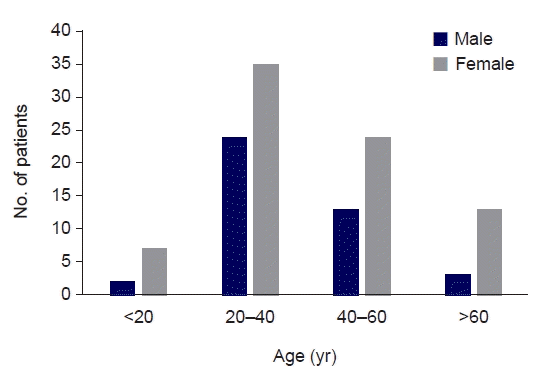
The baseline demographics of the patients are shown in Table 1. Among the 123 patients, the mean age was 40.1±14.8 years (n=121), and 79 (64.2%) patients were female. The most common symptom was abdominal pain (81.3%), followed by chronic diarrhea (66.7%), weight loss (25.2%), GI bleeding (15.4%), and constipation (1.6%). Anemia was the most common sign (75.6%), followed by protein-losing enteropathy (22.8%), obscure bleeding (17.9%), and overt bleeding (0.8%). Other signs, including hair loss and dyslipidemia, were observed in three patients (3.2%). The most common diagnosis was Crohn’s disease (52.0%), followed by ulcerative colitis (16.3%), celiac sprue (10.6%), polyposis (4.9%), intestinal ischemia (4.1%), intestinal tumor (2.4%), and others (7.3%). Two patients (1.6%) were simultaneously diagnosed with Crohn's disease and celiac sprue, and one (0.8%) was diagnosed with Crohn's disease and intestinal ischemia. Fig. 1 shows the distribution of numbers by sex and age of patients who underwent CE. About half of patients (48.0%) were aged 20–40 years.
The technical results of the CE procedure are shown in Table 2. The examination was terminated in the stomach in three patients (2.4%) and in the small intestine in eight patients (6.5%). Excluding these patients, the mean gastric transit time was 57 minutes, and the mean small bowel transit time was 4 hours and 28 minutes. Regarding bowel preparation, two patients (2.4%) used diet control, and the remaining 121 patients (98.4%) used polyethylene glycol. After the examination, the capsules of all patients passed spontaneously, and there was no retention requiring surgical removal. The mean visualization scale analyzed by AI was 84.1%. The visualization scale was poor (<50%) in two cases (1.7%), fair (50%–75%) in 19 cases (15.8%), and excellent (>75%) in 99 cases (82.5%). In the patient group diagnosed with Crohn’s disease (n=64), the mean gastric transit time was 63±79.4 minutes, the mean small bowel transit time was 257±100.9 minutes, and the mean visualization scale was 85.6%±7.8%. In the patient group diagnosed with ulcerative colitis (n=20), the mean gastric transit time was 53±61.8 minutes, the mean small bowel transit time was 250±74.4 minutes, and the mean visualization scale was 84.0%±8.5%. In the patient group diagnosed with celiac sprue (n=13), the mean gastric transit time was 54±50 minutes, the mean small bowel transit time was 259±158 minutes, and the mean visualization scale was 85.5%±6.7% (n=13).
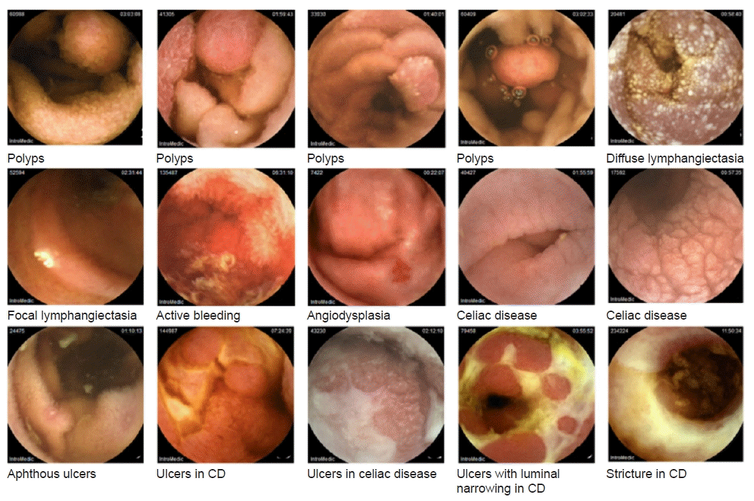
The CE findings are shown in Table 3. Definite lesions were identified in 68 of the 123 patients (55.3%). Through CE, we were able to visually confirm the ulcer category (ulcer, erosion, or aphthous ulcer), as well as mucosal abnormalities, such as active bleeding, celiac mucosa, polyps, and strictures. Crohn’s disease was the most prevalent diagnosis. Of the patients diagnosed with Crohn's disease, 26 (40.6%) had no specific findings, and 30 (46.9%) had definite lesions. Overall, 20 patients with ulcerative colitis were noted, among whom five (25.0%) had significant lesions in ulcer categories. In the entire patient group, significant lesions, including ulcers, polyps, and other lesions, were identified in 68 patients (55.3%) through CE. Typical lesions found in CEs are displayed in Fig. 2.
The CE findings in patients with GI bleeding are shown in Table 4. Among 23 patients with signs of GI bleeding, 22 had obscure GI bleeding. Looking at their findings, ulcers were identified in five patients (22.7%), erosions in two patients (9.1%), and aphthous ulcers in three patients (13.6%). Active bleeding was confirmed alone in three patients (13.6%). Polyps and ulcers, and polyps and active bleeding were simultaneously found in one patient (4.5%) each. No patients had celiac mucosa or stricture, and seven patients (31.8%) had no abnormal findings. No abnormal findings were found in one patient with overt GI bleeding.
Since CE was first applied to the global market in early 2000, its usefulness has been proven through various disease conditions like GI bleeding, tumors, or inflammatory lesions [4-7]. Currently, CE is widely used all over the world; however, it tends to be mainly used in developed countries. Although CE has been used in Kazakhstan since the mid-2000s, the data have not been organized. This is the first report of the Central Asian application of CE performed in Kazakhstan on patients who had various symptoms.
In this study, we were able to confirm the diversity of disease groups applied to CE in Kazakhstan. In a study published in Korea in 2015, 37.2% normal cases, 15.3% cases of vascular lesions including angiodysplasia, telangiectasia, and varices, 28.5% cases of mucosal inflammatory lesions including erosion, ulcer, and ischemic enteritis, and 9.5% other cases were reported. In a multicenter study published in Korea in 2020, the CE tendency was divided into two groups: before 2014 August and after 2014 September; the results demonstrated a decrease in negative results from 38.5% to 28.4% and an increase in the proportions of inflammatory and vascular lesions from 32.9% to 40.4% and 14.4% to 17.5%, respectively [8]. The proportion of tumor lesions decreased from 10.1% to 7.5% [8]. In a study published in Japan, erosion, vascular ectasia, and ulcers were found in 47.9% of cases; polyps, tumors, and lymphomas were found in 14.4% of cases; stenosis was found in 2.3% of cases, and others were found in 4.3% of cases [9]. In this study, ulcer categories were identified in 51 cases (41.4%), active bleeding/venous malformation in 10 cases (8.1%), celiac mucosa in eight cases (6.5%), and polyps in five cases (4.1%). In this study, mucosal inflammatory lesions were observed at a slightly higher frequency than those in previous reported studies [8,9]. It is associated with an increase in IBD in Kazakhstan [1]. Kazakhstan is mixed with Caucasians; therefore, identifying patients with celiac disease that cannot be found in Central Asia was possible.
We used double-head CE, which has the advantage of simultaneously filming both proximal and distal parts of CE. Almost all findings were observed on both sides, but one case of active bleeding was observed only in one direction of CE. This might have been bleeding that occurred while the CE was passing through the distal part in the direction in which the CE progressed. Yung et al. [10] reported that double-headed CE provided different reports compared to single-headed CE in 28.7% of examinations (27/94), and in five cases, active inflammation was missed by the single-headed CE reading. Although clearly presenting the advantages and disadvantages of the bidirectional camera in this study was challenging due to the small number of cases, the advantage of being able to observe both sides is thought to be much more useful in detecting lesions.
In addition to the disease category, the level of perfection of CE has improved over time. In this study, bowel preparation was done with polyethylene glycol in 98.4% of cases and with diet only in 1.6% of cases. In most patients (84.1%), an excellent visualization scale analyzed by the AI algorithm was observed. The proportion of visualized mucosa in the small intestine >75% was 82.3%, that 50%–75% was 16.8%, and that 25%–50% was 0.9%, and that <25% was none. In a previous study, among 397 patients, CE was not completed through small bowel in 15.4% of cases. The mean gastric transit time was 45.7 minutes, and the mean small bowel transit time was 282.1 minutes [9]. In our study, the average gastric transit time was 57 minutes and the small bowel transit time was 268 minutes (4 hours 28 minutes), which was similar to a previous study, while an incomplete study only occurred in 11 cases (8.9%), and no retention required surgery. The high proportion of an excellent visualization scale and the low frequency of small bowel retention prove that the selection of the patient and the CE study preparation were excellent.
According to the European Society of Gastrointestinal Endoscopy position statement [11], competence in small bowel CE (SBCE) can be assessed by considering a minimum of 30 SBCEs and experience in bidirectional endoscopies is desirable for structured training in SBCE. The experience of more than 60 cases of CE using bidirectional endoscopies at two clinics in Kazakhstan was sufficient for training in SBCE to further raise experts in both educational hospitals. It is an appropriate start for SBCE to successfully be implemented in Kazakhstan.
We used AI to analyze CE produced by Captos Co., Ltd. The segment of small intestine, lesion detection, and visualization scale was analyzed and presented by an AI program and reviewed and approved by GI endoscopy experts. This helped reduce the reading time and ensure no lesions were missed. However, this study did not separately analyze the CE reading time or AI reading accuracy. The expected value of the AI reading of SBCE is expected to be comparable to that of experienced endoscopists for lesion detection, reducing the analysis time of the operator [12,13]. Further analysis of the efficacy of using an AI program in SBCE is needed and will follow.
In conclusion, we were the first among Central Asian countries to identify various diseases, such as Crohn’s disease, ulcerative colitis, celiac sprue, and polyposis, through CE. In addition, it is the first study of CE application in Kazakhstan, and technical data like the transit time and visualization scale were obtained from CE performed on patients in Kazakhstan. In particular, there was no case for which surgery was required due to excellent patient selection. Although the data are retrospective and not all data were collected under controlled conditions, our findings are valuable as the first report on the experience of CE in real clinical settings in Kazakhstan.
Notes
Conflicts of interest
Jeongwoo Ju, Haejin Lee, and Yeoun Joo Lee are employees of Captos Co., Ltd. No other potential conflicts of interest relevant to this article were reported.
Author contributions
Conceptualization: KB, JK, YJL. Methodology: KB, JK, YJL. Formal analysis: KB, JK, YJL. Investigation: KB, JK, AG, LY, JJ, HL, YJL. Data curation: KB, AG, LY, JK, JJ, HL, YJL. Writing – original draft preparation: SJS. Writing – review and editing: KB, JK, YJL. Supervision: KB, JK, YJL. All authors have read and agreed to the published version of the manuscript.
References
1. Kaibullayeva J, Ualiyeva A, Oshibayeva A, Dushpanova A, Marshall JK. Prevalence and patient awareness of inflammatory bowel disease in Kazakhstan: a cross-sectional study. Intest Res. 2020; 18:430–7.
2. Enns RA, Hookey L, Armstrong D, Bernstein CN, Heitman SJ, Teshima C, et al. Clinical practice guidelines for the use of video capsule endoscopy. Gastroenterology. 2017; 152:497–514.
3. Ju J, Oh HS, Lee YJ, Jung H, Lee JH, Kang B, et al. Clean mucosal area detection of gastroenterologists versus artificial intelligence in small bowel capsule endoscopy. Medicine (Baltimore). 2023; 102:e32883.
4. Leandri C, Bordacahar B, Ribiere S, Oudjit A, Guillaumot MA, Brieau B, et al. Usefulness of small-bowel capsule endoscopy in gastrointestinal bleeding. Presse Med. 2017; 46:903–10.
5. Cheung DY, Kim JS, Shim KN, Choi MG; Korean Gut Image Study Group. The usefulness of capsule endoscopy for small bowel tumors. Clin Endosc. 2016; 49:21–5.
6. Robertson AR, Yung DE, Douglas S, Plevris JN, Koulaouzidis A. Repeat capsule endoscopy in suspected gastrointestinal bleeding. Scand J Gastroenterol. 2019; 54:656–61.
7. Pennazio M, Rondonotti E, Despott EJ, Dray X, Keuchel M, Moreels T, et al. Small-bowel capsule endoscopy and device-assisted enteroscopy for diagnosis and treatment of small-bowel disorders: European Society of Gastrointestinal Endoscopy (ESGE) guideline-update 2022. Endoscopy. 2023; 55:58–95.
8. Kim SH, Lim YJ, Park J, Shim KN, Yang DH, Chun J, et al. Changes in performance of small bowel capsule endoscopy based on nationwide data from a Korean Capsule Endoscopy Registry. Korean J Intern Med. 2020; 35:889–96.
9. Tominaga K, Sato H, Yokomichi H, Tsuchiya A, Yoshida T, Kawata Y, et al. Variation in small bowel transit time on capsule endoscopy. Ann Transl Med. 2020; 8:348.
10. Yung DE, Robertson AR, Davie M, Sidhu R, McAlindon M, Rahman I, et al. Double-headed small-bowel capsule endoscopy: real-world experience from a multi-centre British study. Dig Liver Dis. 2021; 53:461–6.
11. Sidhu R, Chetcuti Zammit S, Baltes P, Carretero C, Despott EJ, Murino A, et al. Curriculum for small-bowel capsule endoscopy and device-assisted enteroscopy training in Europe: European Society of Gastrointestinal Endoscopy (ESGE) position statement. Endoscopy. 2020; 52:669–86.