Introduction
Case
Case 1
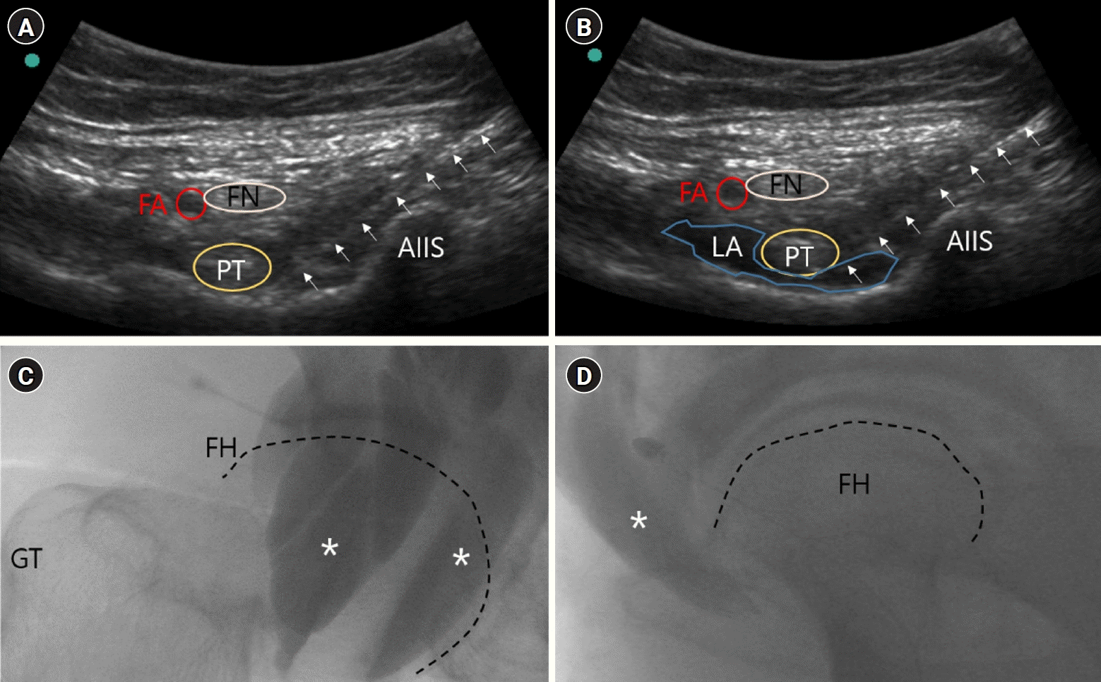
 | Fig. 1.Ultrasound and fluoroscopic images of case 1. (A) The pubic ramus was identified between the iliopubic eminence and the anterior inferior iliac spine (AIIS), and a catheter was mounted between the pubic ramus and the psoas tendon. (B) In total, 20 mL of 0.375% ropivacaine was injected between the psoas tendon and the pubic ramus. Fluid filling is seen between the psoas tendon and the pubic ramus. (C) Posteroanterior image of the hip joint after administering a contrast agent using fluoroscopy. The contrast medium is spread along the iliopsoas muscle passing around the hip joint. (D) Lateral image of the hip joint after administering a contrast agent using fluoroscopy. It was confirmed that the iliopsoas muscle running along the anterior side of the hip joint was imaged. FA, femoral artery; FN, femoral nerve; PT, psoas tendon; LA, local anesthetics; GT, greater trochanter; FH, femoral head (dashed line); asterisk, contrast medium; arrow, catheter. |
Case 2
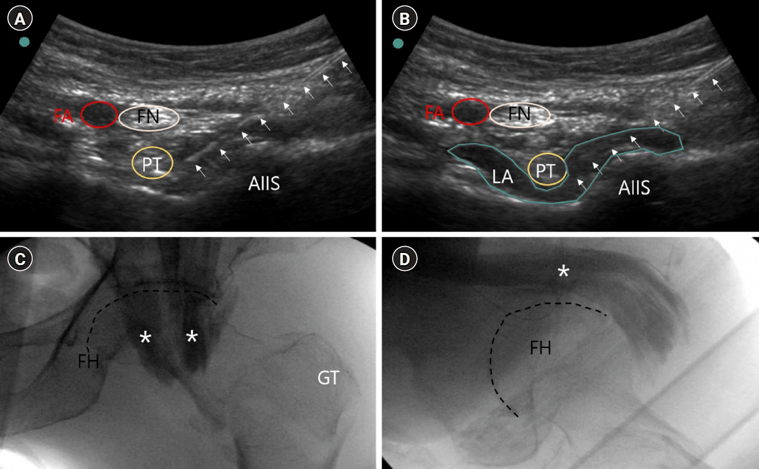
 | Fig. 2.Ultrasound and fluoroscopic images of case 2. (A) The pubic ramus was identified between the iliopubic eminence and the anterior inferior iliac spine (AIIS), and the catheter was mounted between the pubic ramus and psoas tendon. (B) Ropivacaine (0.375%) was injected between the psoas tendon and the pubic ramus. As the fluid fills between the psoas tendon and the pubic ramus, it is seen that the interspace expands. (C) Posteroanterior image of the hip joint after administering a contrast agent using fluoroscopy. The contrast medium is spread along the iliopsoas muscle passing around the hip joint. (D) A lateral image of the hip joint after administering a contrast agent using fluoroscopy. It was confirmed that the iliopsoas muscle running along the anterior side of the hip joint was imaged. FA, femoral artery; FN, femoral nerve; PT, psoas tendon; LA, local anesthetics; GT, greater trochanter; FH, femoral head (dashed line); asterisk, contrast medium; arrow, catheter. |


 PDF
PDF Citation
 Print
Print


 XML Download