Abstract
This is a unique case of metastatic pheochromocytoma of the cervical spine treated with preoperative embolization and subsequent en bloc resection. A 65-year-old man with metastatic pheochromocytoma presented with two weeks of worsening neck pain, left arm and leg weakness and paresthesia, and urinary incontinence. Magnetic resonance imaging showed a metastatic osseous lesion at C6 with severe stenosis and spinal cord compression. The patient underwent successful preoperative angiographic embolization with a liquid embolic agent followed by C5-C7 laminectomy, en bloc tumor resection, and C3-T2 posterior spinal fusion. Six weeks postoperatively, the patient reported improving strength and resolving neck pain and paresthesias. While there is no standard paradigm for the treatment of metastatic pheochromocytomas of the cervical spine, preoperative embolization may minimize intraoperative blood loss and hemodynamic instability during subsequent surgical resection.
Pheochromocytomas are rare, metabolically active, catecholamine secreting tumors that arise from chromaffin cells of the adrenal medulla [3,5]. Pheochromocytoma typically manifests as paroxysmal attacks of headaches, palpations, diaphoresis, and hypertension [3,7]. Long-term endogenous exposure to elevated catecholamine levels can lead to various cardiac complications, including arrhythmias and cardiomyopathy [7,13]. The vast majority of pheochromocytomas are benign and have five-year survival rates ranging from 84% to 96% [6,7,13]. Malignant forms account for roughly 10% of all cases and have five-year survival rates of approximately 40% [4,13]. While metastasis to the lymph nodes, liver, and lungs is relatively common, metastasis to the spine is rare [3,4,7,13]. Furthermore, metastasis to the cervical spine is extremely rare, with only 5 cases of surgical treatment for metastatic pheochromocytomas of the cervical spine having been reported in the literature [2,4,6,9,13]. While the medical management of metastatic pheochromocytomas of the cervical spine is well discussed in the literature, there is limited literature on the surgical and endovascular management of such cases. We report a rare case of malignant pheochromocytoma with cervical metastases treated with preoperative endovascular embolization and subsequent decompression with arthrodesis.
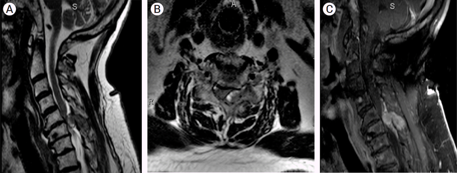
A 65-year-old gentleman with a known history of hypertension, diabetes mellitus, and metastatic pheochromocytoma presented with two weeks of worsening neck pain, left arm and leg weakness with paresthesia, and urinary incontinence. The patient reported falling twice in the preceding two weeks due to worsening weakness. He reported minimal radicular pain. Magnetic resonance imaging (MRI) of the cervical spine showed a metastatic osseous lesion at C6 on the left side, with severe stenosis and compression of the cervical spinal cord (Fig. 1). The patient underwent an adrenalectomy 1 year prior for resection of his primary lesion. The patient also underwent 2 cycles of I-MIBG (Azedra) infusion and external beam radiation therapy (XRT) at that time. Since the resection of his primary adrenal lesion, his autonomic symptoms have been well controlled, with the exception of mild increased perspiration.
Neurologic examination was notable for clonus in the bilateral lower extremities with positive Babinski. Motor examination revealed weakness in left upper and lower extremity with all muscle groups having antigravity strength with moderate resistance except for the deltoid, which had only antigravity strength. He was started on dexamethasone 4mg every 6 hours and admitted to the medicine service for pre-operative anesthesia and endocrine evaluation. His pre-operative hydrochlorothiazide and angiotensin-converting enzyme (ACE) inhibitor was held for post-operative hypotension and was started on a rapid titration of alpha blockade with sodium and fluid loading.
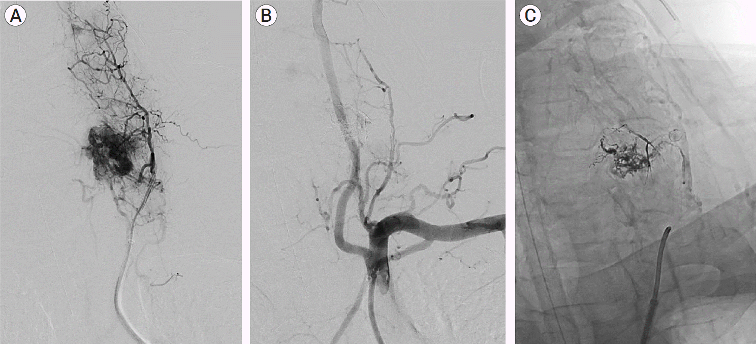
Three days after presenting to the hospital, the patient underwent preoperative formal angiography to determine blood supply to the tumor. Femoral access was obtained and angiography was performed of the bilateral carotid arteries, subclavian arteries and their branches. Robust tumor blush was identified from branches of a left deep cervical artery (Fig. 2A). In order to minimize blood loss and the lower the risk of significant catecholamine release during tumor resection, the decision was made to pursue preoperative embolization. A Neuron MAX (Penumbra, Alameda, CA, USA) 088 guide was positioned in the left subclavian artery, with an 044 Distal Access Catheter (Stryker, Freemont, CA, USA) used to maintain support and access in the left deep cervical artery. The Headway Duo (MicroVention, Alisco Viejo, CA, USA) microcatheter was navigated over an 0.014 inch microwire into a pedicle arising from the deep cervical artery and directly supporting the tumor (Fig. 2A, arrow). Onyx 18 (Medtronic, Minneapolis, MN, USA) was used to embolize the tumor from this position. Follow up angiography demonstrated the Onyx cast with excellent devascularization of the tumor (Fig. 2B, 2C). The patient was extubated postoperatively and remained neurologically stable with no evidence of significant catecholamine release or associated hemodynamic changes.
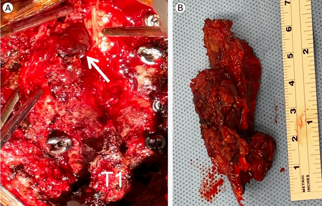
One day post-embolization, C5-C7 bilateral laminectomies and en bloc tumor resection was performed with a C3-T2 posterior spinal fusion. Following the C5-C7 laminectomies, the tumor mass was clearly seen compressing the cervical spinal cord (Fig. 3A).The embolysate was visualized permeating the well-encapsulated tumor. Tumor encroachment into the foramina at the C5-6 and C6-7 levels was observed, and extensive foraminotomies were performed at both levels. Since the tumor had grossly invaded the C6 facet on the left, the facet was drilled extensively close to the depth of the pedicle to facilitate tumor detachment and removal en bloc (Fig. 3B). The estimated blood loss was 500ml and final frozen pathology confirmed metastatic pheochromocytoma.
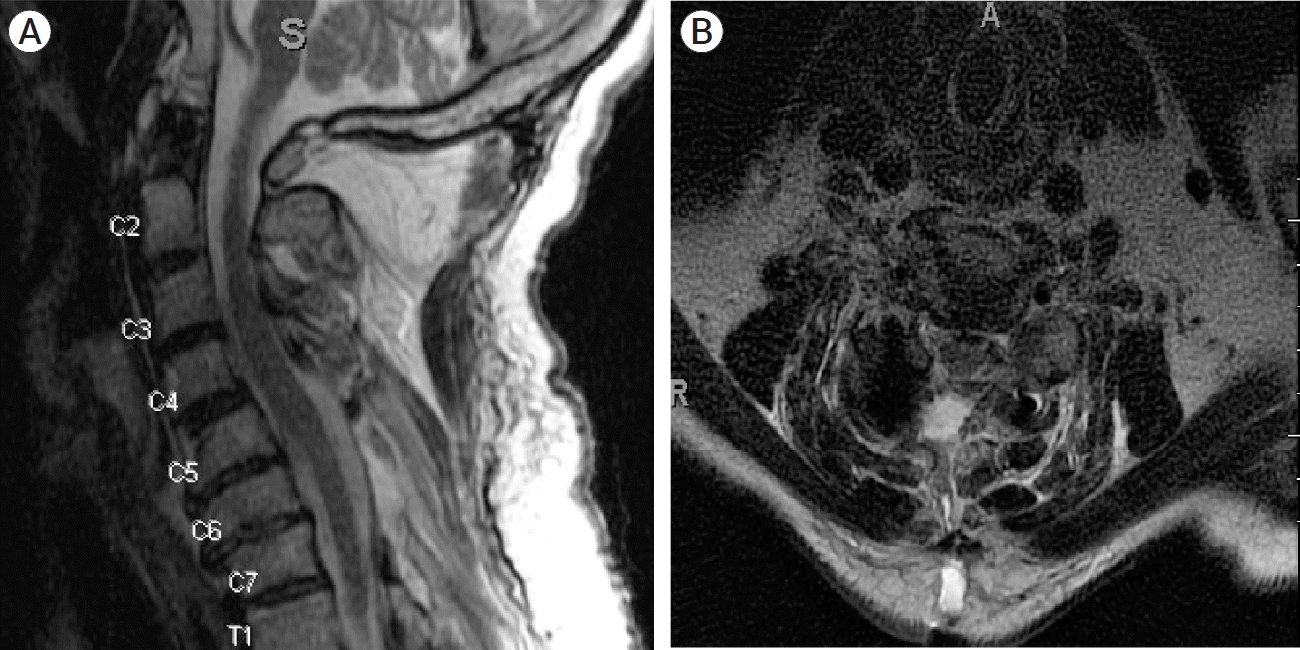
Post-operative imaging revealed complete resection of the cervical lesion and decompression of the cervical cord (Fig. 4A, 4B). Six weeks post-operatively, the patient reported resolving numbness in his fingertips bilaterally, and improving strength in his left upper and lower extremities. He reported a steadier gait and was ambulating without assistance. Motor examination showed significantly improved strength in his left upper and lower extremities with full strength throughout other than in the left deltoid, which was near full strength with resistance against gravity. His neck pain improved, and he only experienced discomfort from his cervical thoracic orthosis (CTO) brace, which he wore at all times post-operatively. At this point he, was cleared to wean from the CTO brace, has re-started radiation therapy, and participated in physical and occupational therapy at home.
At three months post-operatively, his left sided symptoms have entirely resolved and he is without any pain. He has had no further falls and has returned to his normal daily living. Additionally, his systemic symptoms after well controlled on his pre-operative medication regimen.
Pheochromocytoma is a grave neuroendocrine diagnosis due to the well-documented cardiovascular and hemodynamic complications that result from long-term exposure to elevated catecholamine levels. Persistent hemodynamic instability and sustained elevated catecholamine levels have been shown to cause enduring vascular damage and remodeling [4,7,10]. Despite the potential severity and life-altering consequences of this disorder, no optimum therapy has been established for vertebral metastases of pheochromocytoma. While medical management of these tumors is well documented, surgical management of these tumors is poorly understood, and only five cases of surgical resection of cervical pheochromocytomas have been published in the literature [1,2,4,6,7,9,12,13]. Although the survival benefit of resection of spinal metastases is unproven, it does have the benefit of decompressing the spinal cord, reducing tumor burden, and facilitating subsequent adjuvant therapies. Additionally, there is well-documented evidence that surgical resection of spinal metastases leads to postoperative improvements in the typical paroxysmal symptoms associated with pheochromocytomas [4,6,7].
When considering resection, it is important to consider the well-documented intra- and postoperative hemodynamic complications in cases of surgical resection of spinal metastases of pheochromocytoma [3,4,7,9,12]. Numerous studies have shown that perioperative antiadrenergic blockade can help minimize intraoperative hemodynamic instability, as manipulation of the tumor during surgery may result in significant heart rate and blood pressure fluctuations [1,4,7,12]. Furthermore, there is recent evidence that preoperative embolization of the feeding arteries may help to reduce intraoperative blood loss as well as hemodynamic instability due to uncontrolled catecholamine release during resection of spinal metastases of pheochromocytoma [1,3,4,11].
Only five previously reported cases of cervical metastasis of pheochromocytoma have been treated via surgical resection of the tumor with subsequent instrumentation to stabilize the cervical spine (Table 1) [2,4,6,9,13]. Of the five cases, two reported preoperative use of antiadrenergic blockade and one recommended the use of antiadrenergic medication to manage intraoperative blood pressure fluctuations in cases of preoperative hemodynamic instability [2,4,9]. Preoperative embolization was only performed in one case, in which they reported embolizing bilateral ascending cervical feeding arteries with polyvinyl alcohol particles [13]. Although no post-embolization imaging was included, they report minimal intraoperative blood loss which likely suggests a successful preoperative embolization. Despite the limited use of preoperative embolization in cases of metastatic pheochromocytoma of the cervical spine, there is well documented evidence of the benefits of preoperative embolization in cases of metastatic pheochromocytomas of the thoracic, lumbar, and sacral spine [3,4,8,12].
We describe the second case of embolization of a metastatic pheochromocytoma to the cervical spine, and the first case using a liquid embolic agent. Liquid embolic agents are felt to provide more durable embolization than particles and have become increasingly popular amongst neurointerventionalists. Furthermore, liquid embolic agents are more easily visualized, which can be particularly important in the cervical spine due to the known collaterals between the deep cervical arteries, the vertebral arteries, and the anterior spinal artery. Furthermore, due to these known collaterals, care was taken to attempt to catheterize as distally within each feeding branch as possible, to ensure penetration directly into the tumor. Additionally, special attention was taken to ensure there was no non-target penetration of Onyx towards the vertebral artery during injection.
When performed safely, preoperative embolization of spinal metastases can substantially reduce intraoperative blood loss during resection and stabilization procedures. In the case of pheochromocytomas it may also decrease catecholamine release and the likelihood of significant hemodynamic instability.
We describe the first documented case of using a liquid embolic agent for preoperative endovascular embolization, and only the second documented case of preoperative embolization, of a metastatic pheochromocytoma of the cervical spine. Our case provides further evidence that preoperative embolization of the vascular supply to these tumors should be used to minimize intraoperative bleeding and decrease the likelihood of hemodynamic instability due to uncontrolled catecholamine release during subsequent tumor reresection. While surgical approaches for tumor resection are highly variable based on the nature of the spinal lesion, the use of preoperative endovascular embolization should be further investigated, given its well-documented benefits, and expanded for cases of metastatic pheochromocytomas of the cervical spine.
REFERENCES
1. Ahlman H. Malignant pheochromocytoma: State of the field with future projections. Annals of the New York Academy of Sciences. 2006; Aug. 1073(1):449–64.
2. Cross GO, Pace JW. Malignant pheochromocytoma with paroxysmal hypertension and metastasis to the cervical spine. JAMA. 1950; 142(14):1068–70.
3. Kaloostian PE, Zadnik PL, Awad AJ, McCarthy E, Wolinsky JP, Sciubba DM. En bloc resection of a pheochromocytoma metastatic to the spine for local tumor control and for treatment of chronic catecholamine release and related hypertension. J Neurosurg Spine. 2013; Jun. 18(6):611–6.

4. Kaloostian PE, Zadnik PL, Kim JE, Groves ML, Wolinsky JP, Gokaslan ZL, et al. High incidence of morbidity following resection of metastatic pheochromocytoma in the spine: Report of 5 cases. J Neurosurg Spine. 2014; Jun. 20(6):726–33.
5. Kasliwal MK, Sharma MS, Vaishya S, Sharma BS. Metachronous pheochromocytoma metastasis to the upper dorsal spine-6-year survival. Spine Journal. 2008; Sep. 8(5):845–8.

6. Kheir E, Pal D, Mohanlal P, Shivane A, Chakrabarty A, Timothy J. Cervical spine metastasis from adrenal pheochromocytoma. Acta Neurochir (Wien). 2006; Nov. 148(11):1219–20.

7. Liu S, Song A, Zhou X, Kong X, Li WA, Wang Y, et al. Malignant pheochromocytoma with multiple vertebral metastases causing acute incomplete paralysis during pregnancy: Literature review with one case report. Medicine (Baltimore). 2017; Nov. 96(44):e8535.
8. Liu S, Zhou X, Song A, Li WA, Rastogi R, Wang Y, et al. Successful treatment of malignant pheochromocytoma with sacrum metastases: A case report. Medicine (Baltimore). 2018; Aug. 97(35):e12184.
9. Olson JJ, Loftus CM, Hitchon PW. Briefly noted: Metastatic pheochromocytoma of the cervical spine. Spine. 1989; Mar. 14(3):349–51.
10. Rizzoni D, Porteri E, Castellano M, Bettoni G, Muiesan ML, Muiesan P, et al. Vascular hypertrophy and remodeling in secondary hypertension. Hypertension. 1996; Nov. 28(5):785–90.

11. Takahashi K, Ashizawa N, Minami T, Suzuki S, Sakamoto I, Hayashi K, et al. Malignant pheochromocytoma with multiple hepatic metastases treated by chemotherapy and transcatheter arterial embolization. Internal Medicine. 1999; Apr. 38(4):349–54.

12. Visani J, Mongardi L, Cultrera F, de Bonis P, Lofrese G, Ricciardi L, et al. Surgical treatment of metastatic pheochromocytomas of the spine: A systematic review. J Integr Neurosci. 2021; Jun. 20(2):499–507.
13. Yamaguchi S, Hida K, Nakamura N, Seki T, Iwasaki Y. Multiple vertebral metastases from malignant cardiac pheochromocytoma: Case report. Neurol Med Chir (Tokyo). 2003; Jul. 43(7):352–5.
Fig. 1.
(A) Sagittal MRI of extradural metastatic cervical pheochromocytoma at the C6 level with epidural extension (B) Axial MRI at the C6 level demonstrating severe cord compression. (C) Sagittal contrasted MRI demonstrating a diffusely enhancing lesion extending from the posterior elements into the epidural space.

Fig. 2.
(A) Pre-embolization digital subtraction angiography of the deep cervical branch of the left costocervical trunk, demonstrating multiple pedicles contributing to a robust tumor blush. A Headway Duo microcatheter was navigated over an 0.014 inch microwire into a pedicle arising from the deep cervical artery and directly supporting the tumor (arrow). Onyx 18 was used to embolize the tumor from this position. (B) Digital subtraction angiography demonstrating infiltration of Onyx cast throughout the previously visualized tumor blood supply. (C) Digital subtraction angiography of the left subclavian artery demonstrating substantial reduction in tumor blush following Onyx embolization.

Fig. 3.
(A) The lamina of C4-6 has been detached from the right side and the extradural lesion (white arrow) can be visualized beneath the lamina. The grey color of the lesion due to pre-operative onyx embolization and the demarcated capsule of the lesion from the dura can be appreciated. (B) En bloc resection of the C4-6 lamina with the lesion attached to the undersurface.

Fig. 4.
(A) Sagittal MRI post-operatively with complete resection of the lesion and decompression of the cord. (B) Axial MRI at the C6 level showing laminectomies and complete resection.

Table 1.
Review of metastatic pheochromocytomas of the cervical spine treated surgically


 PDF
PDF Citation
 Print
Print


 XML Download