Journal List > Restor Dent Endod > v.49(3) > 1516088327

Rocha, Cardoso, Goebel, Santos, dos Anjos, Ribeiro, Santana, and Cardoso: A global overview of enamel microabrasion for white spot lesions: a bibliometric review

Abstract

This study aimed to identify and analyze articles on enamel microabrasion for the treatment of white spot lesions. A search was conducted on the Web of Science. The following parameters were recorded and analyzed: number of citations, year, journal, impact factor, study design, theme, country and continent, institution, authors, and keywords. Data was analyzed using VOSviewer software. The initial search resulted in 1,126 documents, of which 94 articles were included. The highest number of citations an article received was 65. The oldest article was published in 1975, and the most recent in 2023. The most frequent study design was case report (n = 42). Regarding the themes, it was observed that the main objective of the studies was to evaluate the clinical performance of enamel microabrasion (n = 75), primarily using Opalustre (Ultradent Products Inc., South Jordan, UT, USA) (n = 37) for treating white stains caused by dental fluorosis (n = 41). Most articles originated from Latin America (n = 31), mainly from Brazil (n = 26). The most frequent author was Sundfeld RH (n = 10). This study reveals research trends in the field of enamel microabrasion. The publications were mainly case reports/series using Opalustre for the removal of fluorosis stains.

INTRODUCTION

Dental fluorosis, molar incisor hypomineralization, initial dental caries lesions, and enamel hypoplasia are the primary white spot lesions that affect the dental enamel surface, compromising esthetics [1]. When these lesions result in discoloration of only the subsurface layer of the enamel, microabrasion alone or in conjunction with other minimally invasive procedures such as tooth whitening is indicated. This approach prevents the enamel from being subjected to a repetitive restorative cycle [23].
Enamel microabrasion was developed in the mid-1970s as an alternative to diminish or remove tooth enamel discoloration defects and improve the aesthetic appearance of teeth [4]. This technique involves the mechanical friction of abrasives on the enamel surface and is indicated for the removal of irregularities and white or brown surface stains caused by intrinsic or extrinsic discoloration [56]. It is considered a simple, safe, low-cost, and minimally invasive technique, as it removes only a superficial layer of enamel, effectively addressing the patient's concerns with few drawbacks [57]. Despite its relative simplicity, the literature describes various protocols and materials used for enamel abrasion. Notably, incorrect use of these materials can lead to irreversible damage to the tooth structure [7]. Therefore, enamel microabrasion is contraindicated in uncooperative patients, those with dentin sensitivity, deep staining, tetracycline staining, and staining associated with loss of tooth structure [8].
To date, literature reviews have analyzed the scientific status of dental enamel microabrasion after 10 and 18 years of its development [8910]. However, no bibliometric analysis has been identified. Bibliometric analyses have become common and relevant in the health field, as they help identify global trends and knowledge gaps [1112]. This bibliometric study provides a global overview of publications related to enamel microabrasion over more than 40 years, highlighting the scientific profile of these publications. Therefore, the objective of this study is to analyze all articles related to enamel microabrasion for the treatment of white spot lesions through a bibliometric analysis.

MATERIALS AND METHODS

An electronic search was conducted in July 2023 using the Web of Science Core Collection (WoS-CC) database (https://www.webofscience.com). The following search strategy was employed to select the articles: [TS=(“Enamel Microabrasion” OR Microabrasion, Enamel OR (White Spot* AND Microabrasion) OR (Fluorosis AND Stains) OR (White Spot Lesions AND Treatment) OR Dental Microabrasion OR (Microabrasion AND “Dental Caries”) “Microabrasion Enamel” OR “Microabrasions, Enamel” OR “Microabrasion, Enamel” OR “Dental Microabrasions” OR “Microabrasion, Dental” (Molar Incisor Hypomineralization OR MIH OR Enamel Hypoplasia AND Microabrasion) OR “Microabrasions, Dental” OR “Dental Microabrasion” OR “Enamel Microabrasions”)]. To refine the search strategy, Medical Subject Headings (MeSH), synonyms, and relevant terms related to enamel microabrasion were considered. No filters, time, or language restrictions were applied.
To select the studies, 2 researchers independently reviewed the titles, abstracts, and full texts when necessary (AOR and KC). Any disagreements were resolved by consensus with a third researcher involved in the study (MC). Articles related to enamel microabrasion for white spot lesions were included, while conference articles and editorials were excluded.
The following bibliometric data were extracted from each article: number of citations, year of publication, journal, impact factor (IF 2021) (Journal Citation Reports), study design, theme (main objective, protocol used, and type of white spot), country and continent, institution (based on corresponding author affiliation), authors, and keywords. The study design was classified as follows: systematic review, literature review, case report/series, laboratory study, observational, and interventional study.
Based on the subject of the study, the articles were grouped according to the most prevalent themes: clinical performance of the procedures, effects of microabrasion on dental enamel, and description of the technique. Topics addressed in only a single study were classified as “other.” The types of white spot lesions considered were dental caries, dental fluorosis, molar incisor hypomineralization, and enamel hypoplasia. All extracted data were transferred to Microsoft Excel 2010 (Microsoft, Redmond, WA, USA) for categorization.
The software Visualization of Similarities Viewer (VOSviewer, version 1.6.17.0; Leiden University, Leiden, Netherlands) was used to create figures that identify connections between authors and the most prevalent keywords (3 or more occurrences). In the generated networks, terms corresponding to the largest nodes represent higher frequency. The lines connecting the terms indicate collaboration between them. Additionally, Google Trends was used to investigate the global search popularity of “enamel microabrasion.” Initially, data analysis was performed using the Kolmogorov-Smirnov test to verify the normality of the data distribution – due to the non-normal distribution, the Spearman correlation coefficient test was applied to determine whether there was a correlation between the number of citations, the year of publication and impact factor (2021) of the journal. Data analysis was performed using the SPSS for Windows statistical software (SPSS, version 24.0, IBM, Armonk, NY, USA).

RESULTS

Search results

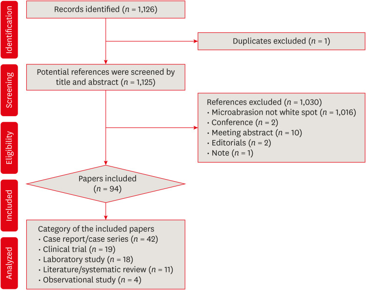
The initial search yielded 1,126 documents. Exclusions included 2 conference documents, 2 editorial documents, 10 meeting abstracts, 1 duplicate, and 1 note, as well as other documents that did not meet the study's objectives (Figure 1). Consequently, 94 articles were included in this bibliometric analysis (Supplementary Table 1).
Figure 1

Study selection protocol flowchart.

rde-49-e29-g001

Citation analysis

The selected articles received a total of 1,110 citations in the Web of Science Core Collection (WoS-CC), with self-citations accounting for 55% of these citations. The most cited article, “The effects of etching, microabrasion, and bleaching on surface enamel,” authored by Tong LSM and collaborators and published in the Journal of Dental Research, received 65 citations. Spearman’s correlation analysis showed a weak positive correlation between the number of citations and the journal’s impact factor (rho = 0.328) and a moderate negative correlation between the number of citations and the year of publication (rho = −0.615).

Publication year

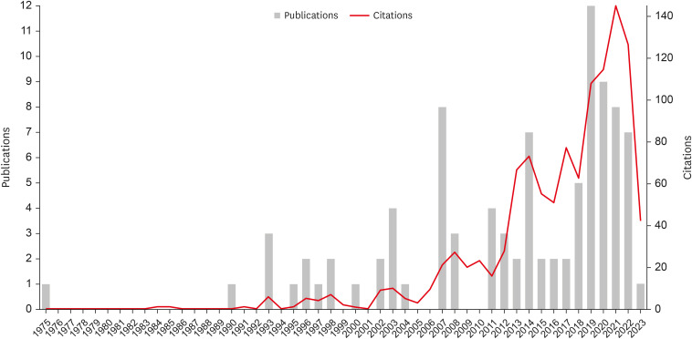
The earliest article, published in April 1975, was titled “Clinical evaluation of the sandpaper disk method for removing fluorosis stains from teeth” by Chandra S and Chawla TN in the Journal of The American Dental Association. The most recent article, “Microabrasion in the management of enamel discolorations in pediatric dentistry: a systematic review,” by Blanchet I and collaborators, was published in January 2023 in the Journal of Clinical Pediatric Dentistry. Most articles (n = 56) were published in the last decade (2014–2023). Figure 2 provides a detailed breakdown of the number of publications and citations per year.
Figure 2

Distribution of the number of publications over the years.

rde-49-e29-g002

Main journals and impact factor

Table 1 lists the main journals and their respective impact factors (2021) that published articles on enamel microabrasion. Operative Dentistry (n = 10) had the most publications, followed by Quintessence International (n = 7), Journal of the American Dental Association (n = 6), and Journal of Esthetic and Restorative Dentistry (n = 6). The journals with the highest impact factors in 2021 were the Journal of Dental Research (IF: 8.924) with 1 article, the Journal of Dentistry (IF: 4.991) with 2 articles, and the Journal of Prosthetic Dentistry (IF: 4.148) with 3 articles.
Table 1

Main journals that published the studies about enamel microabrasion

rde-49-e29-i001
Source title Number of articles Impact factor
Operative Dentistry 10 2,937
Quintessence International 7 2,175
Journal of the American Dental Association 6 3,681
Journal of Esthetic and Restorative Dentistry 6 3,040
British Dental Journal 5 2,727
Journal of Clinical Pediatric Dentistry 5 1,338
American Journal of Dentistry 4 1,748
Journal of Prosthetic Dentistry 3 4,148
Angle Orthodontist 3 2,684

Study design and themes

The most common study design was case reports/series (n = 42), followed by interventional studies (n = 19), laboratory studies (n = 18), literature reviews (n = 6), observational studies (n = 4), and systematic reviews (n = 5). The predominant themes were: “Clinical performance of microabrasive materials” (n = 75), “Effects of microabrasion on dental enamel” (n = 9), “Description of microabrasion technique” (n = 3), and other themes (n = 7).
Regarding the microabrasion protocol, Opalustre (Ultradent Products Inc., South Jordan, UT, USA) was the most used material (n = 37), followed by comparisons between different materials (n = 17), pumice stone and 37% phosphoric acid (n = 13), Prema (Premiere Dental Prod. USA) (n = 8), hydrochloric acid and pumice stone (n = 6), hydrochloric acid alone (n = 3), acid and sandpaper disk (n = 1), diamond tip and acid (n = 1), and laser (n = 1). Seven articles did not test a specific microabrasion material or describe a clinical protocol. The most discussed white spot lesion was dental fluorosis (n = 41), followed by initial dental caries (n = 15), comparisons between different spots (n = 15), molar incisor hypomineralization (n = 8), unidentified stains (n = 10), and enamel hypoplasia (n = 5).

Countries and continents

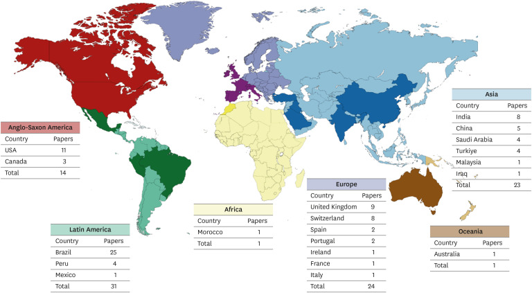
A total of 20 countries contributed to the literature on enamel microabrasion. The top 5 countries were Brazil (26 articles), the United States (11 articles), the United Kingdom (9 articles), Switzerland (8 articles), and India (8 articles). Latin America (n = 31) was the leading continent, followed by Europe (n = 24) and Asia (n = 23) (Figure 3).
Figure 3

Worldwide distribution of origin of publications on enamel microabrasion.

rde-49-e29-g003

Institutions

Fifty-nine institutions contributed to the articles on enamel microabrasion. Table 2 lists the top 10 institutions. The top 3 were Paulista State University (Brazil) with 10 articles, the University of Geneva (Switzerland) with 5 articles, and the University of Pennsylvania (United States) with 4 articles.
Table 2

Main institutions associated with research on enamel microabrasion

rde-49-e29-i002
Institution Country Number of articles
Paulista State University Brazil 10
University of Geneva Switzerland 5
University of Pennsylvania USA 4
Suleyman Demirel University Turkey 3
Campinas State University Brazil 3
University of London England 3
University of Sheffield United Kingdom 3
University of Zurich Switzerland 3

Contributing authors

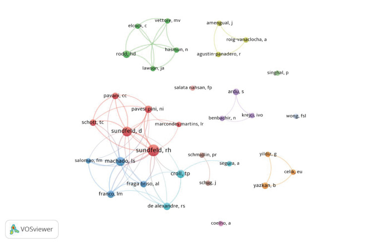
A total of 344 authors contributed to articles on dental microabrasion. Table 3 presents the authors with the highest number of publications. Sundfeld RH led with 10 articles, followed by Sundfeld D with 7 articles and Machado LS with 6 articles. Figure 4 shows the frequency and co-authorship relationships among the authors.
Table 3

Authors with the highest number of publications on enamel microabrasion

rde-49-e29-i003
Authors Number of articles Number of citations
Sundfeld RH 10 173
Sundfeld D 7 132
Machado LS 6 73
Croll TP 5 121
De Alexandre RS 4 95
Rood HD 4 89
Franco LM 4 69
Pavesi Pini NI 4 46
Figure 4

Main groups/authors who researched enamel microabrasion. A minimum number of 2 occurrences (2 studies) per author was considered. The names associated with the highest points correspond to the most frequent authors. On the other hand, the names associated with the lowest points correspond to the authors with the lowest occurrence. The lines and the same color between the dots indicate collaboration and groups of authors.

rde-49-e29-g004

Keywords

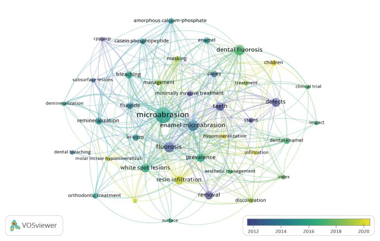
A total of 268 keywords were identified. The most prevalent were “microabrasion” (37 occurrences), “enamel microabrasion” (17 occurrences), “fluorosis” and “dental fluorosis” (16 occurrences each). Figure 5 illustrates the most prevalent keywords and their collaborative relationships.
Figure 5

Frequency and interaction of the main keywords associated with the study. Minimum number of occurrence of keywords: 3 studies. In the highest points are the keywords that appeared more often, on the other hand, the words that appear less often are associated with the lowest points. The lines that unite these points indicate the relationship and use of these words in the same studies. Regarding the colors, the words associated with the foci with colors close to blue, are older terms and closer to yellow, more recent terms, as described in the image scale.

rde-49-e29-g005
Based on Google Trends data, the search popularity of “enamel microabrasion” has remained constant over the past 20 years. High interest was noted in Mexico, followed by Colombia, Peru, Australia, and Argentina. Common related search topics included “dentistry,” “tooth,” “therapy,” and “cost.” Popular search queries were “microabrasion,” “dental microabrasion,” “enamel,” “enamel microabrasion,” and “abrasion.”

DISCUSSION

Bibliometric analyses have gained significant popularity in research in recent years [13]. This notoriety can be primarily attributed to their methodological approach, which encompasses large volumes of data and has a substantial impact on research [14]. The objective of this bibliometric analysis was to examine all articles related to enamel microabrasion for the treatment of white spot lesions. Scientific research on enamel microabrasion primarily includes case reports/series, focusing on the clinical performance of abrasive materials. Regarding the microabrasion protocols used in the studies, it was observed that the most commonly used material was Opalustre (Ultradent Products Inc.), especially for treating white spots caused by dental fluorosis.
Citation analysis is a commonly recognized metric for assessing the relative impact of published research within a scientific field. Articles with a higher number of citations in a given area are considered “citation classics” [15]. In broad fields of knowledge, articles typically need to exceed 100 or 200 citations to be considered classics [11]. However, as observed in this bibliometric analysis, due to the specific nature of this topic within dentistry and the relatively low number of published articles, the most cited article had 65 citations, despite being published 30 years ago. Thus, this study can be considered a classic within the topic of enamel microabrasion for the treatment of white spots [16]. This can be justified by the study’s evaluation of conservative techniques for treating enamel white spots, a topic of great concern in microabrasion. Improperly performed microabrasion can cause irreversible loss of tooth enamel.
Considering the exclusion criteria of this study, we excluded conference and editorial articles, which do not undergo a peer review process before publication, so it is questionable whether the findings of these studies can be used to guide clinical practice [1718]. The inconsistency of conference articles before and after publication also reduces their authenticity and reliability [19]. Thus, considering that these articles could contain inadequate and unreliable information, they were excluded from this bibliometric analysis.
Created by Kane in 1926, one of the first scientific reports on enamel microabrasion describes this technique for removing stains caused by dental fluorosis through the application of an acid solution composed of a mixture of hydrogen peroxide, hydrochloric acid, and anesthetic ether. Following this, a fine-grit sandpaper disk was used on a straight handpiece. This disk was positioned and driven on the discolored enamel surface, which was previously moistened with the acid solution [20]. Due to its pioneering role in scientifically documenting a mechanical technique for the microabrasion of dental enamel to treat white spots, this was the oldest article identified in WoS-CC. However, given the increased risk of excessive wear on the tooth structure associated with the combination of an acid solution and an abrasive sandpaper disk, this technique was not reproduced in subsequent scientific studies.
Most studies were clinical case reports or case series. According to Goebel et al. [21] in their bibliometric study on dental fluorosis, the diverse presentation of white spot lesions makes it difficult to standardize samples for conducting randomized clinical studies. Nonetheless, intervention studies were the second most prevalent study design. However, a considerably low number of systematic reviews were identified. Systematic reviews play an important critical role, providing syntheses of the state of scientific knowledge on a specific topic and addressing issues insufficiently clarified by primary studies [22]. Therefore, systematic reviews on dental enamel microabrasion for the treatment of white spot lesions should be encouraged.
Several protocols have been developed to assist in the treatment of white spot lesions to aesthetically satisfy patients [5]. This justifies the great emphasis on the theme “clinical performance of microabrasive materials.” Enamel microabrasion was first performed in the 1970s using an acid solution combined with a sandpaper disk [20]. In 1982, the combination of pumice stone with 36% hydrochloric acid was proposed, creating a paste that was applied using a rubber cup at low rotation [23]. Due to the high concentration of acid and the associated risks to the patient and dental enamel structure, the use of this mixture was modified to incorporate 18% hydrochloric acid [4]. Additionally, the use of a diamond bur with extra fine grain was recommended before using microabrasive agents to reduce the clinical time of this procedure [24].
The use of 35% phosphoric acid combined with abrasive agents (pumice stone) as a replacement for hydrochloric acid was proposed by Kamp in 1989, being considered an advantageous and accessible mixture since these materials are commonly used in clinical practice for other procedures [25]. However, a higher concentration of acids and the number of abrasives can increase the amount of enamel removed [26]. As a result, pre-dosed commercial products with lower acid and abrasive content were developed. The mixture of 10% hydrochloric acid with silicon carbide particles led to the creation of Prema Compound, the first commercially available abrasive paste [5]. Considering the growing concern of minimal intervention dentistry to preserve healthy tooth structure, Opalustre (Ultradent Products Inc.) was developed, a new commercial paste with an even lower concentration of hydrochloric acid, approximately 6.6% [5]. Consequently, this was the material most used in research on enamel microabrasion.
Different types of white spot lesions were treated using dental enamel microabrasion in the scientific studies identified in this review. However, it was observed that most studies utilized this procedure for the aesthetic treatment of dental fluorosis. This may be associated with the high prevalence of this condition (65%) [27].
The continent responsible for the greatest scientific development in enamel microabrasion was Latin America. Corroborating this data, it was observed that the most prominent country was Brazil and consequently the most prevalent institution was Paulista State University. Another fact that highlights these data is that the 3 authors who published the most on enamel microabrasion came from Paulista State University. Sundfeld RH, Sundfeld D, and Machado LS worked together, dedicating their research to primarily addressing the clinical performance of different enamel microabrasion protocols in various types of white spots, mainly through case reports and series.
This bibliometric analysis has several strengths, notably its exclusive focus on enamel microabrasion, a topic not previously addressed in the literature through this type of analysis. Additionally, no year or language filters were applied, ensuring the most comprehensive coverage of relevant studies. However, a limitation of this study is the use of a single database (WoS-CC). Despite this, the WoS-CC is regarded as one of the primary databases for conducting such studies and has been utilized in numerous other published bibliometric analyses in dentistry [282930].

CONCLUSIONS

Bibliometric analysis can make it possible to observe trends in the literature, draw conclusions about the impact of studies on clinical practice, and, above all, direct research and funding efforts. Thus, it can be concluded that the scientific background of enamel microabrasion for the treatment of white spots is based mainly on case reports and series. The most addressed white spot lesion in the studies was dental fluorosis, and the main objective of the studies was to evaluate the clinical performance of this procedure. The most frequent protocol uses Opalustre (Ultradent Products Inc.) as the material of choice for this procedure. In addition, it was observed that the Paulista State University, located in Brazil, was the main identified research center on enamel microabrasion.

Notes

Funding: This study was financed in part by the Coordenação de Aperfeiçoamento de Pessoal de Nivel Superior – Brazil (CAPES), grant number 001.

Conflict of Interest: No potential conflict of interest relevant to this article was reported.

Author Contributions:

  • Conceptualization: De Oliveira Rocha A, Cardoso K, Goebel MC, Santos PS, Dos Anjos LM.

  • Data curation: Cardoso K.

  • Formal analysis: De Oliveira Rocha A, Dos Anjos LM.

  • Investigation: Goebel MC.

  • Methodology: De Oliveira Rocha A, Cardoso K, Santos PS, Ribeiro JS, Santana CM, Cardoso M.

  • Software: De Oliveira Rocha A, Goebel MC, Dos Anjos LM.

  • Writing - original draft: De Oliveira Rocha A, Cardoso K, Goebel MC, Santos PS, Dos Anjos LM, Ribeiro JS, Santana CM, Cardoso M.

  • Writing - review & editing: Santos PS, Ribeiro JS, Santana CM, Cardoso M.

References

1. Perdigão J. Resin infiltration of enamel white spot lesions: an ultramorphological analysis. J Esthet Restor Dent. 2020; 32:317–324. PMID: 31742888.
2. Gupta A, Dhingra R, Chaudhuri P, Gupta A. A comparison of various minimally invasive techniques for the removal of dental fluorosis stains in children. J Indian Soc Pedod Prev Dent. 2017; 35:260–268. PMID: 28762354.
3. Bernardi LG, Favoreto MW, de Souza Carneiro T, Borges CP, Pulido C, Loguercio AD. Effects of microabrasion association to at-home bleaching on hydrogen peroxide penetration and color change. J Esthet Restor Dent. 2022; 34:335–341. PMID: 34723443.
4. Croll TP, Cavanaugh RR. Enamel color modification by controlled hydrochloric acid-pumice abrasion. II. Further examples. Quintessence Int. 1986; 17:157–164. PMID: 3458266.
5. Pini NI, Sundfeld-Neto D, Aguiar FH, Sundfeld RH, Martins LR, Lovadino JR, et al. Enamel microabrasion: an overview of clinical and scientific considerations. World J Clin Cases. 2015; 3:34–41. PMID: 25610848.
6. Bahadir HS, Haberal M, Çelik Ç. Effect of microabrasion on the staining susceptibility of enamel: an in vitro study. J Dent Res Dent Clin Dent Prospect. 2022; 16:95–100.
7. Sundfeld D, Pavani CC, Pini N, Machado LS, Schott TC, Sundfeld RH. Enamel microabrasion and dental bleaching on teeth presenting severe-pitted enamel fluorosis: a case report. Oper Dent. 2019; 44:566–573. PMID: 30702410.
8. Elkhazindar MM, Welbury RR. Enamel microabrasion. Dent Update. 2000; 27:194–196. PMID: 11218456.
9. Croll TP. Enamel microabrasion: 10 years experience. Asian J Aesthet Dent. 1995; 3:9–15. PMID: 9063104.
10. Sundfeld RH, Croll TP, Briso AL, de Alexandre RS, Sundfeld Neto D. Considerations about enamel microabrasion after 18 years. Am J Dent. 2007; 20:67–72. PMID: 17542197.
11. Karobari MI, Maqbool M, Ahmad P, Abdul MS, Marya A, Venugopal A, et al. Endodontic microbiology: a bibliometric analysis of the top 50 classics. BioMed Res Int. 2021; 2021:6657167. PMID: 34746305.
12. Al-Moraissi EA, Christidis N, Ho YS. Publication performance and trends in temporomandibular disorders research: a bibliometric analysis. J Stomatol Oral Maxillofac Surg. 2023; 124:101273. PMID: 36057419.
13. Donthu N, Kumar S, Mukherjee D, Pandey N, Lim WM. How to conduct a bibliometric analysis: an overview and guidelines. J Bus Res. 2021; 133:285–296.
14. Ahmad P, Slots J. A bibliometric analysis of periodontology. Periodontol 2000. 2021; 85:237–240. PMID: 33226679.
15. Van Noorden R, Maher B, Nuzzo R. The top 100 papers. Nature. 2014; 514:550–553. PMID: 25355343.
16. Tong LS, Pang MK, Mok NY, King NM, Wei SH. The effects of etching, micro-abrasion, and bleaching on surface enamel. J Dent Res. 1993; 72:67–71. PMID: 8418110.
17. Schmucker CM, Blümle A, Schell LK, Schwarzer G, Oeller P, Cabrera L, et al. Systematic review finds that study data not published in full text articles have unclear impact on meta-analyses results in medical research. PLoS One. 2017; 12:e0176210. PMID: 28441452.
18. Scherer RW, Saldanha IJ. How should systematic reviewers handle conference abstracts? A view from the trenches. Syst Rev. 2019; 8:264. PMID: 31699124.
19. Wong FS, Winter GB. Effectiveness of microabrasion technique for improvement of dental aesthetics. Br Dent J. 2002; 193:155–158. PMID: 12213009.
20. Chandra S, Chawla TN. Clinical evaluation of the sandpaper disk method for removing fluorosis stains from teeth. J Am Dent Assoc. 1975; 90:1273–1276. PMID: 1056394.
21. Goebel MC, Rocha AO, Santos PS, Bolan M, Martins-Júnior PA, Santana CM, et al. A bibliometric analysis of the top 100 most-cited papers concerning dental fluorosis. Caries Res. 2023; 57:509–515. PMID: 37100040.
22. Page MJ, McKenzie JE, Bossuyt PM, Boutron I, Hoffmann TC, Mulrow CD, et al. The PRISMA 2020 statement: an updated guideline for reporting systematic reviews. BMJ. 2021; 372:n71. PMID: 33782057.
23. Murrin JR, Barkmeier WW. Chemical treatment of endemic dental fluorosis. Quintessence Int Dent Dig. 1982; 13:363–369. PMID: 6955830.
24. Croll TP. Hastening the enamel microabrasion procedure eliminating defects, cutting treatment time. J Am Dent Assoc. 1993; 124:87–90.
25. Kamp AA. Removal of white spot lesions by controlled acid-pumice abrasion. J Clin Orthod. 1989; 23:690–693. PMID: 2639881.
26. Donly KJ, O’Neill M, Croll TP. Enamel microabrasion: a microscopic evaluation of the “abrosion effect”. Quintessence Int. 1992; 23:175–179. PMID: 1641458.
27. Neurath C, Limeback H, Osmunson B, Connett M, Kanter V, Wells CR. Dental fluorosis trends in US oral health surveys: 1986 to 2012. JDR Clin Trans Res. 2019; 4:298–308. PMID: 30931722.
28. Rocha AO, Anjos LM, Vitali FC, Santos PS, Bolan M, Santana CM, et al. Tooth bleaching: a bibliometric analysis of the top 100 most-cited papers. Braz Dent J. 2023; 34:41–55. PMID: 37194856.
29. Baldiotti AL, Amaral-Freitas G, Barcelos JF, Freire-Maia J, Perazzo MF, Freire-Maia FB, et al. The top 100 most-cited papers in cariology: a bibliometric analysis. Caries Res. 2021; 55:32–40. PMID: 33341798.
30. Dos Anjos LM, Rocha AO, Magrin GL, Kammer PV, Benfatti CA, Matias de Souza JC, et al. Bibliometric analysis of the 100 most cited articles on bone grafting in dentistry. Clin Oral Implants Res. 2023; 34:1198–1216. PMID: 37577958.

SUPPLEMENTARY MATERIAL

Supplementary Table 1

Identified papers about white spot enamel microabrasion
rde-49-e29-s001.xls
TOOLS
Similar articles