Journal List > Korean Circ J > v.54(9) > 1516088318

Kim, Ko, Lee, Ahn, Rha, Choi, Min, Park, Jang, Youn, Kang, Yoon, and Choi: Korean Multicenter Registry of ELUVIA Stent for Femoropopliteal Artery Disease: K-ELUVIA Registry

Author's summary

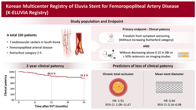
The K-ELUVIA study assessed Eluvia™, a paclitaxel-eluting stent, in treating femoropopliteal artery disease across 7 Korean centers. Enrolling 105 patients undergoing endovascular treatment, our prospective 2-year study revealed promising outcomes. Clinical patency at 2 years stood at 76.3%, with freedom from target lesion revascularization at 79.1%. Noteworthy predictors of clinical patency loss included chronic total occlusion and stent diameter. This study affirms Eluvia’s favorable clinical outcomes in real-world femoropopliteal interventions.

Abstract

Background and Objectives

The K-ELUVIA study aimed to investigate the clinical effectiveness and safety of Eluvia™, a polymer-coated, paclitaxel-eluting stent, for femoropopliteal artery disease using data from a prospective Korean multicenter registry.

Methods

A total of 105 patients with femoropopliteal artery disease who received endovascular treatment (EVT) with Eluvia™ stents at 7 Korean sites were enrolled in a prospective cohort and followed for 2 years. The primary endpoint was the 2-year clinical patency. The secondary endpoint was 2-year freedom from clinically driven target lesion revascularization (TLR).

Results

Mean patient age was 68.2±10.4 years, and most patients (82.7%) were male. Mean lesion length was 168.3±117.6 mm. Chronic total occlusion was found in 57.7% of patients. Trans-Atlantic Inter-Society Consensus for the Management of Peripheral Arterial Disease (TASC II) type C or D lesions were present in 46.1% of patients. Procedural success was achieved in 99.0% of patients. The clinical patency rate was 84.4% at 1 year after EVT and 76.3% at 2 years post-EVT. The freedom from TLR rate was 89.1% at 1 year after EVT and 79.1% at 2 years post-EVT. Chronic total occlusion (hazard ratio [HR], 3.53; 95% confidence interval [CI], 1.08–11.67; p=0.039) and smaller mean stent diameter (HR, 0.40; 95% CI, 0.16–0.98; p=0.044) were identified as independent predictors of loss of clinical patency at 2 years.

Conclusions

The K-ELUVIA study demonstrated favorable 2-year clinical effectiveness and safety outcomes of Eluvia stent for femoropopliteal artery lesions in real-world practice.

Graphical Abstract

kcj-54-565-abf001.jpg

INTRODUCTION

The femoropopliteal artery is the most commonly affected vessel in patients with peripheral artery disease undergoing endovascular treatment (EVT).1) However, EVT using conventional uncoated balloons and bare metal stents (BMS) is associated with relatively high rates of restenosis, especially in complex femoropopliteal lesions. Various forces generated by leg movement, such as axial and radial compression, bending, and torsion, can affect post-EVT outcomes.2)3) To reduce restenosis after EVT for femoropopliteal disease, paclitaxel-based drug-eluting stents (DES) and drug-coated balloons were developed and introduced for clinical use.4)5) Zilver PTX™ (Cook Medical, Bloomington, IN, USA), a polymer-free paclitaxel-coated nitinol DES, was the first DES demonstrating superior primary patency, compared to conventional balloon angioplasty or BMS, in a randomized controlled trial.5)6) Despite favorable outcomes reported in several registry studies, the effectiveness of Zilver PTX™ in real-world settings remains inconsistent.7)8)9)10)
The Eluvia™ (Boston Scientific, Marlborough, MA, USA) DES is a recently introduced and was designed to release paclitaxel over 6 months, facilitated by a biocompatible fluoropolymer coating. In the IMPERIAL study (a randomized controlled trial), Eluvia for femoropopliteal artery lesions demonstrated non-inferiority to Zilver PTX for both efficacy and safety at 12 months, as well as superiority over Zilver PTX for primary patency in a post-hoc analysis.11) However, the lesions included in the IMPERIAL study were relatively short, with a mean length of approximately 8 cm. According to a Korean registry study, more than 50% of femoropopliteal lesions were classified as Trans-Atlantic Inter-Society Consensus for the Management of Peripheral Arterial Disease (TASC II) type C or D.1) Whether Eluvia is still able to achieve favorable clinical outcomes in complex femoropopliteal lesions during real-world practice remains uncertain. Thus, the objective of the present study (called K-ELUVIA) was to investigate the clinical efficacy and safety of the Eluvia stent for femoropopliteal artery disease using data from a prospective multicenter registry.

METHODS

Ethical statement

The study was conducted in accordance with the ethical principles of the Declaration of Helsinki (2013) and was approved by the Institutional Review Board (IRB) at each participating center (Severance Hospital IRB number: 1-2015-0075). It was registered at www.clinicaltrials.gov (NCT03505931). All patients provided written informed consent before enrollment.

Study population and enrollment

The K-ELUVIA was designed as a single-arm, prospective, multicenter trial to assess the efficacy and safety of Eluvia stents for the treatment of femoropopliteal artery disease. The study was conducted at 7 endovascular centers in South Korea. Patients were eligible for participation if all of the following criteria were met: age ≥19 years, Rutherford category 2 to 5, stenotic (≥50% diameter stenosis) or occlusive femoropopliteal artery lesions, and an ankle-brachial index (ABI) <0.9. The exclusion criteria were age >85 years; acute severe limb ischemia or severe chronic limb-threatening ischemia (CLTI; Rutherford category 6); in-stent restenosis of a previously implanted stent; lesion previously treated with a bypass graft; untreated proximal inflow disease of the ipsilateral iliac artery (>50% stenosis or occlusion); uncontrolled congestive heart failure or left ventricular ejection fraction ≤40%; severe hepatic dysfunction (aspartate aminotransferase or alanine aminotransferase ≥3-fold higher than the upper limit of normal); significant leukopenia, neutropenia, thrombocytopenia, anemia, or risk of bleeding; pregnant or fertile female; life expectancy <1 year; or known allergic reaction to heparin, aspirin, clopidogrel, or contrast agent. The following was obtained for each patient prior to study enrollment: medical history, baseline medications, Rutherford classification, ABI, the results of ≥1 lower extremity imaging study (e.g., computed tomography [CT], magnetic resonance imaging, duplex ultrasonography, catheter angiography), TASC II type, and standard laboratory test results.

Endovascular treatment

Loading doses of aspirin 300 mg and clopidogrel 300 mg were administered ≥12 hours before EVT to all patients who were not receiving aspirin and clopidogrel for ≥5 days prior to the procedure. During EVT, all patients received local anesthesia, which was supplemented with intravenous sedation and analgesia, as needed. After insertion of the introducer sheath, unfractionated heparin 5,000 U was administered intravenously, and the activated coagulation time was maintained above 250 seconds with additional doses of heparin, if required. Vascular access was achieved percutaneously using either the ipsilateral antegrade approach or the contralateral cross-over approach from the common femoral artery. A 6 or 7-Fr. TERUMO Inc. sheath (TERUMO Inc., Tokyo, Japan) was employed for the ipsilateral approach, and a 6 or 7-Fr. Balkin or Ansel sheath (Cook Inc., Bloomington, IN, USA) was used for the contralateral approach. A 0.018 or 0.035-inch guidewire was used to cross the target lesion. When occlusion was complete, either an intraluminal or subintimal approach was used for revascularization, at the operator’s discretion. After successful passage of the guidewire, target lesions were routinely predilated using a balloon with a diameter equal to or smaller than the reference diameter. Thereafter, Eluvia stent(s) were implanted to cover the target lesion. The stent diameter was selected based on the target lesion reference diameter. Post-stent dilation was performed when the residual stenosis was >30%. After EVT, dual antiplatelet therapy (aspirin 100 mg/day and clopidogrel 75 mg/day) was recommended for ≥6 months, after which single antiplatelet therapy with aspirin or clopidogrel was used.

Follow-up

Following EVT, patients underwent regular assessment of symptoms based on Rutherford classification at 1 month after the procedure and then every 6 months for 2 years. ABI measurements were obtained at 6, 12, and 24 months post-procedure or whenever deterioration in symptom status was observed. Imaging evaluation (e.g., duplex ultrasonography, CT angiography, invasive arteriography) was recommended at 12 months post-EVT or whenever symptoms worsened by ≥1 Rutherford category or ABI decreased by >0.15.

Endpoints and definitions

The primary study endpoint was initially defined as 12-month primary patency, determined by imaging studies such Duplex ultrasound, CT angiography, or catheter angiography. However, due to the challenges posed by the coronavirus disease 2019 (COVID-19) pandemic and patient apprehension regarding attendance for required imaging studies, the primary endpoint was modified to best reflect the clinical status of the trial subjects. Consequently, the primary endpoint was revised to 2-year clinical patency. This was defined as freedom from symptom worsening, indicated by an increase in Rutherford category accompanied by a ≥0.15 decrease in ABI or >50% restenosis on imaging studies. A lesion or adjacent-segment peak systolic velocity ratio >2.4, as measured by duplex ultrasonography, was considered indicative of >50% restenosis. The secondary study endpoint was the 2-year freedom from clinically driven target lesion revascularization (TLR), defined as any repeat intervention or surgical treatment for restenotic lesions characterized by both worsening symptoms and a ≥0.15 decrease in ABI. Procedural success was defined as <30% residual stenosis plus no evidence of arterial dissection causing flow limitation. Angiography findings were used to categorize arterial calcification severity according to the Peripheral Arterial Calcium Scoring System (PACSS)12): grade 1, mild calcification; grade 2 or 3, moderate calcification; and grade 4, severe calcification.

Statistical analysis

Continuous variables are presented as mean ± standard deviation, and categorical variables are shown as frequency and percentage. Baseline clinical and lesion characteristics were analyzed using the Student’s t-test for continuous data and the χ2 and Fisher’s exact test for categorical data. Primary and secondary endpoints were assessed using Kaplan-Meier survival analysis, with the log-rank test used to compare groups, as appropriate. Predictors of loss of clinical patency were evaluated using Cox proportional hazard regression analyses. Sex, body mass index, age, and variables with a p value <0.2 in univariable Cox regression analysis were included in the multivariable Cox regression model. The p values <0.05 were considered statistically significant. All statistical analyses were performed using SPSS 25.0 software (IBM Corp., Armonk, NY, USA) and R version 4.2.0 (R Foundation for Statistical Computing, Vienna, Austria).

RESULTS

Study population and baseline characteristics

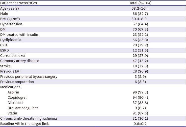
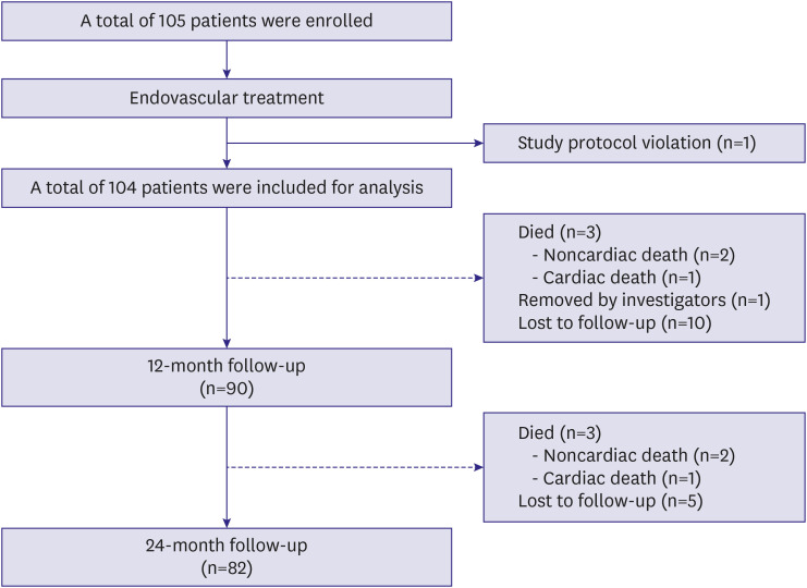
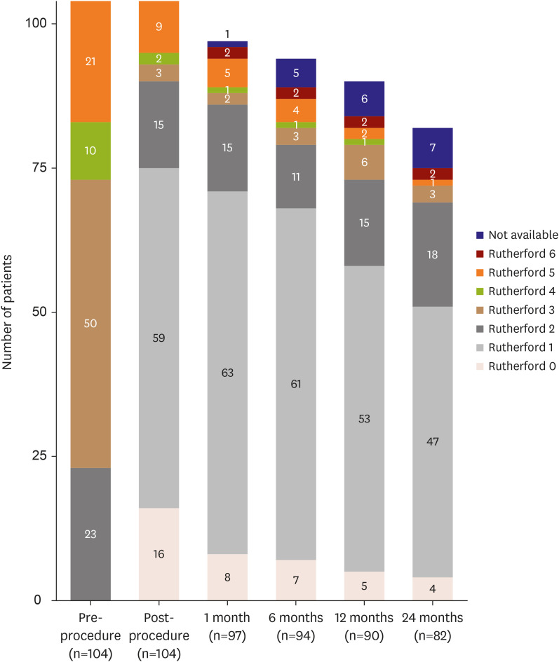
A total of 105 patients were enrolled in the K-ELUIVA study from May 2018 to March 2021. One patient was excluded because inclusion criteria were not met, with the remaining 104 patients included in the final analysis (Figure 1). Baseline patient characteristics are summarized in Table 1. The mean patient age was 68.2±10.4 years, and the majority of patients (82.7%) were male. Hypertension and diabetes were present in 64.4% and 67.3% of patients, respectively. Chronic kidney disease was present in 19.2% of patients, and 11.5% of all patients required hemodialysis for end-stage renal disease (ESRD). The most frequent clinical presentation of femoropopliteal artery disease was severe claudication (Rutherford category 3, 48.5%; Figure 2). The pre-procedural mean ABI in the treated extremity was 0.6±0.2.
Figure 1

Patient flow chart of the K-ELUVIA study.

kcj-54-565-g001
Table 1

Baseline patient characteristics

kcj-54-565-i001
Patient characteristics Total (n=104)
Age (years) 68.2±10.4
Male 86 (82.7)
BMI (kg/m2) 30.4±8.9
Hypertension 67 (64.4)
DM 70 (67.3)
DM treated with insulin 23 (22.1)
Dyslipidemia 56 (53.8)
CKD 20 (19.2)
ESRD 12 (11.5)
Current smoker 29 (27.9)
Coronary artery disease 47 (45.2)
Stroke 18 (17.3)
Previous EVT 28 (26.9)
Previous peripheral bypass surgery 3 (2.9)
Previous amputation 6 (5.8)
Medications
Aspirin 96 (92.3)
Clopidogrel 94 (90.4)
Cilostazol 37 (35.6)
Oral anticoagulant 9 (8.7)
Statin 91 (87.5)
Chronic limb-threatening ischemia 31 (30.1)
Baseline ABI in the target limb 0.6±0.2
Data are mean ± standard deviation or number (percentage).
ABI = ankle-brachial index; BMI = body mass index; CKD = chronic kidney disease; DM = diabetes mellitus; ESRD = end-stage renal disease; EVT = endovascular treatment.
Figure 2

Distribution of Rutherford categories before endovascular treatment, after the procedure, and during follow-up.

kcj-54-565-g002

Lesion characteristics and procedural data

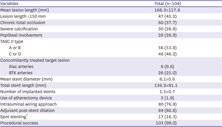
Lesion and procedural characteristics are summarized in Table 2. The mean lesion length was 168.3±117.6 mm. A totally occluded lesion was treated in 57.7% of patients. Complex lesions (TASC II type C or D lesions) were present in 46.1% of patients, and 26.9% of patients had concurrent popliteal artery involvement. The procedural success rate was 99.0%. Only 1 patient had a suboptimal result, characterized by slow antegrade flow after stent implantation because of distal thrombus embolization.
Table 2

Baseline lesion characteristics and procedural data

kcj-54-565-i002
Variables Total (n=104)
Mean lesion length (mm) 168.3±117.6
Lesion length >150 mm 47 (45.2)
Chronic total occlusion 60 (57.7)
Severe calcification 30 (28.8)
Popliteal involvement 28 (26.9)
TASC II type
A or B 56 (53.8)
C or D 48 (46.2)
Concomitantly treated target lesion
Iliac arteries 6 (9.6)
BTK arteries 26 (25.0)
Mean stent diameter (mm) 6.1±0.6
Total stent length (mm) 136.2±81.1
Number of implanted stents 1.5±0.7
Use of atherectomy device 2 (1.9)
Intraluminal wiring approach 80 (76.9)
Adjuvant post-stent dilation 84 (80.8)
Spot stenting* 17 (16.3)
Procedural success 103 (99.0)
Data are mean ± standard deviation or number (percentage).
BTK = below the knee; TASC II = Trans-Atlantic Inter Society Consensus for the Management of Peripheral Arterial Disease.
*Total length of stents/length of lesion <0.5.
The intraluminal wiring approach was utilized in 76.9% of patients. Mean total stent length and diameter were 136.2±81.1 mm and 6.1±0.6 mm, respectively. Adjuvant post-stent dilation was performed in 80.8% of patients. One patient developed arterial rupture with nonfatal bleeding, which was successfully managed by implanting a covered stent. During hospitalization, 1 patient died because of sepsis secondary to an infected foot wound.

Clinical outcomes

The median duration of clinical follow-up was 530 days (interquartile range, 369–718 days). Of the 104 patients included in this study, 82 completed the 2-year follow-up. There were 6 mortalities (2 cardiovascular and 4 noncardiovascular deaths), and 15 patients were lost to follow-up. Two of participants required minor amputation due to the deterioration of foot wounds despite undergoing repeated EVT, but there were no cases of major amputation. Imaging studies were conducted in 42.3% of patients at 12 months. Patient symptom status improved from a median Rutherford category of 3 pre-procedure to 1 after EVT. This improvement was sustained for 24 months (Figure 2).
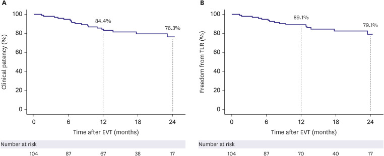
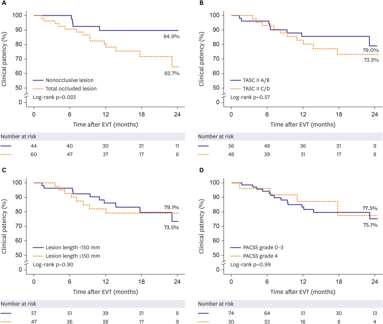
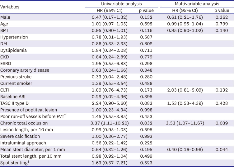
The primary outcome of clinical patency was 84.4% at 1 year after EVT and 76.3% at 2 years post-EVT (Figure 3A). The secondary outcome of freedom from clinically driven TLR was 89.1% at 1 year after EVT and 79.1% at 2 years post-EVT (Figure 3B). The clinical patency rate was higher in nonocclusive lesions than in chronic total occlusions (89.7% vs. 64.5%, p=0.023; Figure 4A). However, complex lesions (TASC II type C/D vs. type A/B; Figure 4B), long lesions (≥150 mm vs. <150 mm; Figure 4C), and severely calcified lesions (PACSS grade 4 vs. grade 0–3; Figure 4D) were not significantly associated with clinical patency during follow-up. In multivariable Cox regression analysis, chronic totally occluded lesions (hazard ratio [HR], 3.53; 95% confidence interval [CI], 1.08–11.67; p=0.039) and mean stent diameter (HR, 0.40; 95% CI, 0.16–0.98; p=0.044) were identified as independent predictors of loss of clinical patency at 2 years (Table 3).
Figure 3

Kaplan-Meier curves and estimates of clinical patency over time (A) and TLR free–survival (B).

EVT = endovascular treatment; TLR = target lesion revascularization.
kcj-54-565-g003
Figure 4

Kaplan-Meier curves and estimates of clinical patency over time according to lesion characteristics. Total occluded lesion (A), TASC II classification (B), lesion length (C), and calcification (D).

EVT = endovascular treatment; PACSS = Peripheral Arterial Calcium Scoring System; TASC II = Trans-Atlantic Inter-Society Consensus for the Management of Peripheral Arterial Disease.
kcj-54-565-g004
Table 3

Univariable and multivariable Cox proportional hazard analyses for predictors of loss of clinical patency

kcj-54-565-i003
Variables Univariable analysis Multivariable analysis
HR (95% CI) p value HR (95% CI) p value
Male 0.47 (0.17–1.32) 0.152 0.61 (0.21–1.76) 0.362
Age 1.01 (0.97–1.05) 0.695 0.99 (0.95–1.04) 0.799
BMI 0.95 (0.90–1.01) 0.116 0.95 (0.90–1.02) 0.140
Hypertension 0.78 (0.31–1.93) 0.587
DM 0.88 (0.33–2.33) 0.802
Dyslipidemia 0.84 (0.34–2.08) 0.711
CKD 0.84 (0.24–2.89) 0.779
ESRD 1.95 (0.55–6.83) 0.298
Coronary artery disease 0.63 (0.24–1.66) 0.348
Previous stroke 0.33 (0.04–2.48) 0.280
Current smoker 1.39 (0.55–3.54) 0.488
CLTI 1.89 (0.76–4.73) 0.173 2.03 (0.81–5.09) 0.132
Baseline ABI 0.29 (0.02–4.96) 0.395
TASC II type D 2.24 (0.90–5.60) 0.083 1.53 (0.53–4.39) 0.428
Presence of popliteal lesion 1.00 (0.23–4.34) 0.998
Poor run-off vessels before EVT* 1.45 (0.55–3.85) 0.453
Chronic total occlusion 3.37 (1.11–10.20) 0.032 3.53 (1.07–11.67) 0.039
Lesion length, per 10 mm 0.99 (0.95–1.03) 0.595
Severe calcification 1.00 (0.36–2.77) 0.993
Intraluminal approach 0.56 (0.22–1.42) 0.222
Mean stent diameter, per 1 mm 0.64 (0.32–1.26) 0.195 0.40 (0.16–0.98) 0.044
Total stent length, per 10 mm 0.98 (0.92–1.04) 0.499
Spot stenting 1.63 (0.37–7.21) 0.523
ABI = ankle-brachial index; BMI = body mass index; CI = confidence interval; CKD = chronic kidney disease; CLTI = chronic limb-threatening ischemia; DM = diabetes mellitus; ESRD = end-stage renal disease; EVT = endovascular treatment; HR = hazard ratio; TASC II = Trans-Atlantic Inter-Society Consensus for the Management of Peripheral Arterial Disease.
*>2 tibial arterial lesions with >50% stenosis.
Total length of stents/length of lesion <0.5.
One patient treated with subintimal angioplasty and Eluvia implantation developed an asymptomatic aneurysmal change in the target lesion, as detected on follow-up CT (Supplementary Figure 1). Three patients were found to have a hypoechoic halo surrounding the Eluvia stent on 1-year follow-up duplex ultrasonography (Supplementary Figure 2), which also was not associated with any symptoms.

DISCUSSION

In this single-arm prospective registry study, Eluvia, a self-expandable polymer-based paclitaxel-eluting stent, provided excellent clinical outcomes in terms of 2-year clinical patency (76.3%) and freedom from clinically driven TLR (79.1%).
The Eluvia is a fluoropolymer-based low-dose paclitaxel-eluting stent designed to release paclitaxel over 6 months. The clinical efficacy of Eluvia for femoropopliteal artery disease has been investigated in 2 randomized controlled trials. The EMINENT trial demonstrated superior 1-year primary patency with Eluvia stents than with BMS. At 1-year follow-up, Eluvia was associated with a primary patency rate of 83.2% and a TLR rate of 8.9%.13) In the IMPERIAL study, Eluvia was not only noninferior to but also superior to Zilver PTX (a nonpolymer-based paclitaxel-eluting DES) in terms of 12-month primary patency rate (86.8% vs. 81.5%).11)14) The results of IMPERIAL study suggested that even with a low paclitaxel dose, the polymer-based slow drug-releasing Eluvia stent more effectively prevents restenosis than the nonpolymer Zilver PTX stent.15)
Importantly, the mean lesion length in these 2 previous randomized controlled trials was <100 mm (75.6 mm in the EMINENT trial; 86.5 mm in the IMPERIAL trial), and the proportion of totally occluded lesions was relatively low (42.3% in the EMINENT trial; 31.2% in the IMPERIAL trial), compared with the proportion in real-world registries.1)15)16) Several prospective or retrospective registry studies have investigated the efficacy of the Eluvia stent for the treatment of more complex lesions. The largest study (in terms of sample size) was the CAPSICUM study, which was a prospective multicenter single-arm registry. It included a total of 1,204 limbs and documented 1-year clinical outcomes after femoropopliteal EVT with Eluvia of 87.1% primary patency rate and 93.8% TLR-free survival rate. These rates were comparable to those observed in the IMPERIAL trial, despite the mean lesion length being 186 mm, which was substantially longer than the length in the 2 randomized controlled trials.15) In a retrospective registry study, Stavroulakis et al.16) reported primary patency rates of 90% at 1 year and 71% at 2 years, as well as TLR-free survival rates of 94% at 1 year and 80% at 2 years, using Eluvia for femoropopliteal artery lesions with a mean length of 194 mm. The ULTIMATE study, a retrospective cohort study comparing the Viabahn stent graft (W L Gore & Associates Inc., Flagstaff, AZ, USA) and the Eluvia stent for long femoropopliteal artery lesions (mean lesion length, 230 mm) also found higher primary patency rates with Eluvia (75.9%) than with Viabahn (69.5%) at 24 months.17) Our current study, which included more complex femoropopliteal lesions than the randomized trials in terms of longer mean lesion length (168.3 mm) and a higher proportion of totally occluded lesions (57.7%), demonstrated clinical patency and clinically driven freedom from TLR rates similar to those reported in the previous real-world registry studies. Thus, Eluvia appears to also be an effective endovascular device for complex femoropopliteal lesions.
Previous studies reported CLTI, long lesion length, small reference vessel diameter, totally occluded lesions, and ESRD as risk factors for loss of patency or need for TLR after treatment with DES for femoropopliteal artery disease.15)18) Similarly, we also identified chronic total occlusion and smaller stent diameter as independent predictors of loss of patency at 2 years post-EVT in the present study.
Several studies reported cases of aneurysmal degeneration after Eluvia implantation,11)19)20) and 2 studies found incidences of 16.8% and 20% for dark halos around the stent (observed on duplex ultrasonography).15)16) Nevertheless, this halo sign has also been observed after implantation of BMS and stent grafts.21) In the EMINENT study, the incidence of halo signs was similar in the BMS and Eluvia stent groups.13) A study investigating EMINENT and IMPERIAL studies also concluded that there was no difference in the occurrence of halo signs across femoropopliteal arterial stent types.22) In the CAPSICUM study, subintimal angioplasty and use of intravascular ultrasonography were described as predictors of aneurysmal change.15) Thus, vessel injury from aggressive procedures may have led to the development of aneurysmal degeneration of the target lesions. However, the halos observed in the EMINENT study were not associated with aneurysm formation. Thus, the pathologic mechanism and long-term effects of the halo sign remain unclear. In the present study, one patient was observed to have aneurysmal changes on follow-up CT images, and 3 patients exhibited a hypoechoic halo sign around the stent on follow-up duplex ultrasonography. However, we cannot report the true incidence of aneurysmal change or halo sign because only a minority of patients underwent duplex ultrasonography during follow-up. Furthermore, transverse ultrasonography images, which provide the best view for diagnosing aneurysmal changes and hypoechoic halos surrounding stents, were not available for all of the patients evaluated with ultrasonography.
This study has limitations. First, it is a single-arm, nonrandomized study with a relatively small sample size. Second, the COVID-19 pandemic prevented some patients from returning to the hospital for follow-up visits, despite our best efforts. This resulted in a loss to follow-up rate of approximately 15%, with almost half of the participants being unable to undergo scheduled imaging assessments. Third, routine transverse ultrasonography was not incorporated into the follow-up assessments, which limited our ability to detect a halo sign or aneurysmal degeneration following Eluvia implantation.
The K-ELUVIA study demonstrated favorable 2-year clinical efficacy and safety outcomes of the Eluvia stent for femoropopliteal artery lesions in real-world practice.

ACKNOWLEDGMENTS

We would like to thank Editage and ChatGPT for their linguistic assistance in improving the grammar and spelling of this manuscript.

Notes

Funding: This study was supported by grants from Boston Scientific (Marlborough, MA, USA); the Korea Health Technology R&D Project through the Korea Health Industry Development Institute (KHIDI) (No. HI20C1566) and the Bio & Medical Technology Development Program of the National Research Foundation & MSIT (No.2020M3A9I4038455); the Patient-Centered Clinical Research Coordinating Center (PACEN), funded by the Ministry of Health & Welfare, Republic of Korea (HC20C0081); and the Korea Medical Device Development Fund, funded by the Korean government (202011B29-03, 202011D12-02) and the Cardiovascular Research Center (Seoul, Korea). Dr. Ko and Dr. Choi received research grants from Medtronic, Boston Scientific, Samjin Pharm, Korea United Pharm, Dong-A Pharm, and Otsuka, Korea. None of these companies, including Boston Scientific, were involved in developing the study protocols or procedures for this K-Eluvia study.

Conflict of Interest: The authors have no financial conflicts of interest.

Data Sharing Statement: The data generated in this study is available from the corresponding authors upon reasonable request.

Author Contributions:

  • Conceptualization: Kim J, Lee SJ, Ahn CM, Rha SW, Choi CU, Min PK, Park JK, Jang JY, Youn YJ, Kang TS, Yoon CH, Choi D, Ko YG.

  • Data curation: Kim J, Lee SJ, Ahn CM, Rha SW, Choi CU, Min PK, Park JK, Jang JY, Youn YJ, Kang TS, Yoon CH, Choi D, Ko YG.

  • Formal analysis: Kim J, Lee SJ, Ko YG.

  • Funding acquisition: Choi D, Ko YG.

  • Investigation: Kim J, Lee SJ, Rha SW, Choi CU, Min PK, Park JK, Jang JY, Youn YJ, Kang TS, Yoon CH, Choi D, Ko YG.

  • Methodology: Kim J, Choi D, Ko YG.

  • Project administration: Choi D, Ko YG.

  • Resources: Ko YG.

  • Software: Ko YG.

  • Supervision: Lee SJ, Ahn CM, Ko YG.

  • Validation: Kim J, Ahn CM, Ko YG.

  • Visualization: Kim J, Ko YG.

  • Writing - original draft: Kim J, Ko YG.

  • Writing - review & editing: Kim J, Ahn CM, Ko YG.

References

1. Ko YG, Ahn CM, Min PK, et al. Baseline characteristics of a retrospective patient cohort in the Korean Vascular Intervention Society Endovascular Therapy in Lower Limb Artery Diseases (K-VIS ELLA) Registry. Korean Circ J. 2017; 47:469–476. PMID: 28765738.
2. Poulson W, Kamenskiy A, Seas A, Deegan P, Lomneth C, MacTaggart J. Limb flexion-induced axial compression and bending in human femoropopliteal artery segments. J Vasc Surg. 2018; 67:607–613. PMID: 28526560.
3. Acin F, de Haro J, Bleda S, Varela C, Esparza L. Primary nitinol stenting in femoropopliteal occlusive disease: a meta-analysis of randomized controlled trials. J Endovasc Ther. 2012; 19:585–595. PMID: 23046322.
4. Tepe G, Zeller T, Albrecht T, et al. Local delivery of paclitaxel to inhibit restenosis during angioplasty of the leg. N Engl J Med. 2008; 358:689–699. PMID: 18272892.
5. Dake MD, Ansel GM, Jaff MR, et al. Paclitaxel-eluting stents show superiority to balloon angioplasty and bare metal stents in femoropopliteal disease: twelve-month Zilver PTX randomized study results. Circ Cardiovasc Interv. 2011; 4:495–504. PMID: 21953370.
6. Dake MD, Ansel GM, Jaff MR, et al. Sustained safety and effectiveness of paclitaxel-eluting stents for femoropopliteal lesions: 2-year follow-up from the Zilver PTX randomized and single-arm clinical studies. J Am Coll Cardiol. 2013; 61:2417–2427. PMID: 23583245.
7. Dake MD, Ansel GM, Bosiers M, et al. Paclitaxel-coated Zilver PTX drug-eluting stent treatment does not result in increased long-term all-cause mortality compared to uncoated devices. Cardiovasc Intervent Radiol. 2020; 43:8–19. PMID: 31502026.
8. Kichikawa K, Ichihashi S, Yokoi H, et al. Zilver PTX post-market surveillance study of paclitaxel-eluting stents for treating femoropopliteal artery disease in Japan: 2-year results. Cardiovasc Intervent Radiol. 2019; 42:358–364. PMID: 30411151.
9. Iida O, Takahara M, Soga Y, et al. The characteristics of in-stent restenosis after drug-eluting stent implantation in femoropopliteal lesions and 1-year prognosis after repeat endovascular therapy for these lesions. JACC Cardiovasc Interv. 2016; 9:828–834. PMID: 27101908.
10. Iida O, Takahara M, Soga Y, et al. 1-Year results of the ZEPHYR registry (Zilver PTX for the femoral artery and proximal popliteal artery): predictors of restenosis. JACC Cardiovasc Interv. 2015; 8:1105–1112. PMID: 26117463.
11. Gray WA, Keirse K, Soga Y, et al. A polymer-coated, paclitaxel-eluting stent (Eluvia) versus a polymer-free, paclitaxel-coated stent (Zilver PTX) for endovascular femoropopliteal intervention (IMPERIAL): a randomised, non-inferiority trial. Lancet. 2018; 392:1541–1551. PMID: 30262332.
12. Rocha-Singh KJ, Zeller T, Jaff MR. Peripheral arterial calcification: prevalence, mechanism, detection, and clinical implications. Catheter Cardiovasc Interv. 2014; 83:E212–E220. PMID: 24402839.
13. Gouëffic Y, Torsello G, Zeller T, et al. Efficacy of a drug-eluting stent versus bare metal stents for symptomatic femoropopliteal peripheral artery disease: primary results of the EMINENT randomized trial. Circulation. 2022; 146:1564–1576. PMID: 36254728.
14. Müller-Hülsbeck S, Benko A, Soga Y, et al. Two-year efficacy and safety results from the IMPERIAL randomized study of the Eluvia polymer-coated drug-eluting stent and the Zilver PTX polymer-free drug-coated stent. Cardiovasc Intervent Radiol. 2021; 44:368–375. PMID: 33225377.
15. Iida O, Takahara M, Soga Y, et al. 1-Year outcomes of fluoropolymer-based drug-eluting stent in femoropopliteal practice: predictors of restenosis and aneurysmal degeneration. JACC Cardiovasc Interv. 2022; 15:630–638. PMID: 35331454.
16. Stavroulakis K, Torsello G, Bosiers M, Argyriou A, Tsilimparis N, Bisdas T. 2-Year outcomes of the Eluvia drug-eluting stent for the treatment of complex femoropopliteal lesions. JACC Cardiovasc Interv. 2021; 14:692–701. PMID: 33736776.
17. Ichihashi S, Takahara M, Yamaoka T, et al. Drug eluting versus covered stent for femoropopliteal artery lesions: results of the ULTIMATE study. Eur J Vasc Endovasc Surg. 2022; 64:359–366. PMID: 35671936.
18. Dake MD, Fanelli F, Lottes AE, et al. Prediction model for freedom from TLR from a multi-study analysis of long-term results with the Zilver PTX drug-eluting peripheral stent. Cardiovasc Intervent Radiol. 2021; 44:196–206. PMID: 33025243.
19. Tsujimura T, Iida O, Asai M, et al. Aneurysmal degeneration of fluoropolymer-coated paclitaxel-eluting stent in the superficial femoral artery: a rising concern. CVIR Endovasc. 2021; 4:56. PMID: 34216312.
20. Bisdas T, Beropoulis E, Argyriou A, Torsello G, Stavroulakis K. 1-Year all-comers analysis of the Eluvia drug-eluting stent for long femoropopliteal lesions after suboptimal angioplasty. JACC Cardiovasc Interv. 2018; 11:957–966. PMID: 29798772.
21. Soga Y, Ando K.. “Low Echoic Area” around stent after bare and drug-coated stenting or stent graft placement for superficial femoral artery disease. SAGE Open Med Case Rep. 2021; 9:2050313X211014519.
22. Holden A, Gouëffic Y, Gray WA, Davis EJ, Weinberg I, Jaff MR. Hypoechoic halo imaging findings following femoropopliteal artery stent implantation: risk factors and clinical outcomes. JACC Cardiovasc Interv. 2023; 16:1654–1664. PMID: 37438033.
SUPPLEMENTARY MATERIALS

Supplementary Figure 1

Representative computed tomography images of aneurysmal dilatation of superficial femoral artery at 9 months (A) and at 16 months (B) after Eluvia implantation.
kcj-54-565-s001.ppt

Supplementary Figure 2

Representative duplex ultrasound images of “Halo sign” of superficial femoral artery after Eluvia implantation.
kcj-54-565-s002.ppt
TOOLS
Similar articles