Abstract
Improving liver regeneration (LR) remains a medical issue, and there is currently a lack of safe and effective drugs for LR. Rhizoma Dioscoreae (SanYak, SY) is a traditional Chinese medicine. However, the underlying action mechanism of SY treatment for LR is yet to be fully elucidated. To explore the mechanism by which SY affects LR, we have conducted a series of methods for network pharmacological analysis, molecular docking, and in vivo experimental validation in mice. Overall, 9 compounds and 30 predicted target genes of SY were found to be associated with the therapeutic effects of LR. Compared with the model group, hematoxylin and eosin staining revealed that the mice with preoperative drug intervention possessed fewer postoperative hepatocyte bubbles and relatively regular morphology. Furthermore, the serum alanine transaminase and aspartate aminotransferase levels were reduced, immunohistochemistry revealed elevated proliferating cell nuclear antigen positivity rate, and Western blotting demonstrated that the phospho-protein kinase B (AKT)/AKT ratio was downregulated and that vascular endothelial growth factor A (VEGFA) expression levels were upregulated. This study explored dioscin, the main active ingredient of SY, and its potential therapeutic effects on LR. It repairs damaged liver following surgery and promotes liver cell proliferation. The action mechanism comprises reducing AKT phosphorylation levels and upregulating VEGFA expression levels. Thus, this study provides a new direction for further research on the mechanism of SY promoting LR.
Primary liver cancer is one of the most common malignant tumors observed worldwide. In 2020, 905,677 new cases of primary liver cancer and 830,180 deaths associated with it were reported globally, accounting for 4.7% of all new cancer cases (ranking sixth) and 8.3% of all cancer-related deaths (ranking third) [1]. When liver resection or transplantation is used to treat liver diseases, small-for-size syndrome due to postoperative small liver volume can lead to liver failure and regeneration disorders and increase the mortality of patients [2]. Therefore, the promotion of liver regeneration is of great clinical significance. However, the regeneration mechanism of the remaining liver following hepatectomy (PHx) remains unclear, and thus, it is necessary to explore this mechanism [3]. The minimization of intraoperative bleeding events is of utmost importance during liver resection, as such bleeding events constitute the main issue during liver resection [4]. Pringle maneuver is a widely used method in liver resection that can reduce intraoperative bleeding [5]. However, ischemia-reperfusion (IR) injury of residual liver tissue due to ischemia and hypoxia followed by reflow after vascular clamping is a known disadvantage of this surgical procedure [6]. Therefore, based on the classic PHx regeneration animal model [7-10], we designed an IR combined with 85% hepatectomy in mouse (IR + 85%PHx group) to simulate clinical surgery and investigate the potential mechanism of liver regeneration.
Rhizoma Dioscoreae (SanYak, SY) is the dried rhizome of the Dioscoreaceae plant. It is flat in appearance and sweet in taste. Recently, several studies have reported the anti-inflammatory, antitumorigenic, and hepatoprotective effects of SY [11-13]. However, the active ingredients in SY that are involved in protecting the liver and whether SY is beneficial for liver regeneration remain unclear.
Network pharmacology is a promising approach that can be employed to discover of new medicines of traditional Chinese medicine (TCM) prescriptions and understand their scientific basis and therapeutic mechanisms. With the rapid development of bioinformatics and pharmacology, network pharmacology can comprehensively explore the relation between drugs, targets, and diseases and visualize the drug–target–disease network [14,15]. This method delineates the effect of drugs on diseases through targets, which considerably helps understand and explore the complex relation among the three [16].
Therefore, this study aimed to predict the target of SY for liver regeneration from the perspective of network pharmacology and further analyze the potential and action mechanism of SY in liver regeneration alongside animal models to provide a theoretical and reasonable basis for further research regarding SY for liver regeneration. Our findings have implications for clinical application.
The chemical constituents of SY were screened using the Traditional Chinese Medicine Systems Pharmacology Database and Analysis Platform (TCMSP) database (https://old.tcmsp-e.com/tcmsp.php) [17]. The pharmacokinetic properties of drugs in the human body determine the success or failure of drug development. Oral bioavailability (OB) and drug-likeness (DL) are key parameters for drug screening and are used to evaluate whether a compound can be developed as a drug. Generally, OB is ≥ 30% and DL is ≥ 0.18 in the screening conditions for candidate active ingredients. We used “Rhizoma Dioscoreae” as the keyword to query and screen the active ingredients and corresponding targets under the above conditions. After deleting the redundant searches, we queried the gene name of the target protein in the UniProt database (https://www.uniprot.org/) to obtain the SY-related target genes [18,19].
We downloaded the human liver regeneration–related genes from the public databases GeneCards and TCMSP [20], mapped and screened the disease target genes of liver regeneration and those corresponding to the active ingredients of SY, and matched them to generate a Venn diagram to obtain the common target genes. The point gene is a potential target of the SY active ingredient in the treatment of liver regeneration. We used Cytoscape3.7.1 software to build a “drug–active ingredient–common target–disease” network diagram, wherein each node represented a gene, protein, or active ingredient, and the connection between the nodes represented the active ingredient and its effect on the connection between targets [21].
The potential targets of SY for liver regeneration were imported into the String database (https://cn.string-db.org/) to construct the target protein–protein interaction (PPI) network of SY for liver regeneration [22]. The species was limited to “homo sapiens,” the minimum interaction threshold was a medium confidence level of 0.4 “medium confidence,” and the rest of the parameters remained unchanged; the protein interaction relationship was obtained, and the result was saved in the TSV format. The node1, node2, and combined score information was stored in the file and imported into Cytoscape 3.7.1 software to draw the interaction network and analyze it. The network analysis results were then saved. The size of the node reflected the size of the degree value, and the thickness of the edge reflected the size of the combined score to obtain the final PPI network. The results were saved in the TSV format and imported into Cytoscape 3.7.1 for visualization, and the PPI network diagram of SY for liver regeneration was obtained.
We introduced the interacting drug–disease common targets into Metascape for gene enrichment analysis [23]. Considering p < 0.01 as the screening condition for statistically significant differences, the min overlap was 3, min enrichment was 1.5, and GO functions and KEGG pathway enrichment analysis were performed to acquire the targets. We used the online mapping tool in the online platform of bioinformatics (http://www.Bioinformatics.com.cn/) to visually analyze the enrichment results of the GO and KEGG pathways. GO enrichment mainly analyzed the biological process (BP), cellular composition, and molecular function (MF) of the target. KEGG pathway enrichment analyzed the important biological pathways of the targets.
We imported the drug–disease targets and the major signaling pathways enriched by KEGG into the Cytoscape 3.7.1 database to build a “target–pathway” network.
We downloaded the three-dimensional structure of the target protein from the Research Collaboration for Structural Bioinformatics, Protein Data Bank (RSCB PDB) (http://www.rcsb.org) [24,25]. Water and protein impurities were removed using PyMOL (https://pymol.org/) software [26]. Then, we downloaded the two-dimensional structure of the active ingredient from the PubChem database (https://pubchem.ncbi.nlm.nih.gov/) and imported it into Chem3D software (https://www.chemdraw.com.cn/) to convert it into a three-dimensional structure. The hydrogenation of the target proteins and small molecules was performed using AutoDockTools 1.5.6 software (https://autodock.scripps.edu/). The grid box was centered on the original ligand, and the data was derived followed by molecular docking using the AutoDock Vina application [27]. The target proteins and small molecules were visualized using PyMoL software.
Ethical approval: The study was conducted according to the guidelines of the Care and Use of Laboratory Animals issued by the Chinese Council on Animal Research and approved in 2019 by the Institutional Animal Care and Use Committee (IACUC) of Dalian Medical University (Dalian, China) (Approval Number: AEE19042).
Grouping and administration: Specific pathogen free (SPF) C57BL/6J male mice of 8–10 or 7–9 weeks (administration group) were purchased from the SPF Animal Experiment Center of Dalian Medical University to prepare surgical models. Male C57BL/6J mice were randomly divided into three groups (with four excluded in each group due to anesthesia accidents and surgical operation failures): Sham operation group (Sham group), IR combined with 85% partial hepatectomy group (IR + 85%PHx group), and diosgenin (DIO) (40 mg/kg, ID0350, Solarbio; suspended in 0.5% sodium carboxymethyl cellulose, Yuanye) intervention IR combined with 85% partial hepatectomy group (DIO IR + 85%PHx group). Mice are provided with edible SPF-grade maintenance feed and water. The DIO IR + 85%PHx group were orally administered with 0.2 ml (use a 1 ml syringe to aspirate 0.2 ml of suspension after removing the needle and replacing it with a gavage needle) DIO suspension solution, and the Sham and IR + 85%PHx groups were intragastrically administered with the same amount of 0.5% sodium carboxymethyl cellulose for 7 consecutive days. Following the intragastric administration on the 7th day and fasting for 12 h, then an IR + 85%PHx model was prepared and the liver was collected.
Modeling, material collection, and sample processing: After the mice were anesthetized via Sevoflurane (4.0% v/v; Maruishi Pharmaceutical), the liver was exposed via laparotomy, and the hepatic portal vein was clamped using fibrous vascular clips. Subsequently, the liver lobes were sequentially ligated and resected, leaving only the superior right lobe to construct a DIO IR + 85%PHx mouse model. The maximum time for one mouse to complete 85%PHx was 20 min; hence, the uniform clamping time was considered 20 min to exclude differences owing to different clamping times [28]. Following the operation, the mice were placed on a constant-temperature electric blanket at 37°C to recover, and the samples were collected after 24 h. The DIO IR + 85%PHx model was prepared in the same way as the IR + 85%PHx model was prepared. In the Sham group, only the abdominal cavity of the mice was opened and the liver lobes were not removed. The samples were collected at 0 h. The obtained liver samples were immediately stored in a liquid nitrogen tank or neutral formalin, 24 h following which they were stored in a −80°C refrigerator. After the mice were anesthetized, blood was collected from their eyeballs. All mice were sacrificed by overdose anesthesia. After placing the blood samples at room temperature at about 25°C, wait for the serum to precipitate, centrifuge at 3,000 rpm of a 805 g g-force for 15 min at 4°C , collect the serum, and then put them in a –80°C freezer for later use.
Determination of serum liver function: The levels of alanine transaminase (ALT) and aspartate aminotransferase (AST) were detected using an automatic biochemical analyzer (Hitachi 7600 automatic biochemical analyzer).
Immunohistochemical detection of proliferating cell nuclear antigen (PCNA): Liver tissue was dewaxed step by step. Briefly, antigen retrieval was performed with sodium citrate. Then, diluted PCNA (1:200,10205-2-AP, Proteintech) was incubated overnight at 4°C, following which appropriate proportion of biotin-labeled goat anti-mouse/rabbit IgG secondary antibody was added to it and incubated for 30 min. The mixture was washed with phosphate-buffered saline and then subjected to diaminobenzidine color development, routine dehydration, and sealing. Photographs were captured under a digital slide scanner (Pannoramic DESK, P-MIDI 3D Histech) and scan software (Pannoramic Scanner, 3D Histech), analyzed using Image J-fiji 1.53 software.
Detection of related protein expression using Western blotting: The liver tissues of the mice in each group were collected from the −80°C refrigerator, and the total protein was extracted from the liver tissues on ice to determine protein concentration using the bicinchoninic acid method. Western blotting samples and sodium dodecyl sulfate–polyacrylamide gel electrophoresis gels were prepared. After separating the proteins using electrophoresis, they were transferred to a membrane. Nitrocellulose (NC) membranes were incubated overnight at 4°C with diluted antibodies against protein kinase B (AKT; 1:2,000, 10176-2-AP, Proteintech), phospho-AKT (1:5,000, 66444-1-lg, Proteintech), vascular endothelial growth factor A (VEGFA; 1:500, R26073, Zenbio), and glyceraldehyde 3-phosphate dehydrogenase (GAPDH; 1:4,000, 10494-1-AP, Proteintech). After washing, the NC membrane was incubated with diluted horseradish peroxidase (HRP)-goat anti-rabbit IgG (1:4,000, SA00001-2, Proteintech) or HRP-goat anti-mouse IgG (1:4,000, SA00001-1, Proteintech) for 2 h. After washing again, Western blotting was developed using a gel imaging system and the pictures were stored.
GraphPad Prism 9.0 software was used for statistical analysis, and all measurement data were expressed as x :40 using one-way analysis of variance and statistical p-value of *p < 0.05, **p < 0.01, ***p < 0.001, ****p < 0.0001, ns p > 0.05, and the experimental results were repeated thrice or more, with four mice in each group.
We prepared a flow chart to refine the main aim of this study (Fig. 1). A total of 71 known components of yam were screened in the TCMSP database, with OB ≥ 30% and DL ≥ 0.18 set as the screening thresholds. SY active ingredients and drug targets, including denabutyrin B, hanciol, doragluxanthin, and methyl promotin; Acteoside's four active ingredients could not be screened for relevant target information; As a result, they are discarded when target forecasting is performed. The 12 chemical components screened corresponded to 144 related target proteins (Table 1), and 67 target genes were found in the UniProt database. The acquisition results of liver regeneration disease targets were searched in the GeneCards database, and 4,546 human-derived liver regeneration–related targets were obtained. A relevance score of ≥ 5.0 was considered the screening threshold, and finally, 1,069 candidate liver regeneration disease target genes were obtained.
Venn analysis was performed on 67 target genes corresponding to the active ingredients of SY and 1,069 target genes of liver regeneration, and 30 drug–disease common target genes corresponding to 9 active ingredients of SY were obtained (Fig. 2). Using Cytoscape 3.7.1 software, the network structure was visualized, and the “drug–active ingredient–common target–disease” network diagram was constructed. Compounds with high degree values reflected from the network included DIO, AIDS180907, stigmasterol, and hancinone C. These compounds may be the active components of SY for liver regeneration (Fig. 3).
We imported 30 drug–disease common targets into the String database, hidden in the nodes without interaction, obtained the target protein interaction relation, and imported the results into the Cytoscape 3.7.1 software to draw the target PPI network diagram. There were 28 nodes in the PPI map (two target proteins were not involved in the interaction) and 160 interaction lines. The potential target information for SY was ranked and analyzed according to the degree value, and six key targets in the PPI network were screened with the degree of freedom of ≥ 18 as the threshold, namely AKT1, VEGFA, TP53, HIF1A, PTGS2, and ESR1; suggesting that these targets may be potential key targets of SY for liver regeneration (Fig. 4).
GO functional annotation and KEGG pathway enrichment analysis were performed on the intersection targets based on the Metascape database. Among them, there were 14 entries for cellular component (CC), 481 entries for BP, and 51 entries for MF. We plotted the results of each class. The GO enrichment analysis results revealed that BPs mainly included responses to decreased oxygen levels, response to the hormone, and cellular response to chemical stress. CCs mainly included transcription regulator complex, endocytic vesicle, and peroxisome. MFs mainly included nuclear receptor activity, protein kinase binding, and protein homodimerization activity (Fig. 5A). KEGG signal pathway analysis was conducted, and 101 pathways were screened. Pathway enrichment analysis was performed on the potential SY targets in the treatment of liver regeneration, and the signal pathways enriched to the top nine in reliability were pathways in cancer, Kaposi sarcoma–associated herpesvirus infection, HIF-1 signaling pathway, PI3K–AKT signaling pathway, thyroid hormone signaling pathway, apelin signaling pathway, pathways of neurodegeneration (multiple diseases), VEGF signaling pathway, and longevity regulating pathway (multiple species). Among them, the number of targets of the HIF-1, PI3K–AKT, and VEGF signaling pathways were more than four, which can be considered as the core pathways of SY for liver regeneration. The HIF-1 signaling pathway is jointly regulated by eight potential targets, namely CDKN1A, NOS2, NOS3, AKT1, HIF1A, RELA, MTOR, and VEGFA. The PI3K–AKT signaling pathway is jointly regulated by 10 potential targets, namely GSK3B, CDKN1A, RXRA, NOS3, AKT1, TP53, RELA, MTOR, PIK3CG, and VEGFA. The VEGF signaling pathway is jointly regulated by four potential targets, namely AKT1, VEGFA, NOS3, and PTGS2 (Fig. 5B).
Select the KEGG signal enrichment pathway, screen out the common drug-disease targets that interact with it, and use the Cytascape 3.7.1 software to build the "target-path" network diagram. AKT1 and VEGFA are closely related to the liver regeneration process, which may be the key target of SY acting on liver regeneration. SY is therefore thought to function primarily through several pathways, including the PI3K-AKT and VEGF signaling pathways, for liver regenerative therapy (Fig. 6).
Molecular docking is a theoretical simulation method that is used to study the interactions between molecules and predict their binding modes and affinities. The first two target proteins and small molecules were screened for docking. In general, the lower the energy required for a ligand to bind to a molecule, the more likely docking will be successful. If the binding energy is < 0 kcal/mol−1, the molecule will combine with itself, and if the binding energy is < −5 kcal/mol−1, the molecular docking result will be good. The hydrophobic small molecule and active cavity of the target protein form a stable complex via hydrogen bonding. Herein, DIO exhibited strong binding activity with AKT and VEGFA, among which the AKT binding energy was < −8.05 kcal/mol−1 and that of VEGFA was < −8.36 kcal/mol−1, indicating that the two molecules bind well to DIO and that DIO may exert biological effects (Table 2, Fig. 7).
Effect of DIO on the pathological structure of liver tissue in IR + 85%PHx mice: The Sham group had neatly arranged hepatocytes, the sinus endothelium was complete, smooth and non-hyperemic, and the hepatocytes were normal in morphology. IR + 85%PHx group had severe liver tissue damage, obvious edema in the hepatic sinus space, a large number of vacuolar changes in liver cells, concentrated and degenerated hepatocyte nuclei, and incomplete or disappeared hepatic sinus structure. In the DIO + IR + 85%PHx group, a small amount of vacuolar change in hepatocytes was significantly better than that in the IR + 85%PHx group (Fig. 8).
Effect of DIO on serological indexes of IR + 85%PHx mice: Following DIO treatment, the ALT and AST levels in the DIO IR + 85%PHx group were significantly lower than those in the IR + 85%PHx group by 61.78% (p < 0.001) and 53.48% (p < 0.0001), respectively (Fig. 9).
Preoperative intervention of dioscin in liver regeneration in IR + 85%PHx mice: We performed PCNA detection in the Sham, IR + 85%PHx, and DIO IR + 85%PHx groups to obtain the mean optical density (AOD) value. The AOD value of PCNA in the DIO IR + 85%PHx group was 29.67%, which was significantly higher than that in the Sham group (p < 0.001). The AOD value of PCNA in the DIO IR + 85%PHx group was 42.77%, which was significantly higher than that in the IR + 85%PHx group (p < 0.0001) (Fig. 10).
Effect of DIO on the expression of AKT and VEGFA in IR + 85%PHx mice: Compared with the Sham group, the protein expression of p-AKT/AKT was significantly increased by 128.25% (p < 0.01) in the IR + 85%PHx group and significantly decreased by 40.72% (p < 0.05) in the DIO + IR + 85%PHx group. Compared with the Sham group, the VEGFA protein expression level of the IR + 85%PHx group showed no significant difference, and there was no statistical significance. However, compared with the IR + 85%PHx group, the expression level of VEGFA protein in the DIO IR + 85%PHx group increased by 28.65% (p < 0.05) (Fig. 11).
Since the conception of the myth of Prometheus stealing fire, there have been numerous fantasies regarding the mysteries behind liver regeneration. In the ancient East, Chinese medicine played an important role in the prevention and treatment of diseases for thousands of years. The role and mechanism of Chinese herbal medicine in enhancing the immune function of the body and promoting cell division and proliferation have been gradually revealed by modern science [29,30]. The effective application of TCM in the treatment of liver regeneration also has a certain theoretical basis. According to the five elements theory of TCM, the liver belongs to wood and the kidney belongs to water. Enriching water (kidney) to nourish wood (liver) is an important method for treating liver diseases. SY, the dried rhizome of Dioscorea opposita, family Dioscoreaceae, invigorates the spleen and strengthens the kidney. Thus, SY may promote liver function, which is wood, by promoting kidney function, which is water. Recently, studies have reported that SY, an important Chinese herbal medicine, plays an essential role in reversing cell transformation, promoting immunity, and regulating the intestinal tract [31-33]. In addition, SY exhibits anti-inflammatory, antitumor, and liver-protective effects.
Herein, 12 active ingredients of SY were screened from the TCMSP database, which corresponded to 144 related target proteins. In total, 67 target genes were found in the UniProt database, and 1,069 liver regeneration targets were obtained from the GeneCards database. The 67 target genes of SY active ingredients were intersected with 1,069 liver regeneration targets to obtain 30 common target genes, corresponding to 9 active ingredients of SY. Based on the “degree value” in the drug–ingredient–target–disease network, the main active ingredients of SY were DIO, AIDS180907, stigmasterol, and hancinone C. Studies have reported that DIO may protect the liver and promote liver regeneration by upregulating the expression of nuclear factor erythroid 2–related factor 2, thus increasing the activity of superoxide dismutase and inhibiting the production of oxygen free radicals [34]. By inputting the common targets of GO and KEGG analyses, we found that the effect of SY on liver regeneration was primarily associated with the regulation of gene expression, cytosol, enzyme binding, and protein kinase binding. Additionally, SY likely plays a key role through the HIF-1 and PI3K–AKT signaling pathways. Afterwards, we consulted the literature and used Cytoscape 3.7.1 to perform network analysis, and concluded that SY may act on the HIF-1 and PI3K–AKT signaling pathways through its main active ingredient, DIO, and play a key role in the treatment of liver regeneration [35,36]. This may be an important molecular mechanism of SY in treating liver regeneration. The potential joint targets of the HIF-1 and P13K–AKT signaling pathways are VEGFA and AKT1, being the first two interacting proteins, which were comprehensively considered the central verification proteins in this study.
AKT1 is one of three closely related serine/threonine protein kinases (AKT1, AKT2, and AKT3), known as AKT kinases, that regulate numerous BPs, including metabolism, proliferation, cell survival, growth, and angiogenesis. AKT is activated and inactivated through the regulation of phosphorylation and dephosphorylation, respectively [37,38]. Inflammation is closely associated with injury-induced liver regeneration, including surgery and drugs. Studies have reported that reducing inflammation can promote liver regeneration [39,40]. By inhibiting the phosphorylation activation of the PI3K–AKT pathway, which can inhibit the maturation of dendritic cells, reduce the secretion of the inflammation-related factor IL-12, and alleviate liver injury and fibrosis in patients with liver fibrosis [41]. In acute liver failure, by inhibiting the activation of the PI3K–AKT pathway, which can inhibit the expression of oxidative stress–related proteins and reduce inflammation to protect the liver [42]. Another study reported that by inhibiting the phosphorylation of PI3K–AKT, which can inhibit the expression of PCSK6 and its downstream-related inflammatory genes, suppress the production of inflammatory factors, and protect against acute liver injury [43]. Autophagy is an evolutionarily conserved process that facilitates the turnover of proteins and organelles, clearing subcellular debris and maintaining cell homeostasis [44]. Studies have reported that apigenin inhibits cell proliferation and induces autophagy by inhibiting the PI3K/AKT/mTOR pathway [45]. In our experimental results, the expression levels of p-AKT/AKT in the liver was significantly increased in the IR + 85%PHx group and decreased in the DIO IR + 85%PHx group. This shows that DIO can inhibit the phosphorylation of AKT, thereby inhibiting the BPs related to AKT. Several studies have reported that DIO can reduce inflammation and promote autophagy through the AMPK-mTOR, PGC1-α/ERα, and other pathways [46,47]. Some studies suggest that DIO can inhibit the activation of the phosphorylation of AKT and related pathways to reduce inflammation and promote autophagy [48]. Excessive oxidative stress and inflammation following PHx hinder liver regeneration, and IR strengthens this damaging effect [49]. In acute kidney injury, by reducing phosphorylation of AKT and REK1/2, autophagy levels are promoted, damaged organelles in IR damage are removed, and kidneys are protected [50]. In a proteomics study involving the rat brain, DIO reportedly prevented and reduced cerebral IR injury [51,52]. An in vivo and in vitro study involving rats reported that following DIO treatment, the combination of HIKESHI and HSP70 weakened the transcriptional activity of nuclear factor kappa-light-chain-enhancer of activated B cells, thereby inhibiting the release of proinflammatory factors, such as TNF-α and IL-6, and reducing cerebral IR injury [53]. Liver regeneration can be divided into three phases: initiation, proliferation, and termination phases. Hepatocytes enter the S phase and reach the peak of DNA synthesis 24 h following PHx [9]. Based on the findings of the above studies, DIO may inhibit the activation of phosphorylation of AKT and its pathways, inhibit inflammation and oxidative stress, and promote autophagy to protect the liver 24 h after IR when combined with PHx.
VEGF is a major regulator of angiogenesis [54] and a potent and specific growth factor for vascular endothelial cells that can promote endothelial cell proliferation, angiogenesis, and vascular permeability. The VEGF protein family includes VEGFA, B, C, D, E, F, and placental growth factor, among which VEGFA is the most characteristic family member and the most powerful angiogenesis stimulator. Hepatic vascular network reconstruction is an important part of the liver regeneration process. This network does not only provide blood and oxygen to liver cells but also promote the reconstruction of the liver structure. Following PHx, the mRNA level of VEGF gradually increased, eventually returning to the baseline level. The activation of VEGFA is critical for initiating liver sinusoidal endothelial cell proliferation, which promotes liver regeneration following PHx [55]. In our experimental results, the protein expression levels of VEGFA in the liver of the IR + 85%PHx group was not statistically significant; however, they were significantly increased in the DIO IR + 85%PHx group, further confirming that DIO may promote the expression levels of VEGFA as well as liver vascular remodeling and liver regeneration.
Certain studies have also reported that AKT and VEGFA are involved in angiogenesis and participate in promoting this process by forming the VEGFA/VEGFR2-PI3K/AKT axis or the AKT1/GSK3β/VEGFA pathway [56], further suggesting that there may be an unknown loop between AKT and VEGFA, which warrants further research.
Network pharmacology for disease treatment has attracted global attention. Based on modern computer simulation technology, it provides an effective method for component screening and drug target prediction. However, we are aware that liver regeneration cannot be fully explained or promoted by a single molecule, pathway, or drug. It involves a dynamic balance wherein the molecular network fluctuates within the allowable range, similar to homeostasis in the human body. This understanding is further consistent with the balance of yin and yang in TCM.
This study is a preliminary exploration of the dominant components and targets and effective pathways of SY using network pharmacology. DIO in SY was further determined to exhibit pharmacological effects on liver regeneration, reducing serum ALT and AST levels following IR combined with partial PHx, repairing damaged liver, and regulating VEGFA and AKT expression levels to promote liver regeneration. Thus, the potential role of DIO in the treatment of liver regeneration diseases can be determined, which may drive changes in clinical postoperative medication, thereby improving patient outcomes.
Notes
REFERENCES
1. Sung H, Ferlay J, Siegel RL, Laversanne M, Soerjomataram I, Jemal A, Bray F. 2021; Global cancer statistics 2020: GLOBOCAN estimates of incidence and mortality worldwide for 36 cancers in 185 countries. CA Cancer J Clin. 71:209–249. DOI: 10.3322/caac.21660. PMID: 33538338.
2. Clavien PA, Oberkofler CE, Raptis DA, Lehmann K, Rickenbacher A, El-Badry AM. 2010; What is critical for liver surgery and partial liver transplantation: size or quality? Hepatology. 52:715–729. DOI: 10.1002/hep.23713. PMID: 20683967.
3. Forbes SJ, Newsome PN. 2016; Liver regeneration - mechanisms and models to clinical application. Nat Rev Gastroenterol Hepatol. 13:473–485. DOI: 10.1038/nrgastro.2016.97. PMID: 27353402.
4. Imai D, Maeda T, Wang H, Shimagaki T, Sanefuji K, Kayashima H, Tsutsui S, Matsuda H, Yoshizumi T, Mori M. 2021; Risk factors for and outcomes of intraoperative blood loss in liver resection for hepatocellular tumors. Am Surg. 87:376–383. DOI: 10.1177/0003134820949995. PMID: 32993315.
5. Al-Saeedi M, Ghamarnejad O, Khajeh E, Shafiei S, Salehpour R, Golriz M, Mieth M, Weiss KH, Longerich T, Hoffmann K, Büchler MW, Mehrabi A. 2020; Pringle maneuver in extended liver resection: a propensity score analysis. Sci Rep. 10:8847. DOI: 10.1038/s41598-020-64596-y. PMID: 32483357. PMCID: PMC7264345.
6. Teoh NC. 2011; Hepatic ischemia reperfusion injury: contemporary perspectives on pathogenic mechanisms and basis for hepatoprotection-the good, bad and deadly. J Gastroenterol Hepatol. 26 Suppl 1:180–187. DOI: 10.1111/j.1440-1746.2010.06584.x. PMID: 21199530.
7. Martins PN, Theruvath TP, Neuhaus P. 2008; Rodent models of partial hepatectomies. Liver Int. 28:3–11. DOI: 10.1111/j.1478-3231.2007.01628.x. PMID: 18028319.
8. Palmes D, Spiegel HU. 2004; Animal models of liver regeneration. Biomaterials. 25:1601–1611. DOI: 10.1016/S0142-9612(03)00508-8. PMID: 14697862.
9. Mitchell C, Willenbring H. 2014; Addendum: a reproducible and well-tolerated method for 2/3 partial hepatectomy in mice. Nat Protoc. 9:1532. DOI: 10.1038/nprot.2014.122. PMID: 24874817.
10. Ando T, Hoshi M, Tezuka H, Ito H, Nakamoto K, Yamamoto Y, Saito K. 2023; Absence of indoleamine 2,3dioxygenase 2 promotes liver regeneration after partial hepatectomy in mice. Mol Med Rep. 27:24. DOI: 10.3892/mmr.2022.12911. PMID: 36484383. PMCID: PMC9813552.
11. Zhou Q, Yu DH, Zhang N, Liu SM. 2019; Anti-inflammatory effect of total saponin fraction from Dioscorea nipponica Makino on gouty arthritis and its influence on NALP3 inflammasome. Chin J Integr Med. 25:663–670. DOI: 10.1007/s11655-016-2741-5. PMID: 28197935.
12. Lee CY, Chou YE, Hsin MC, Lin CW, Wang PH, Yang SF, Hsiao YH. 2020; Dioscorea nipponica Makino suppresses TPA-induced migration and invasion through inhibition of matrix metalloproteinase-9 in human cervical cancer cells. Environ Toxicol. 35:1194–1201. DOI: 10.1002/tox.22984. PMID: 32519806.
13. Liu C, Liao JZ, Li PY. 2017; Traditional Chinese herbal extracts inducing autophagy as a novel approach in therapy of nonalcoholic fatty liver disease. World J Gastroenterol. 23:1964–1973. DOI: 10.3748/wjg.v23.i11.1964. PMID: 28373762. PMCID: PMC5360637.
14. Zuo H, Zhang Q, Su S, Chen Q, Yang F, Hu Y. 2018; A network pharmacology-based approach to analyse potential targets of traditional herbal formulas: an example of Yu Ping Feng decoction. Sci Rep. 8:11418. DOI: 10.1038/s41598-018-29764-1. PMID: 30061691. PMCID: PMC6065326.
15. Zeng L, Yang K. 2017; Exploring the pharmacological mechanism of Yanghe Decoction on HER2-positive breast cancer by a network pharmacology approach. J Ethnopharmacol. 199:68–85. DOI: 10.1016/j.jep.2017.01.045. PMID: 28130113.
16. Nacher JC, Schwartz JM. 2008; A global view of drug-therapy interactions. BMC Pharmacol. 8:5. DOI: 10.1186/1471-2210-8-5. PMID: 18318892. PMCID: PMC2294115.
17. Ru J, Li P, Wang J, Zhou W, Li B, Huang C, Li P, Guo Z, Tao W, Yang Y, Xu X, Li Y, Wang Y, Yang L. 2014; TCMSP: a database of systems pharmacology for drug discovery from herbal medicines. J Cheminform. 6:13. DOI: 10.1186/1758-2946-6-13. PMID: 24735618. PMCID: PMC4001360.
18. UniProt Consortium. 2021; UniProt: the universal protein knowledgebase in 2021. Nucleic Acids Res. 49:D480–D489.
19. Wang Y, Wang Q, Huang H, Huang W, Chen Y, McGarvey PB, Wu CH, Arighi CN. UniProt Consortium. 2021; A crowdsourcing open platform for literature curation in UniProt. PLoS Biol. 19:e3001464. DOI: 10.1371/journal.pbio.3001464. PMID: 34871295. PMCID: PMC8675915.
20. Stelzer G, Rosen N, Plaschkes I, Zimmerman S, Twik M, Fishilevich S, Stein TI, Nudel R, Lieder I, Mazor Y, Kaplan S, Dahary D, Warshawsky D, Guan-Golan Y, Kohn A, Rappaport N, Safran M, Lancet D. 2016; The GeneCards suite: from gene data mining to disease genome sequence analyses. Curr Protoc Bioinformatics. 54:1. DOI: 10.1002/cpbi.5. PMID: 27322403.
21. Shannon P, Markiel A, Ozier O, Baliga NS, Wang JT, Ramage D, Amin N, Schwikowski B, Ideker T. 2003; Cytoscape: a software environment for integrated models of biomolecular interaction networks. Genome Res. 13:2498–2504. DOI: 10.1101/gr.1239303. PMID: 14597658. PMCID: PMC403769.
22. Szklarczyk D, Gable AL, Nastou KC, Lyon D, Kirsch R, Pyysalo S, Doncheva NT, Legeay M, Fang T, Bork P, Jensen LJ, von Mering C. 2021; Correction to 'The STRING database in 2021: customizable protein-protein networks, and functional characterization of user-uploaded gene/measurement sets'. Nucleic Acids Res. 49:10800. Erratum for: Nucleic Acids Res. 2021;49:D605-D612. DOI: 10.1093/nar/gkaa1074. PMID: 33237311. PMCID: PMC7779004.
23. Zhou Y, Zhou B, Pache L, Chang M, Khodabakhshi AH, Tanaseichuk O, Benner C, Chanda SK. 2019; Metascape provides a biologist-oriented resource for the analysis of systems-level datasets. Nat Commun. 10:1523. DOI: 10.1038/s41467-019-09234-6. PMID: 30944313. PMCID: PMC6447622.
24. Burley SK, Berman HM, Duarte JM, Feng Z, Flatt JW, Hudson BP, Lowe R, Peisach E, Piehl DW, Rose Y, Sali A, Sekharan M, Shao C, Vallat B, Voigt M, Westbrook JD, Young JY, Zardecki C. 2022; Protein Data Bank: a comprehensive review of 3D structure holdings and worldwide utilization by researchers, educators, and students. Biomolecules. 12:1425. DOI: 10.3390/biom12101425. PMID: 36291635. PMCID: PMC9599165.
25. Burley SK, Bhikadiya C, Bi C, Bittrich S, Chen L, Crichlow GV, Christie CH, Dalenberg K, Di Costanzo L, Duarte JM, Dutta S, Feng Z, Ganesan S, Goodsell DS, Ghosh S, Green RK, Guranović V, Guzenko D, Hudson BP, Lawson CL, et al. 2021; RCSB Protein Data Bank: powerful new tools for exploring 3D structures of biological macromolecules for basic and applied research and education in fundamental biology, biomedicine, biotechnology, bioengineering and energy sciences. Nucleic Acids Res. 49:D437–D451. DOI: 10.1093/nar/gkaa1038. PMID: 33211854. PMCID: PMC7779003.
26. DeLano WL. 2002; Unraveling hot spots in binding interfaces: progress and challenges. Curr Opin Struct Biol. 12:14–20. DOI: 10.1016/S0959-440X(02)00283-X. PMID: 11839484.
27. Morris GM, Huey R, Lindstrom W, Sanner MF, Belew RK, Goodsell DS, Olson AJ. 2009; AutoDock4 and AutoDockTools4: automated docking with selective receptor flexibility. J Comput Chem. 30:2785–2791. DOI: 10.1002/jcc.21256. PMID: 19399780. PMCID: PMC2760638.
28. Liu W, Shi Y, Cheng T, Jia R, Sun MZ, Liu S, Liu Q. 2021; Weighted gene coexpression network analysis in mouse livers following ischemia-reperfusion and extensive hepatectomy. Evid Based Complement Alternat Med. 2021:3897715. DOI: 10.1155/2021/3897715. PMID: 35003298. PMCID: PMC8736699.
29. Wang S, Long S, Deng Z, Wu W. 2020; Positive role of Chinese herbal medicine in cancer immune regulation. Am J Chin Med. 48:1577–1592. DOI: 10.1142/S0192415X20500780. PMID: 33202152.
30. Chen F, Zhong Z, Tan HY, Guo W, Zhang C, Tan CW, Li S, Wang N, Feng Y. 2020; Uncovering the anticancer mechanisms of Chinese herbal medicine formulas: therapeutic alternatives for liver cancer. Front Pharmacol. 11:293. DOI: 10.3389/fphar.2020.00293. PMID: 32256363. PMCID: PMC7093640.
31. Yu S, Han B, Xing X, Li Y, Zhao D, Liu M, Wang S. 2021; A protein from Dioscorea polystachya (Chinese Yam) improves hydrocortisone-induced testicular dysfunction by alleviating leydig cell injury via upregulation of the Nrf2 pathway. Oxid Med Cell Longev. 2021:3575016. DOI: 10.1155/2021/3575016. PMID: 34887997. PMCID: PMC8651383.
32. Meng X, Hu W, Wu S, Zhu Z, Lu R, Yang G, Qin C, Yang L, Nie G. 2019; Chinese yam peel enhances the immunity of the common carp (Cyprinus carpio L.) by improving the gut defence barrier and modulating the intestinal microflora. Fish Shellfish Immunol. 95:528–537. DOI: 10.1016/j.fsi.2019.10.066. PMID: 31678187.
33. Zhang N, Liang T, Jin Q, Shen C, Zhang Y, Jing P. 2019; Chinese yam (Dioscorea opposita Thunb.) alleviates antibiotic-associated diarrhea, modifies intestinal microbiota, and increases the level of short-chain fatty acids in mice. Food Res Int. 122:191–198. DOI: 10.1016/j.foodres.2019.04.016. PMID: 31229072.
34. Mohamadi-Zarch SM, Baluchnejadmojarad T, Nourabadi D, Khanizadeh AM, Roghani M. 2020; Protective effect of diosgenin on LPS/D-Gal-induced acute liver failure in C57BL/6 mice. Microb Pathog. 146:104243. DOI: 10.1016/j.micpath.2020.104243. PMID: 32389705.
35. Feng X, Zhang Q, Li J, Bie N, Li C, Lian R, Qin L, Feng Y, Wang C. 2022; The impact of a novel Chinese yam-derived polysaccharide on blood glucose control in HFD and STZ-induced diabetic C57BL/6 mice. Food Funct. 13:2681–2692. DOI: 10.1039/D1FO03830C. PMID: 35170609.
36. Wu MM, Wang QM, Huang BY, Mai CT, Wang CL, Wang TT, Zhang XJ. 2021; Dioscin ameliorates murine ulcerative colitis by regulating macrophage polarization. Pharmacol Res. 172:105796. DOI: 10.1016/j.phrs.2021.105796. PMID: 34343656.
37. Liu P, Wang Z, Wei W. 2014; Phosphorylation of Akt at the C-terminal tail triggers Akt activation. Cell Cycle. 13:2162–2164. DOI: 10.4161/cc.29584. PMID: 24933731. PMCID: PMC4111671.
38. Yang WL, Wu CY, Wu J, Lin HK. 2010; Regulation of Akt signaling activation by ubiquitination. Cell Cycle. 9:487–497. DOI: 10.4161/cc.9.3.10508. PMID: 20081374. PMCID: PMC3077544.
39. Campana L, Esser H, Huch M, Forbes S. 2021; Liver regeneration and inflammation: from fundamental science to clinical applications. Nat Rev Mol Cell Biol. 22:608–624. DOI: 10.1038/s41580-021-00373-7. PMID: 34079104.
40. Engelmann C, Habtesion A, Hassan M, Kerbert AJ, Hammerich L, Novelli S, Fidaleo M, Philips A, Davies N, Ferreira-Gonzalez S, Forbes SJ, Berg T, Andreola F, Jalan R. 2022; Combination of G-CSF and a TLR4 inhibitor reduce inflammation and promote regeneration in a mouse model of ACLF. J Hepatol. 77:1325–1338. DOI: 10.1016/j.jhep.2022.07.006. PMID: 35843375.
41. Xiang M, Liu T, Tian C, Ma K, Gou J, Huang R, Li S, Li Q, Xu C, Li L, Lee CH, Zhang Y. 2022; Kinsenoside attenuates liver fibro-inflammation by suppressing dendritic cells via the PI3K-AKT-FoxO1 pathway. Pharmacol Res. 177:106092. DOI: 10.1016/j.phrs.2022.106092. PMID: 35066108. PMCID: PMC8776354.
42. Zhong W, Qian K, Xiong J, Ma K, Wang A, Zou Y. 2016; Curcumin alleviates lipopolysaccharide induced sepsis and liver failure by suppression of oxidative stress-related inflammation via PI3K/AKT and NF-κB related signaling. Biomed Pharmacother. 83:302–313. DOI: 10.1016/j.biopha.2016.06.036. PMID: 27393927.
43. Wu X, Luo Y, Wang S, Li Y, Bao M, Shang Y, Chen L, Liu W. 2022; AKAP12 ameliorates liver injury via targeting PI3K/AKT/PCSK6 pathway. Redox Biol. 53:102328. DOI: 10.1016/j.redox.2022.102328. PMID: 35576690. PMCID: PMC9118925.
44. Amaravadi RK, Thompson CB. 2007; The roles of therapy-induced autophagy and necrosis in cancer treatment. Clin Cancer Res. 13:7271–7279. DOI: 10.1158/1078-0432.CCR-07-1595. PMID: 18094407.
45. Yang J, Pi C, Wang G. 2018; Inhibition of PI3K/Akt/mTOR pathway by apigenin induces apoptosis and autophagy in hepatocellular carcinoma cells. Biomed Pharmacother. 103:699–707. DOI: 10.1016/j.biopha.2018.04.072. PMID: 29680738.
46. Li H, Pang B, Nie B, Qu S, Zhang K, Xu J, Yang M, Liu J, Li S. 2022; Dioscin promotes autophagy by regulating the AMPK-mTOR pathway in ulcerative colitis. Immunopharmacol Immunotoxicol. 44:238–246. DOI: 10.1080/08923973.2022.2037632. PMID: 35174751.
47. Yang Q, Wang C, Jin Y, Ma X, Xie T, Wang J, Liu K, Sun H. 2020; Corrigendum to "Disocin prevents postmenopausal atherosclerosis in ovariectomized LDLR-/mice through a PGC-1α/ERα pathway leading to promotion of autophagy and inhibition of oxidative stress, inflammation and apoptosis" [Pharmacol. Res. 148 (2019) 104414]. Pharmacol Res. 153:104523. Erratum for: Pharmacol Res. 2019;148:104414. DOI: 10.1016/j.phrs.2019.104523. PMID: 31926782.
48. Bhardwaj N, Tripathi N, Goel B, Jain SK. 2021; Anticancer activity of diosgenin and its semi-synthetic derivatives: role in autophagy mediated cell death and induction of apoptosis. Mini Rev Med Chem. 21:1646–1665. DOI: 10.2174/1389557521666210105111224. PMID: 33402081.
49. Lamanilao GG, Dogan M, Patel PS, Azim S, Patel DS, Bhattacharya SK, Eason JD, Kuscu C, Kuscu C, Bajwa A. 2023; Key hepatoprotective roles of mitochondria in liver regeneration. Am J Physiol Gastrointest Liver Physiol. 324:G207–G218. DOI: 10.1152/ajpgi.00220.2022. PMID: 36648139. PMCID: PMC9988520.
50. Hou X, Huang M, Zeng X, Zhang Y, Sun A, Wu Q, Zhu L, Zhao H, Liao Y. 2021; The role of TRPC6 in renal ischemia/reperfusion and cellular hypoxia/reoxygenation injuries. Front Mol Biosci. 8:698975. DOI: 10.3389/fmolb.2021.698975. PMID: 34307458. PMCID: PMC8295989.
51. Zhang X, Wang X, Khurm M, Zhan G, Zhang H, Ito Y, Guo Z. 2020; Alterations of brain quantitative proteomics profiling revealed the molecular mechanisms of diosgenin against cerebral ischemia reperfusion effects. J Proteome Res. 19:1154–1168. DOI: 10.1021/acs.jproteome.9b00667. PMID: 31940440.
52. Zhang X, Wang X, Xue Z, Zhan G, Ito Y, Guo Z. 2021; Prevention properties on cerebral ischemia reperfusion of medicine food homologous Dioscorea yam-derived diosgenin based on mediation of potential targets. Food Chem. 345:128672. DOI: 10.1016/j.foodchem.2020.128672. PMID: 33352403.
53. Zhang X, Xue Z, Zhu S, Guo Y, Zhang Y, Dou J, Zhang J, Ito Y, Guo Z. 2022; Diosgenin revealed potential effect against cerebral ischemia reperfusion through HIKESHI/HSP70/NF-κB anti-inflammatory axis. Phytomedicine. 99:153991. DOI: 10.1016/j.phymed.2022.153991. PMID: 35217435.
54. 2020; Correction to: heart disease and stroke statistics-2019 update: a report from the American Heart Association. Circulation. 141:e33. Erratum for: Circulation. 2019;139:e56-e528. DOI: 10.1161/CIR.0000000000000746.
55. Ding BS, Nolan DJ, Butler JM, James D, Babazadeh AO, Rosenwaks Z, Mittal V, Kobayashi H, Shido K, Lyden D, Sato TN, Rabbany SY, Rafii S. 2010; Inductive angiocrine signals from sinusoidal endothelium are required for liver regeneration. Nature. 468:310–315. DOI: 10.1038/nature09493. PMID: 21068842. PMCID: PMC3058628.
56. Tian J, Cheng L, Kong E, Gu W, Jiang Y, Hao Q, Kong B, Sun L. 2022; linc00958/miR-185-5p/RSF-1 modulates cisplatin resistance and angiogenesis through AKT1/GSK3β/VEGFA pathway in cervical cancer. Reprod Biol Endocrinol. 20:132. DOI: 10.1186/s12958-022-00995-2. PMID: 36056431. PMCID: PMC9438131.
Fig. 1
Research design flowchart.
DIO, diosgenin; IR, ischemia-reperfusion; PHx, hepatectomy; AST, aspartate aminotransferase; AKT, protein kinase B; VEGFA, vascular endothelial growth factor A; GAPDH, glyceraldehyde 3-phosphate dehydrogenase; LR, liver regeneration; SY, SanYak, Rhizoma Dioscoreae; PPI, protein–protein interaction; KEGG, Kyoto Encyclopedia of Genes and Genomes; IHC, immunohistochemistry. ****p < 0.0001.
Fig. 2
Venn diagram of Rhizoma Dioscoreae and disease corresponding to target at the intersection.
Red represents the number of targets of the active components, and the blue represents the number of liver regeneration genes, which included 30 cross-targeted genes. LR, liver regeneration; SY, SanYak, Rhizoma Dioscoreae.
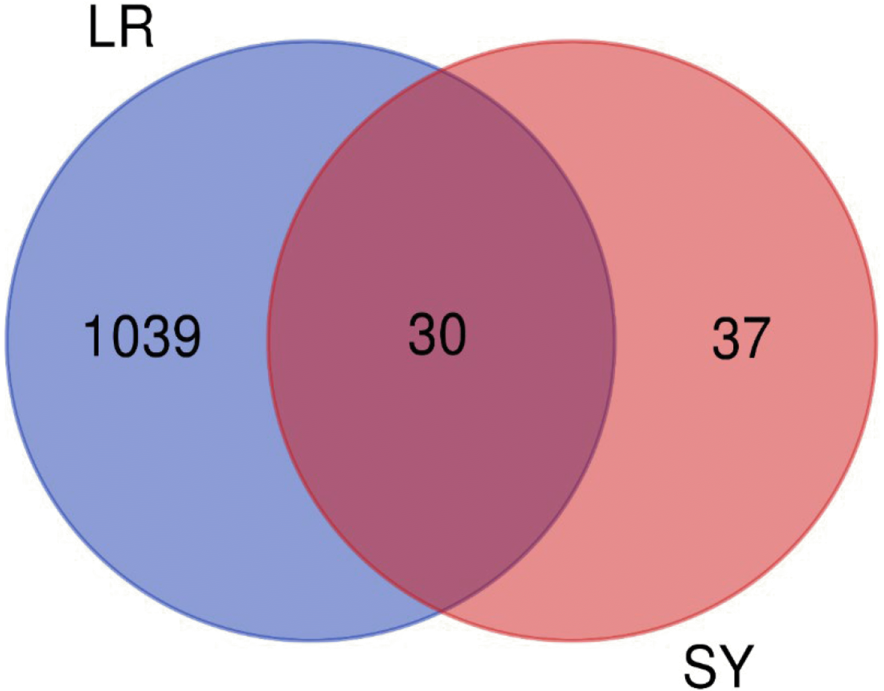
Fig. 3
Gene target network of the effective components of Rhizoma Dioscoreae in the “drug–component–target–disease” network.
Green squares represent Rhizoma Dioscoreae, red hexagons represent active ingredients, yellow circles represent diseases, and blue diamonds represent target proteins. LR, liver regeneration; SY, SanYak, Rhizoma Dioscoreae.
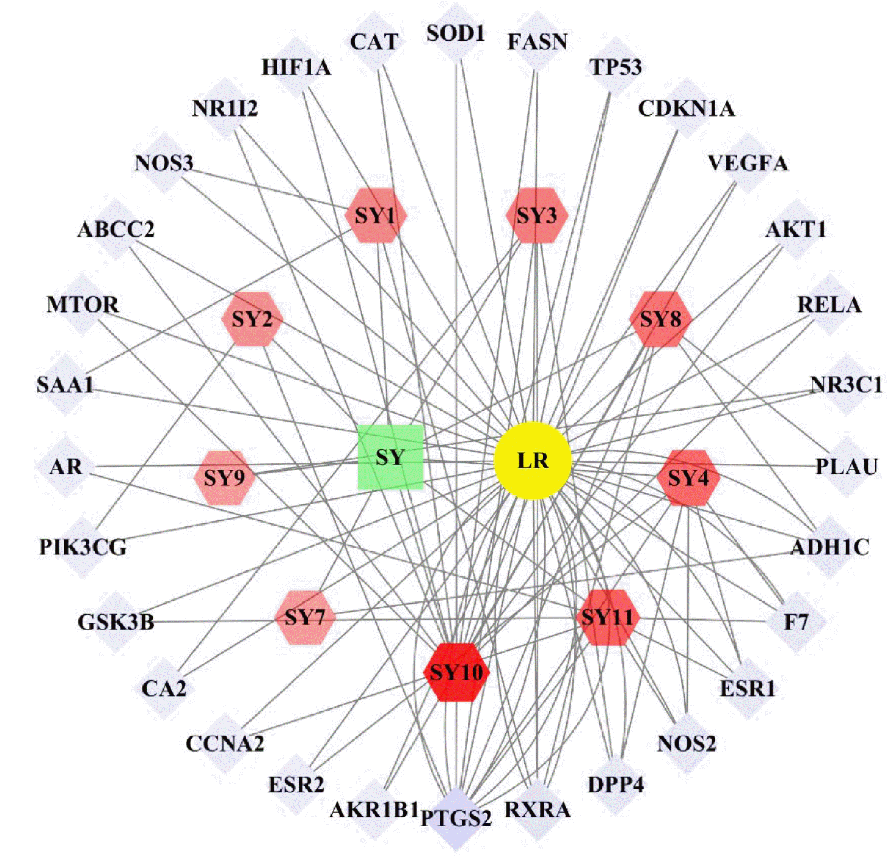
Fig. 4
PPI network diagram of the target proteins.
The size of the point represents the degree, color depth represents the compactness, and thickness of the line represents the intermediateness. Taking a degree of freedom of ≥18 as the line represents target proteins were AKT1, VEGFA, TP53, HIF1A, PTGS2, and ESR1. PPI, protein–protein interaction.
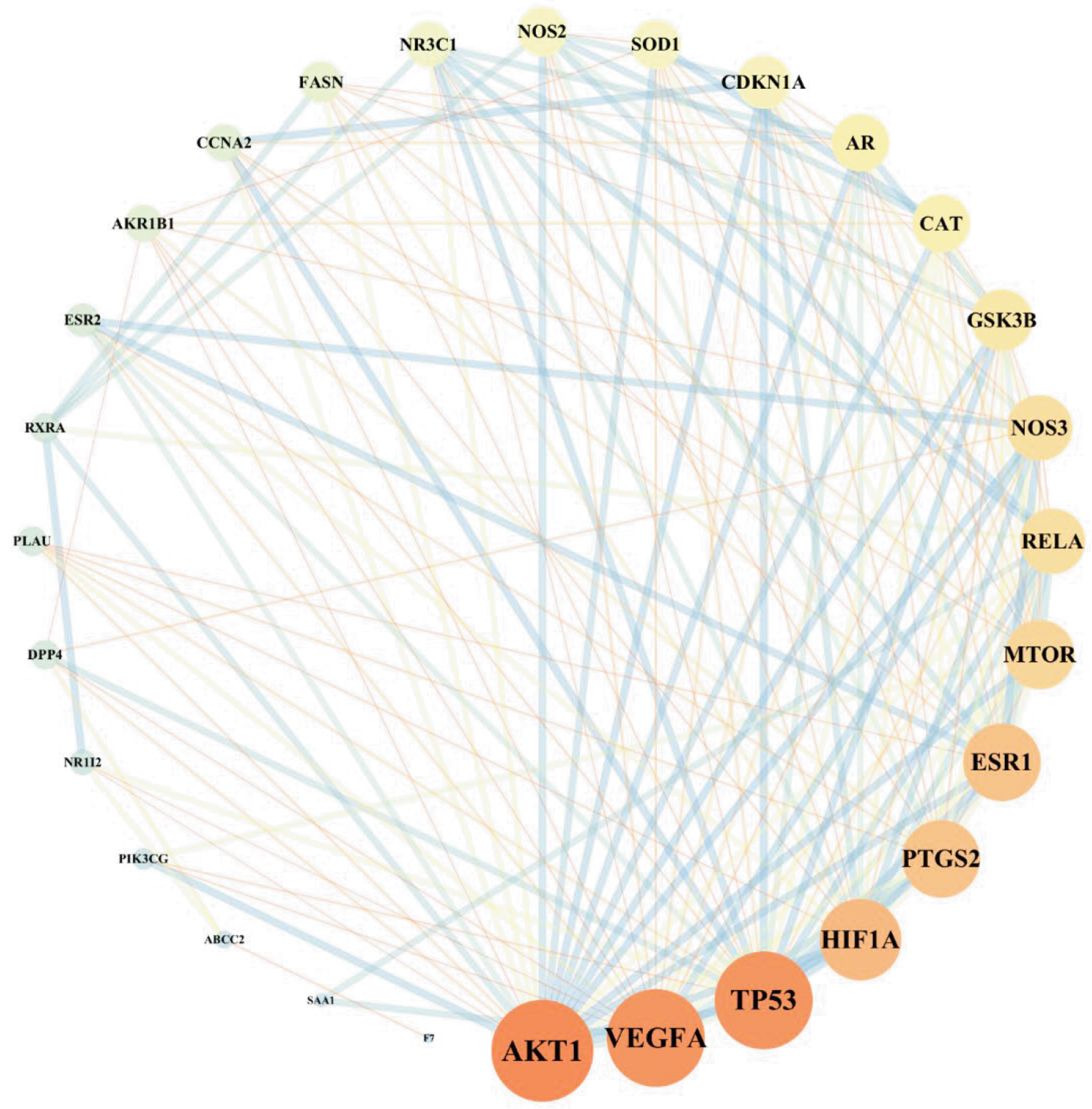
Fig. 5
GO and KEGG enrichment analyses of key target genes.
(A) Top items of biological function are listed on the horizontal axis, including the top eight items of molecular function (MF, blue), top seven items of cell component (CC, orange), and top twenty items of biological process (BP, green). (B) Histogram of the top nine pathways based on KEGG enrichment analysis. GO, Gene Ontology; KEGG, Kyoto Encyclopedia of Genes and Genomes.
Fig. 6
Compound–target–pathway network.
The yellow circle represents the pathway, and the blue diamond represents the target point.
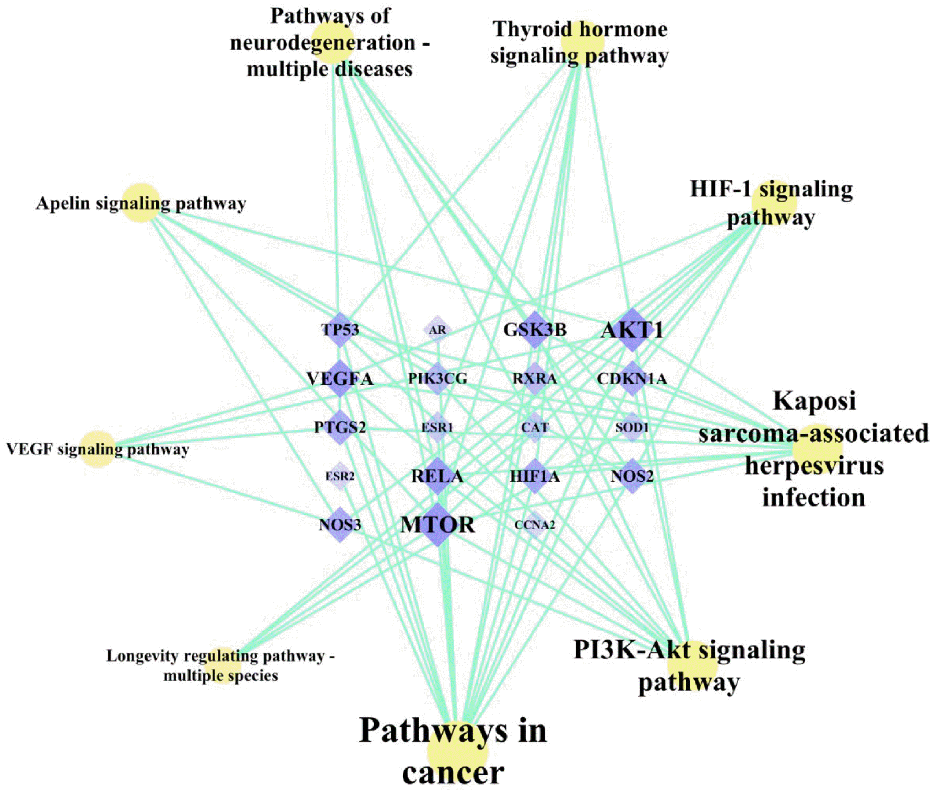
Fig. 7
Molecular docking of SY compounds and hub target proteins.
(A) Binding mode of AKT and diosgenin. (B) Binding mode of AKT and diosgenin. SY, SanYak, Rhizoma Dioscoreae; AKT, protein kinase B.
Fig. 8
Liver tissue stained with H&E.
(A) Sham group. (B) IR + 85%PHx 24 h group. (C) DIO IR + 85%PHx 24 h group (n = 4). Scale bar = 100 µm. IR, ischemia-reperfusion; PHx, hepatectomy; DIO, diosgenin.

Fig. 9
Determination of ALT and AST in serum (n = 4).
(A) Significantly increased ALT levels in IR + 85%PHx 24 h group, ****p < 0.0001 (vs. Sham group); ALT levels were significantly reduced in DIO IR + 85%PHx 24 h group, ***p < 0.001 (vs. IR + 85%PHx 24 h group). (B) Significantly increased AST levels in IR + 85%PHx 24 h group, ****p < 0.0001 (vs. Sham group); AST levels were significantly reduced in the DIO IR + 85%PHx 24 h group, ****P < 0.0001 (vs. IR + 85%PHx 24 h group) (n = 4). Values are presented as mean ± SD. ALT, alanine transaminase; AST, aspartate aminotransferase; IR, ischemia-reperfusion; PHx, hepatectomy; DIO, diosgenin.
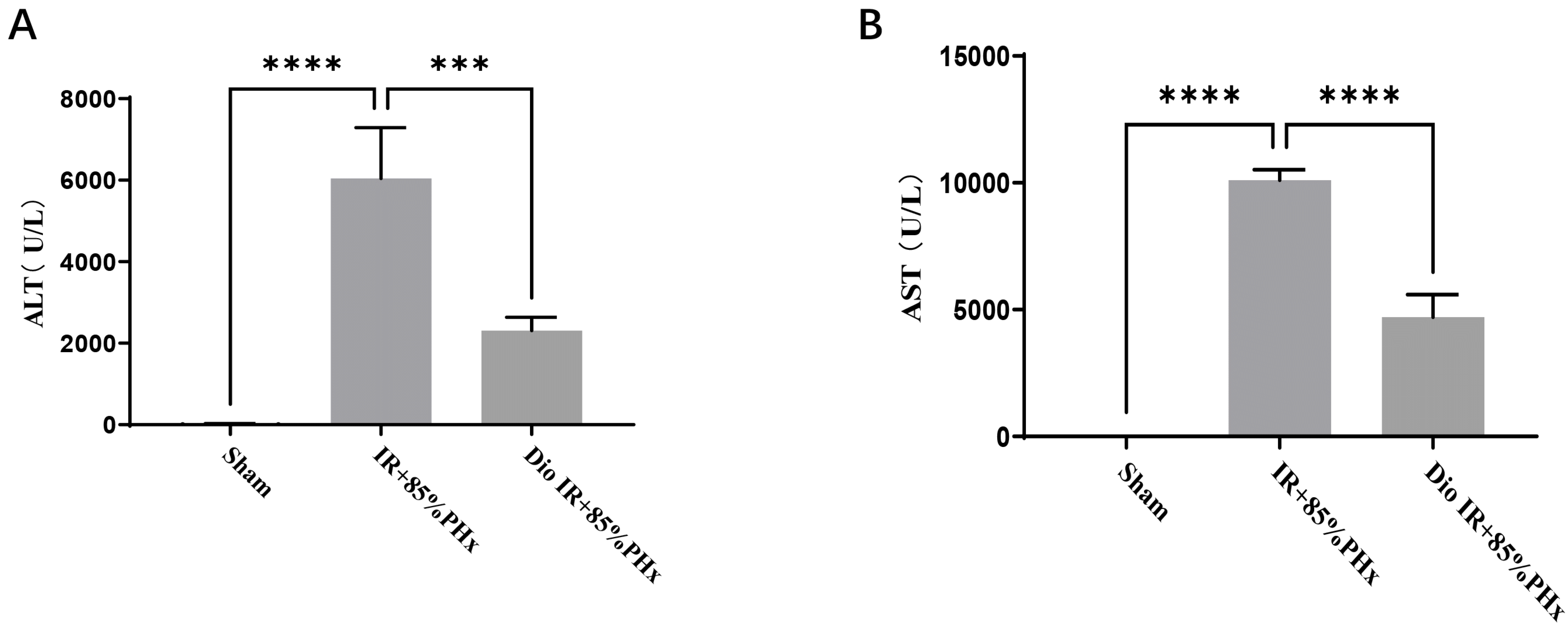
Fig. 10
Immunohistochemistry determines the positive rate of proliferating cell nuclear antigen (PCNA) (n = 4) (200×).
(A) Sham group. (B) IR + 85%PHx 24 h group. (C) DIO IR + 85%PHx 24 h group. (D) Analysis of results: PCNA positivity increased in IR + 85%PHx 24 h group, ***p < 0.001 (vs. Sham group); PCNA positivity increased in the DIO IR + 85%PHx 24 h group, ****p < 0.0001 (vs. IR + 85%PHx 24 h group). Values are presented as mean ± SD. IR, ischemia-reperfusion; PHx, hepatectomy; DIO, diosgenin.

Fig. 11
Effect of DIO on the expression of AKT and VEGFA in IR + 85%PHx mice.
(A) Western blot image showing the expression levels of p-AKT, AKT, and VEGFA in the liver tissue (n = 4). (B) Increased p-AKT/AKT expression levels in the IR + 85%PHx 24 h group, **p < 0.01 (vs. Sham group); decreased p-AKT/AKT expression levels in the DIO IR + 85%PHx 24 h group, *p < 0.05 (vs. IR + 85%PHx 24 h group). (C) No difference in VEGFA expression levels in the IR + 85%PHx 24 h group compared with the Sham group. Increased VEGFA expression levels in the DIO IR + 85%PHx 24 h group, *p < 0.05 (vs. IR + 85%PHx 24 h group). Values are presented as mean ± SD. AKT, protein kinase B; VEGFA, vascular endothelial growth factor A; GAPDH, glyceraldehyde 3-phosphate dehydrogenase; IR, ischemia-reperfusion; PHx, hepatectomy; DIO, diosgenin; ns, > 0.05.
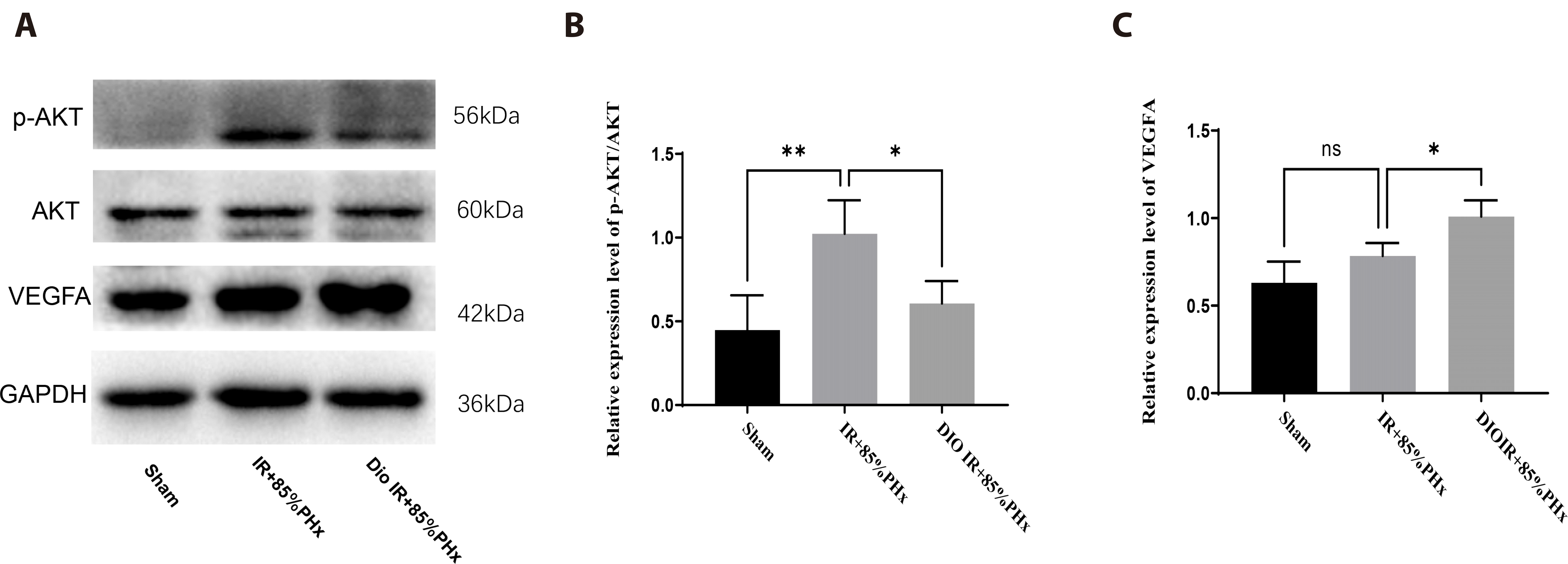
Table 1
Active ingredients in Rhizoma Dioscoreae


 PDF
PDF Citation
 Print
Print


 XML Download