Abstract
Nanobodies derived from camelids and sharks offer unique advantages in therapeutic applications due to their ability to bind to epitopes that were previously inaccessible. Traditional methods of nanobody development face challenges such as ethical concerns and antigen toxicity. Our study presents a synthetic, phage-displayed nanobody library using trinucleotide-directed mutagenesis technology, which allows precise amino acid composition in complementarity-determining regions (CDRs), with a focus on CDR3 diversity. This approach avoids common problems such as frameshift mutations and stop codon insertions associated with other synthetic antibody library construction methods. By analyzing FDA-approved nanobodies and Protein Data Bank sequences, we designed sub-libraries with different CDR3 lengths and introduced amino acid substitutions to improve solubility. The validation of our library through the successful isolation of nanobodies against targets such as PD-1, ATXN1 and STAT3 demonstrates a versatile and ethical platform for the development of high specificity and affinity nanobodies and represents a significant advance in biotechnology.
Nanobodies, derived from the variable domain of heavy chain-only antibodies in camelids and sharks, represent a distinctive and highly adaptable class of antibody-like molecules [1-4]. Their advantages over traditional antibodies are many, including significantly extended complementarity determining regions 3 (CDR3) loops that facilitate high-affinity binding to previously inaccessible epitopes [5-7]. This feature, combined with their single-domain architecture, greatly streamlines the development of multi-specific and multivalent biological therapeutics, particularly in the field of CAR T-cell therapies, by simplifying genetic modifications [8-10].
The process of inducing an immune response to produce heavy chain antibodies in camelids mirrors that of conventional antibody production in other mammals [11,12]. The technique of isolating nanobodies by cloning nanobody genes from immunized camelids is well established, with phage display serving as a key method for generating diverse nanobody libraries [13]. These libraries are capable of maintaining functional diversity and rapidly yielding high-affinity nanobodies [13]. However, certain challenges, such as immunization risks due to antigen toxicity or pathogenicity and misfolding of antigens into inclusion bodies, require alternative strategies [14]. Synthetic antibody libraries, which use synthetic DNA to create optimized scaffolds, are emerging as a viable solution, potentially providing antibodies with a broader range of specificities and higher affinities [15-19].
Recent advances in nanobody engineering have ushered in sophisticated synthetic nanobody libraries that are meticulously designed by analyzing natural nanobody sequences, particularly their complementarity-determining regions (CDRs) [15-19]. This design strategy involves a high degree of randomization in the CDR regions using DNA oligonucleotides with random sequences (NNK or NNB), which promotes the creation of libraries that reflect the natural diversity of nanobodies [20]. Despite these advances, the potential introduction of stop codons poses a challenge, potentially limiting the diversity and functionality of the resulting libraries.
Our research seeks to mitigate these limitations through the application of trinucleotide-directed mutagenesis (TRIM) technology, which allows for precise control of the amino acid composition in the CDRs. This method not only closely mimics the natural diversity found in nanobodies, but also avoids typical pitfalls such as frameshift mutations and the inadvertent introduction of stop codons [21,22]. The goal is to create a synthetic, phage-displayed nanobody library characterized by high functional diversity targeting a broad spectrum of disease-related proteins [2,23,24].
The construction of such nanobody libraries is crucial for the identification of antigen-specific nanobodies. While immune libraries derived from animal immunization typically provide high-affinity nanobodies, they require multiple libraries for different antigens, driving up costs [18,25]. Conversely, naive libraries from non-immunized donors offer an alternative that bypasses immunization but may require significant volumes of blood and additional steps to increase affinity [26]. Synthetic libraries are therefore a more versatile and accessible choice for many laboratories, facilitating the identification of both antigen-specific and conformation-specific nanobodies [27].
This work contributes to the advancement of nanobody technology by developing a synthetic phage-displayed nanobody library enhanced by TRIM technology with a focus on CDR3 diversity. The efficacy of these libraries was confirmed by the successful isolation of nanobodies against specific antigens. This approach not only overcomes the obstacles associated with traditional methods, but also represents a significant advancement in nanobody engineering, enriching both scientific knowledge and practical application of these essential molecular tools.
The synthetic phage-displayed nanobody library was constructed based on an IGHV3-23*4 gene. We randomized CDR1 and CDR2 with degenerated codons, and randomized CDR3 with 10 different lengths (12 residues to 22 residues except 13 residues) using the TRIM method. This method allows a controlled composition of amino acids in the CDR3, with the following distribution 14% Y, 12% G, 10% S, 9% D, 7% T, 6% R, 6% A, 5% L, 5% V, 4% P, 4% N, 3% F, 3% E, 3% I, 3% W, 2% Q, 2% K, and 2% H, specifically excluding C, M, and stop codons to ensure functional diversity. The primers used are listed in Table 1. After amplification of the nanobody library genes, the nanobody library genes were digested with SfiI and purified using a Quick Gel Extraction Kit. The digested nanobody library genes were then cloned into the phagemid vector pADL-10b and electrotransformed into Escherichia coli TG1 cells. E. coli transformants were cultured overnight at 37°C on LB plates supplemented with 100 μg/ml ampicillin. Library size was assessed by counting colony forming units after gradient dilution. To evaluate the diversity and quality of the synthetic phage-displayed nanobody library, 12 individual clones were sequenced to analyze the amino acid distribution and composition of the CDR3 region. Finally, the colonies were scraped from the plates and stored in LB medium with 20% (v/v) glycerol at –80°C, a condition optimal for long-term storage and future use.
The frozen nanobody library cell stock was thawed and cultured in 400 ml SB medium (20 g/l yeast extract, 30 g/l tryptone, and 10 g/l MOPS) supplemented with 2% glucose and 100 μg/ml ampicillin. After reaching an OD600 of approximately 0.5, the cells were harvested by centrifugation at 4,000 rpm for 15 min at room temperature, and the cell pellets were resuspended in fresh 400 ml SB medium supplemented with 100 μg/ml ampicillin. For phage infection, 1012 cfu of M13KO7 helper phages were added and incubated at 37°C at 80 rpm for 1 h. Nanobody library expression was then induced by the addition of 50 μg/ml kanamycin and 200 μM IPTG for overnight at 30°C. The next day, the nanobody library was centrifuged at 4°C for 15 min at 4,000 rpm. The supernatant was then processed for phage precipitation using a 5× PEG/NaCl solution (20% PEG and 2.5M NaCl). The resulting phage pellet was resuspended in 1× PBS containing a protease inhibitor. For phage titration, serial dilution of the phage preparation was performed in SB media. The diluted phages were then incubated with TG1 cells.
Screening of the library was performed by panning on purified programmed cell death protein 1 (PD-1), ataxin 1 (ATXN1), signal transducer and activator of transcription 3 (STAT3) molecules. An immunotube was coated with the antigen (10 μg/ml) for 1 h at 200 rpm at 37°C. The immunotube and phage were washed with deionized water (DIW) and blocked with 0.05% Tween 20 in PBS (PBST) containing 3% skim milk for 1 h at room temperature. After removing the blocking solution in the immunotube, the immunotube was washed with DIW, and the phage particles in a total volume of 1 ml were added to the antigen-coated immunotube for 2 h at 200 rpm at 37°C. After incubation, the supernatants were discarded and the immunotube was rinsed three to ten times with PBST buffer (PBS with 0.05% Tween 20). Bound phages were eluted by adding 100 mM trimethylamine (pH 10) and neutralized with 1 M Tris/HCl (pH 7.4). For further rounds of panning, the eluted phages were amplified by infecting log-phase E. coli TG1 cells followed by their superinfection with helper phages. In addition, input and output phages were titrated by infecting the log-phase TG1 with an aliquot of phage particles and then plating the samples on LB-ampicillin/2% glucose plates. Four consecutive rounds of plating were performed. To evaluate the synthetic phage-displayed nanobody library, 20 individual colonies were randomly selected and sequenced.
Recombinant ATXN1 and STAT3 were purchased from Abbexa and Sino Biological, respectively. Recombinant PD-1 was prepared in house. Briefly, the gene of PD-1 was synthesized at Integrated DNA Technologies and then cloned in-frame into the pcDNA3.4 vector using the Gibson assembly cloning method (NEB) [28-30]. Each target protein expression vector was transfected into Expi293 cells according to the manufacturer's instructions [28-30]. After 5 days, the supernatant was harvested by centrifugation at 4,000 × g for 10 min. The supernatant was passed through a Ni-NTA agarose resin column three times and washed with 40 ml of washing buffer (50 mM sodium phosphate, 300 mM NaCl, and 25 mM imidazole at pH 8.0). The bound proteins were eluted with 15 ml of elution buffer (50 mM sodium phosphate, 300 mM NaCl, and 250 mM imidazole at pH 8.0). Buffer exchange to pH 7.4 PBS was then performed using Amicon Ultra-4 spin columns (Merck Millipore) with a 3 kDa cutoff. The purity of the purified protein samples was assessed by 15% SDS-PAGE gel [28].
The gene of anti-PD-1, anti-ATXN1, and anti-STAT3 nanobody clones were amplified by PCR and then cloned in-frame into the pMopac12 vector using the Gibson assembly cloning method. Plasmids were electrotransformed into E. coli BL21(DE3) cell. E. coli transformants were cultured at 37°C in 500 ml TB medium supplemented with 500 μg/ml chloramphenicol. After reaching an OD600 of 1.0, the nanobody expression was induced by the addition of 500 μM IPTG for overnight at 20°C at 150 rpm. The next day, the culture was centrifuged at 4°C for 20 min at 10,000 × g. After centrifugation, the supernatants were discarded and the cell pellet was resuspended in buffer (10 mM Tris, 0.75 M sucrose at pH 7.5). Resuspended cells were digested their cell walls by the addition of buffer (10 mM Tris, 0.75 M sucrose at pH 7.5) with 20 mg/ml lysozyme and incubated for 20 min at 4°C at 100 rpm. After incubation, cells were added with 1 mM EDTA and incubated for 20 min at 4°C at 100 rpm. The supernatants were harvested by centrifugation at 4°C for 30 min at 10,000 × g. The protein purification process is the same as mentioned above.
The binding activities against target proteins of each nanobody were determined by ELISA, according to the previously reported methods [28-30]. Briefly, 100 ng per well of the respective antigens were coated onto a 96-well polystyrene ELISA plate (Thermo Fisher Scientific) and incubated overnight at 4°C. To prevent nonspecific binding, the plate was blocked with 1× PBS (pH 7.4) containing 3% bovine serum albumin for 1 h at room temperature. Fifty μl of serially diluted recombinant nanobody protein, ranging from 1,000 nM to 10 nM, was prepared and added to the wells at the volume per well. The plate was then incubated at room temperature for 1 h to allow binding. After incubation, the plate was washed four times with PBST buffer. For detection, mouse anti-Myc tag antibody (9E10, diluted 1:1,000, prepared in house) was added and incubated for 1 h and the plate was then washed four times with PBST buffer. After washing, anti-mouse IgG antibody conjugated to HRP (diluted 1:5,000, Abcam) was added and incubated for 1 h. The plate was then washed four times with PBST buffer. To develop the color reaction, 50 μl of 3,3′,5,5′-tetramethylbenzidine (TMB) substrate was added to each well. The reaction was terminated by adding 50 μl of 2 M H2SO4, which stopped the color development by TMB. Finally, absorbance was measured at 450 nm using an Infinite 200 PRO NanoQuant microplate reader (Tecan Trading AG). This absorbance measurement reflects the binding activity of the antibody to the antigen, with higher absorbance correlating with stronger antibody-antigen interactions.
To construct a synthetic phage-displayed nanobody library, we analyzed 342 nanobody sequences from the Protein Data Bank (PDB) (Fig. 1A). We performed multiple sequence alignments using Cluster Omega to characterize the amino acid sequences of these antibodies. The median length of the nanobody sequences was found to be 123.5 amino acids (aa), with a range of 111 to 139 aa (Fig. 1B). This revealed a diversity of amino acid lengths within the nanobody regions, particularly in CDR3. According to Kabat numbering, the most common lengths of CDR1 and CDR2 are 8 and 10 aa, respectively. In contrast to CDR1 and CDR2, the length of CDR3 varies from 5 to 28 aa. The most frequent top 14 lengths of CDR3 are 9 to 22 aa (Fig. 1C). Fig. 1D shows the nanobody sequence logos [31], representing the degree of conservation of each residue of nanobody. Conserved sequence blocks in framework regions (FRs) had high bit scores. Conversely, CDRs had lower bit scores, reflecting their inherent sequence variability.
We designed 10 sub-libraries for the synthetic nanobody library, each with a different length: 8 aa for CDR1, 10 aa for CDR2, and 12 to 22 aa except 13 for CDR3. In addition, we selected the Vicugna pacos (alpaca) IGHV3S39*01, which has a high sequence similarity of 91% to the human IGHV3-23*04 gene, known for its stability in human antibodies. The framework region 2 (FR2) of single domain antibodies such as nanobodies contains hydrophobic residues whose positions correspond to the interface of VH and VL, which often decrease the solubility of antibodies. Therefore, we introduced unique amino acid substitutions in the FR2 of the nanobody compared to camel VH sequences, specifically at positions V37F, G44E, L45R, and W47G, which are critical for nanobody solubility [32]. These substitutions were incorporated into the nanobody template (Fig. 2).
For CDR random mutagenesis of nanobody, we used degenerate codons and TRIM technology, which allows precise control over amino acid incorporation (Table 1). We constructed 10 sub-libraries corresponding to the CDR3 length variations. The amino acid composition of each CDR3 residue was carefully designed to include a specific percentage of different amino acids while excluding cysteine, methionine to avoid unwanted disulfide bonds and methionine oxidation, and stop codons to promote functional diversity (Fig. 3A).
To construct phage-displayed nanobody sub-libraries, nanobody was divided into 4 fragments (FR1, CDR1-FR2, CDR2-FR3, CDR3-FR4-Hisx6-HA tag) and each fragment was amplified by PCR. Amplified fragments were joined after overlap extension PCR and both ends were digested with SfiI to form sticky ends. These sub-library genes were ligated into the pADL-10b phage display vector and transformed into E. coli TG1 cells (Fig. 3B).
The resulting nanobody sub-libraries ranged in size from 107 to 109; 1.18 × 10⁹ for #12, 1.46 × 10⁹ for #14, 2.83 × 10⁹ for #15, 1.75 × 10⁸ for #16, 3.24 × 10⁹ for #17, 1.92 × 10⁸ for #18, 1.18 × 10⁹ for #19, 1.25 × 10⁹ for #20, 3.92 × 10⁸ for #21, and 6.65 × 10⁷ for #22 (Fig. 3C). To assess the quality of our libraries, 10 clones each were sequenced and the CDR3 lengths are shown in Fig. 3D.
To validate the synthetic phage nanobody libraries, we screened the synthetic phage nanobody library against three antigens, such as PD-1 (CD279), ATXN1, and STAT3. Using immunotubes coated with each of these target proteins, we performed biopanning as described in the Methods section. 4–5 rounds of biopanning were performed to enrich nanobody clones, and the concentrations of coated antigens were systematically reduced. The output/input ratio generally increased, indicating successful enrichment of specific clones for PD-1, ATXN1, and STAT3 (Table 2). After these 4–5 rounds, we randomly selected 12 phage clones for each antigen, and then analyzed the sequence of each nanobody. The CDR sequences of four PD-1-specific nanobody clones are shown in Table 3, and clone 1-1 was used for subsequent experiments.
To assess the binding activities of the isolated nanobody clones for their antigen, we then produced recombinant proteins of the anti-PD-1, anti-ATXN1, and anti-STAT3 nanobodies. Nanobody genes were amplified by PCR and then cloned into pMopac12 plasmid vector with genetic fusion of 6×His and Myc tag at the C-terminus. The plasmids were transformed into E. coli BL21(DE3) for periplasmic expression of the protein. The expressed proteins were then purified by affinity chromatography. To confirm the correct expression of the recombinant proteins and the molecular weights of the nanobodies, the same amount of purified nanobodies was resolved by SDS-PAGE in non-reducing and reducing conditions (Fig. 4A). All nanobodies, anti-PD-1, anti-ATXN1, and anti-STAT3 had monomeric structures and molecular weights of approximately 10–15 kDa. We also quantified the expression yield of the nanobodies, which was determined to be 0.06–1 mg/l culture medium. Finally, we evaluated the binding specificity of the soluble nanobodies to their respective antigens by ELISA. Extracellular domain of PD-1, ATXN1, STAT3 were immobilized on immunoplates and nanobodies were treated with concentrations of 1,000 nM, 100 nM and 10 nM. All nanobodies showed dose-dependent binding to each target protein (Fig. 4B).
To predict the epitope of anti-ATXN1 and anti-STAT3 nanobodies with their respective targets, ATXN1 and STAT3, we performed in silico protein structure analysis. First, Schrödinger-based homology modeling for antibody structure prediction was performed with amino acid sequences to obtain the protein structure of each nanobodies (Fig. 5A, left and Fig. 5B, left). The predicted structures of the nanobodies were then docked to their targets: ATXN1 (PDB ID: 1OA8) and STAT3 (PDB ID: 6NJS) to simulate the formation of complex structures. The top 3 cluster size docking models were examined for protein interaction analysis and each interface was highlighted by color (Fig. 5A, right and Fig. 5B, right). First and second docking models of ATXN1/anti-ATXN1 nanobody share α-helix of ATXN1 as epitope, while nanobody binds to β–sheet in third model. The first and third docking models of STAT3/anti-STAT3 nanobodies share a region between the coiled-coil domain and the linker of STAT3 as epitope, but the second model showed that binds to a region between the linker and the SH2 domain. This analysis provides insight into the potential binding modes and interaction sites of these nanobodies with their respective target proteins.
The development of nanobodies has traditionally relied on animal immunization techniques, primarily using camelids, to generate high-affinity antibodies [23,24,33]. While effective, these conventional methods present significant logistical and ethical challenges, including the need for extensive animal use and associated costs. Recognizing these limitations, the scientific community has increasingly turned to synthetic antibody libraries as a more sustainable and scalable solution [34]. In particular, phage display technology has emerged as a cornerstone of this shift, enabling high-throughput screening of antibody variants with desired affinities and specificities [24,27,27].
Our research contributes to this evolving landscape by utilizing TRIM technology to construct a comprehensive synthetic phage-displayed nanobody library. This approach represents a significant advancement over previous methods that predominantly used degenerate codons for CDR diversification. While effective in introducing variability, these traditional techniques often fail to accurately control amino acid composition, leading to the inclusion of stop codons and the potential for frameshift mutations [21,22,35-37]. Our implementation of TRIM technology circumvents these issues and allows for the precise synthesis of CDRs that closely mimic the diversity observed in natural antibodies. This precision engineering facilitated the creation of sub-libraries tailored to specific CDR3 lengths, thereby increasing the functional diversity of our library and aligning it with natural antibody variability.
The isolation of specific nanobodies targeting PD-1 [38], ATXN1 [39], and STAT3 [40] further underscores the practical utility of our synthetic library. These targets, which are associated with critical pathological processes in cancer, neurodegeneration and immune regulation, represent key areas where nanobodies can offer therapeutic and diagnostic advances. Our approach not only facilitates the rapid screening and identification of high-affinity nanobodies, but also provides a more accessible platform for exploring a broader range of disease-related antigens. This flexibility and accessibility distinguish our library from camelid-derived or humanized antibodies, which may be limited by the specificities and affinities of the initial immunization repertoire.
In addition, the expression and characterization of anti-ATXN1 and anti-STAT3 nanobodies using optimized phage display vectors and an E. coli expression system highlights the robustness of our platform. The incorporation of structural analysis through docking model predictions provides an additional layer of insight, enabling a deeper understanding of the molecular interactions involved. Such structural elucidation is rare in the field and provides a comparative advantage by revealing the mechanisms underlying the specificity and affinity of nanobodies to antigens.
In summary, our work not only addresses several limitations of traditional nanobody generation techniques, but also introduces a sophisticated and efficient strategy for the development of nanobodies against a diverse array of disease targets. By advancing the science of nanobody engineering, our study contributes valuable knowledge and tools to the therapeutic and diagnostic applications of these promising biomolecules, positioning our research at the forefront of molecular biotechnology.
Notes
REFERENCES
1. Ingram JR, Schmidt FI, Ploegh HL. 2018; Exploiting nanobodies' singular traits. Annu Rev Immunol. 36:695–715. DOI: 10.1146/annurev-immunol-042617-053327. PMID: 29490163.
2. Könning D, Zielonka S, Grzeschik J, Empting M, Valldorf B, Krah S, Schröter C, Sellmann C, Hock B, Kolmar H. 2017; Camelid and shark single domain antibodies: structural features and therapeutic potential. Curr Opin Struct Biol. 45:10–16. DOI: 10.1016/j.sbi.2016.10.019. PMID: 27865111.
3. Rahbarizadeh F, Ahmadvand D, Sharifzadeh Z. 2011; Nanobody; an old concept and new vehicle for immunotargeting. Immunol Invest. 40:299–338. DOI: 10.3109/08820139.2010.542228. PMID: 21244216.
4. Kang SH, Lee CH. 2021; Development of therapeutic antibodies and modulating the characteristics of therapeutic antibodies to maximize the therapeutic efficacy. Biotechnol Bioprocess Eng. 26:295–311. DOI: 10.1007/s12257-020-0181-8. PMID: 34220207. PMCID: PMC8236339.
5. Liu C, Lin H, Cao L, Wang K, Sui J. 2022; Research progress on unique paratope structure, antigen binding modes, and systematic mutagenesis strategies of single-domain antibodies. Front Immunol. 13:1059771. DOI: 10.3389/fimmu.2022.1059771. PMID: 36479130. PMCID: PMC9720397.
6. Gonzalez-Sapienza G, Rossotti MA, Tabares-da Rosa S. 2017; Single-domain antibodies as versatile affinity reagents for analytical and diagnostic applications. Front Immunol. 8:977. DOI: 10.3389/fimmu.2017.00977. PMID: 28871254. PMCID: PMC5566570.
7. Henry KA, MacKenzie CR. 2018; Antigen recognition by single-domain antibodies: structural latitudes and constraints. MAbs. 10:815–826. DOI: 10.1080/19420862.2018.1489633. PMID: 29916758. PMCID: PMC6260137.
8. Bao C, Gao Q, Li LL, Han L, Zhang B, Ding Y, Song Z, Zhang R, Zhang J, Wu XH. 2021; The application of nanobody in CAR-T therapy. Biomolecules. 11:238. DOI: 10.3390/biom11020238. PMID: 33567640. PMCID: PMC7914546.
9. Safarzadeh Kozani P, Naseri A, Mirarefin SMJ, Salem F, Nikbakht M, Evazi Bakhshi S, Safarzadeh Kozani P. 2022; Nanobody-based CAR-T cells for cancer immunotherapy. Biomark Res. 10:24. DOI: 10.1186/s40364-022-00371-7. PMID: 35468841. PMCID: PMC9036779.
10. Oh J, Warshaviak DT, Mkrtichyan M, Munguia ML, Lin A, Chai F, Pigott C, Kang J, Gallo M, Kamb A. 2019; Single variable domains from the T cell receptor β chain function as mono- and bifunctional CARs and TCRs. Sci Rep. 9:17291. DOI: 10.1038/s41598-019-53756-4. PMID: 31754147. PMCID: PMC6872726.
11. Sun Y, Huang T, Hammarström L, Zhao Y. 2020; The immunoglobulins: new insights, implications, and applications. Annu Rev Anim Biosci. 8:145–169. DOI: 10.1146/annurev-animal-021419-083720. PMID: 31846352.
12. Arbabi-Ghahroudi M. 2022; Camelid single-domain antibodies: promises and challenges as lifesaving treatments. Int J Mol Sci. 23:5009. DOI: 10.3390/ijms23095009. PMID: 35563400. PMCID: PMC9100996.
13. Muyldermans S. 2021; A guide to: generation and design of nanobodies. FEBS J. 288:2084–2102. DOI: 10.1111/febs.15515. PMID: 32780549. PMCID: PMC8048825.
14. Laustsen AH, Greiff V, Karatt-Vellatt A, Muyldermans S, Jenkins TP. 2021; Animal immunization, in vitro display technologies, and machine learning for antibody discovery. Trends Biotechnol. 39:1263–1273. DOI: 10.1016/j.tibtech.2021.03.003. PMID: 33775449.
15. Zimmermann I, Egloff P, Hutter CAJ, Kuhn BT, Bräuer P, Newstead S, Dawson RJP, Geertsma ER, Seeger MA. 2020; Generation of synthetic nanobodies against delicate proteins. Nat Protoc. 15:1707–1741. DOI: 10.1038/s41596-020-0304-x. PMID: 32269381.
16. Gebauer M, Skerra A. 2020; Engineered protein scaffolds as next-generation therapeutics. Annu Rev Pharmacol Toxicol. 60:391–415. DOI: 10.1146/annurev-pharmtox-010818-021118. PMID: 31914898.
17. Ferrari D, Garrapa V, Locatelli M, Bolchi A. 2020; A novel nanobody scaffold optimized for bacterial expression and suitable for the construction of ribosome display libraries. Mol Biotechnol. 62:43–55. DOI: 10.1007/s12033-019-00224-z. PMID: 31720928.
18. Moutel S, Bery N, Bernard V, Keller L, Lemesre E, de Marco A, Ligat L, Rain JC, Favre G, Olichon A, Perez F. 2016; NaLi-H1: a universal synthetic library of humanized nanobodies providing highly functional antibodies and intrabodies. Elife. 5:e16228. DOI: 10.7554/eLife.16228. PMCID: PMC4985285.
19. Valdés-Tresanco MS, Molina-Zapata A, Pose AG, Moreno E. 2022; Structural insights into the design of synthetic nanobody libraries. Molecules. 27:2198. DOI: 10.3390/molecules27072198. PMID: 35408597. PMCID: PMC9000494.
20. Misson Mindrebo L, Liu H, Ozorowski G, Tran Q, Woehl J, Khalek I, Smith JM, Barman S, Zhao F, Keating C, Limbo O, Verma M, Liu J, Stanfield RL, Zhu X, Turner HL, Sok D, Huang PS, Burton DR, Ward AB, et al. 2023; Fully synthetic platform to rapidly generate tetravalent bispecific nanobody-based immunoglobulins. Proc Natl Acad Sci U S A. 120:e2216612120. DOI: 10.1073/pnas.2216612120. PMID: 37276407. PMCID: PMC10268213.
21. Huang CY, Lok YY, Lin CH, Lai SL, Wu YY, Hu CY, Liao CB, Ho CH, Chou YP, Hsu YH, Lo YH, Chern E. 2023; Highly reliable GIGA-sized synthetic human therapeutic antibody library construction. Front Immunol. 14:1089395. DOI: 10.3389/fimmu.2023.1089395. PMID: 37180155. PMCID: PMC10174300.
22. Shim H. 2015; Synthetic approach to the generation of antibody diversity. BMB Rep. 48:489–494. DOI: 10.5483/BMBRep.2015.48.9.120. PMID: 26129672. PMCID: PMC4641231.
23. Muyldermans S. 2021; Applications of nanobodies. Annu Rev Anim Biosci. 9:401–421. DOI: 10.1146/annurev-animal-021419-083831. PMID: 33233943.
24. Liu W, Song H, Chen Q, Yu J, Xian M, Nian R, Feng D. 2018; Recent advances in the selection and identification of antigen-specific nanobodies. Mol Immunol. 96:37–47. DOI: 10.1016/j.molimm.2018.02.012. PMID: 29477934.
25. Kulkarni SS, Falzarano D. 2021; Unique aspects of adaptive immunity in camelids and their applications. Mol Immunol. 134:102–108. DOI: 10.1016/j.molimm.2021.03.001. PMID: 33751993.
26. Ferrara F, Erasmus MF, D'Angelo S, Leal-Lopes C, Teixeira AA, Choudhary A, Honnen W, Calianese D, Huang D, Peng L, Voss JE, Nemazee D, Burton DR, Pinter A, Bradbury ARM. 2022; A pandemic-enabled comparison of discovery platforms demonstrates a naïve antibody library can match the best immune-sourced antibodies. Nat Commun. 13:462. Erratum in: Nat Commun. 2022;13:2097. DOI: 10.1038/s41467-021-27799-z. PMID: 35075126. PMCID: PMC8786865.
27. Moreno E, Valdés-Tresanco MS, Molina-Zapata A, Sánchez-Ramos O. 2022; Structure-based design and construction of a synthetic phage display nanobody library. BMC Res Notes. 15:124. DOI: 10.1186/s13104-022-06001-7. PMID: 35351202. PMCID: PMC8966178.
28. Lee CM, Kim M, Park SW, Kang CK, Choe PG, Kim NJ, Jo HJ, Shin HM, Lee CH, Kim HR, Park WB, Oh MD. 2024; Clinical outcomes and immunological features of COVID-19 patients receiving B-cell depletion therapy during the Omicron era. Infect Dis (Lond). 56:116–127. DOI: 10.1080/23744235.2023.2276784. PMID: 37916860.
29. Kang CK, Kim MG, Park SW, Kim YW, Lee CM, Choe PG, Park WB, Kim NJ, Kim M, Lee S, Kim IS, Lee CH, Shin HM, Kim HR, Oh MD. 2023; Comparable humoral and cellular immunity against Omicron variant BA.4/5 of once-boosted BA.1/2 convalescents and twice-boosted COVID-19-naïve individuals. J Med Virol. 95:e28558. DOI: 10.1002/jmv.28558. PMID: 36755360.
30. Kang CK, Shin HM, Choe PG, Park J, Hong J, Seo JS, Lee YH, Chang E, Kim NJ, Kim M, Kim YW, Kim HR, Lee CH, Seo JY, Park WB, Oh MD. 2022; Broad humoral and cellular immunity elicited by one-dose mRNA vaccination 18 months after SARS-CoV-2 infection. BMC Med. 20:181. DOI: 10.1186/s12916-022-02383-4. PMID: 35508998. PMCID: PMC9067342.
31. Wagih O. 2017; ggseqlogo: a versatile R package for drawing sequence logos. Bioinformatics. 33:3645–3647. DOI: 10.1093/bioinformatics/btx469. PMID: 29036507.
32. Vincke C, Loris R, Saerens D, Martinez-Rodriguez S, Muyldermans S, Conrath K. 2009; General strategy to humanize a camelid single-domain antibody and identification of a universal humanized nanobody scaffold. J Biol Chem. 284:3273–3284. DOI: 10.1074/jbc.M806889200. PMID: 19010777.
33. Jin BK, Odongo S, Radwanska M, Magez S. 2023; Nanobodies: a review of generation, diagnostics and therapeutics. Int J Mol Sci. 24:5994. DOI: 10.3390/ijms24065994. PMID: 36983063. PMCID: PMC10057852.
34. Burkovitz A, Ofran Y. 2016; Understanding differences between synthetic and natural antibodies can help improve antibody engineering. MAbs. 8:278–287. DOI: 10.1080/19420862.2015.1123365. PMID: 26652053. PMCID: PMC4966605.
35. Pack P, Ilag V, Hardt C, Rheinnecker M, Wellnhofer G, Virnekäs B. 1996; Trinucleotide-directed mutagenesis (TRIM) and 2nd generation mini-antibodies. Immunotechnology. 2:70. DOI: 10.1016/1380-2933(96)80679-8.
36. Knappik A, Ge L, Honegger A, Pack P, Fischer M, Wellnhofer G, Hoess A, Wölle J, Plückthun A, Virnekäs B. 2000; Fully synthetic human combinatorial antibody libraries (HuCAL) based on modular consensus frameworks and CDRs randomized with trinucleotides. J Mol Biol. 296:57–86. DOI: 10.1006/jmbi.1999.3444. PMID: 10656818.
37. Prassler J, Thiel S, Pracht C, Polzer A, Peters S, Bauer M, Nörenberg S, Stark Y, Kölln J, Popp A, Urlinger S, Enzelberger M. 2011; HuCAL PLATINUM, a synthetic Fab library optimized for sequence diversity and superior performance in mammalian expression systems. J Mol Biol. 413:261–278. DOI: 10.1016/j.jmb.2011.08.012. PMID: 21856311.
38. Budimir N, Thomas GD, Dolina JS, Salek-Ardakani S. 2022; Reversing T-cell exhaustion in cancer: lessons learned from PD-1/PD-L1 immune checkpoint blockade. Cancer Immunol Res. 10:146–153. DOI: 10.1158/2326-6066.CIR-21-0515. PMID: 34937730.
39. Coffin SL, Durham MA, Nitschke L, Xhako E, Brown AM, Revelli JP, Villavicencio Gonzalez E, Lin T, Handler HP, Dai Y, Trostle AJ, Wan YW, Liu Z, Sillitoe RV, Orr HT, Zoghbi HY. 2023; Disruption of the ATXN1-CIC complex reveals the role of additional nuclear ATXN1 interactors in spinocerebellar ataxia type 1. Neuron. 111:915. Erratum for: Neuron. 2023;111:481-492.e8. DOI: 10.1016/j.neuron.2022.11.016. PMID: 36577402. PMCID: PMC9957872.
40. Zou S, Tong Q, Liu B, Huang W, Tian Y, Fu X. 2020; Targeting STAT3 in cancer immunotherapy. Mol Cancer. 19:145. DOI: 10.1186/s12943-020-01258-7. PMID: 32972405. PMCID: PMC7513516.
Fig. 1
Characterization of nanobody sequences.
(A) Structure of a nanobody using a ribbon diagram, highlighting the CDR1, CDR2, and CDR3 regions in pink, lime, and blue, respectively. Model structure is generated by Schrödinger software. (B) Frequency of 342 nanobody lengths as recorded in the Protein Data Bank (PDB). (C) Graphical representation of the length variability of the CDR1, CDR2, and CDR3 regions within nanobodies. (D) Sequence logo generated from a multiple sequence alignment of nanobody sequences extracted from the PDB, providing insight into conserved motifs. Sequence logo is generated by ggseqlogo of R package.
Fig. 2
Comparative multiple sequence alignment.
Nanobody sequences from Homo sapiens IGHV3-23*4, Vicugna pacos IGHV3S39*01, an FDA-approved nanobody, and the designed synthetic nanobody library were presented. The presence of random mutagenesis in the library is indicated by a “#” symbol, demonstrating the diversity within the synthetic library.
Fig. 3
Construction of the synthetic nanobody library.
(A) Quantification of the amino acid composition in the CDR3 region of the synthetic nanobody library using color coding for different amino acid types: sky blue for glycine, purple for histidine, light green for hydrophobic (aliphatic) amino acids, green for aromatic amino acids, yellow for polar amino acids, red for negatively charged amino acids, and blue for positively charged amino acids. (B) Outlines the cloning strategy used to construct the nanobody library, visualized using BioRender.com. (C) The size of 10 different nanobody sub-libraries. (D) Evaluation of the quality of these 10 nanobody sub-libraries based on sequencing results.
Fig. 4
Binding activities of the isolated nanobody candidates.
(A) SDS-PAGE analysis of the purified recombinant nanobody candidates. (B) ELISA of nanobody candidates. Anti-PD-1, anti-ATXN1, and anti-STAT3 nanobodies were examined with their target. Means ± S.D. were shown. Each experiment was repeated over three times independently. PD-1, programmed cell death protein 1; ATXN1, ataxin 1; STAT3, signal transducer and activator of transcription 3.
Fig. 5
Structural and docking analyses of anti-ATXN1 and anti-STAT3 nanobodies.
(A, B) Model structure of anti-ATXN1 (A, left) and anti-STAT3 nanobodies (B, left), with the CDR regions colored accordingly: pink for CDR1, yellow for CDR2, and purple for CDR3. The right panels of (A) and (B) show surface representations of the ATXN1 (PDB ID: 1OA8) and STAT3 (PDB ID: 6NJS) molecules within the docking models of the ATXN1/anti-ATXN1 and STAT3/anti-STAT3 nanobody interactions. Expected epitope regions are color-coded based on their model-specific or shared presence in the first, second, and third models, which enhances our understanding of the binding interfaces. The epitope regions of the first, second, third models are colored pink, green, and purple, respectively. The epitope regions shared by the first and second models are colored yellow. The epitope regions shared by the second and third models are colored blue. The epitope regions shared by the first and third models are red teal. The nanobody structures of the first, second, third models are colored pink, green, and purple, respectively. ATXN1, ataxin 1; STAT3, signal transducer and activator of transcription 3; CDR, complementarity-determining region.
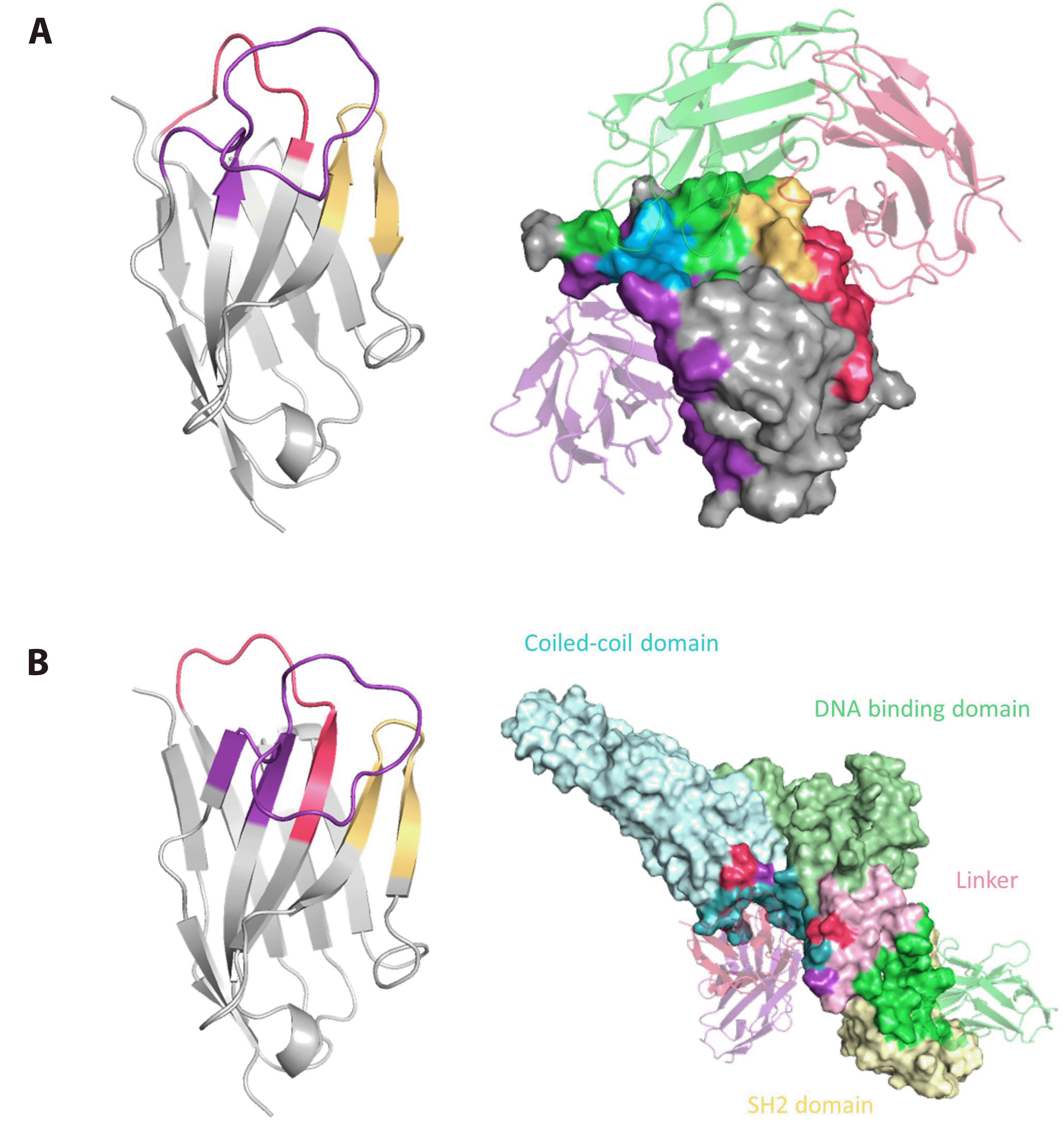
Table 1
Primers with degenerate codon and TRIM used for construction of 10 nanobody sub-libraries
Table 2
CFU during biopanning of phage nanobody libraries


 PDF
PDF Citation
 Print
Print


 XML Download