Abstract
Group I metabotropic glutamate receptors (mGluRs) modulate postsynaptic neuronal excitability and epileptogenesis. We investigated roles of group I mGluRs on low extracellular Mg2+ concentration ([Mg2+]o)-induced epileptiform activity and neuronal cell death in the CA1 regions of isolated rat hippocampal slices without the entorhinal cortex using extracellular recording and propidium iodide staining. Exposure to Mg2+-free artificial cerebrospinal fluid can induce interictal epileptiform activity in the CA1 regions of rat hippocampal slices. MPEP, a mGluR 5 antagonist, significantly inhibited the spike firing of the low [Mg2+]o-induced epileptiform activity, whereas LY367385, a mGluR1 antagonist, did not. DHPG, a group 1 mGluR agonist, significantly increased the spike firing of the epileptiform activity. U73122, a PLC inhibitor, inhibited the spike firing. Thapsigargin, an ER Ca2+-ATPase antagonist, significantly inhibited the spike firing and amplitude of the epileptiform activity. Both the IP3 receptor antagonist 2-APB and the ryanodine receptor antagonist dantrolene significantly inhibited the spike firing. The PKC inhibitors such as chelerythrine and GF109203X, significantly increased the spike firing. Flufenamic acid, a relatively specific TRPC 1, 4, 5 channel antagonist, significantly inhibited the spike firing, whereas SKF96365, a relatively non-specific TRPC channel antagonist, did not. MPEP significantly decreased low [Mg2+]o DMEM-induced neuronal cell death in the CA1 regions, but LY367385 did not. We suggest that mGluR 5 is involved in low [Mg2+]o-induced interictal epileptiform activity in the CA1 regions of rat hippocampal slices through PLC, release of Ca2+ from intracellular stores and PKC and TRPC channels, which could be involved in neuronal cell death.
Reduction of extracellular Mg2+ concentration ([Mg2+]o) has been reported to induce excitatory patterns of electrical activity in cultured rat hippocampal neurons [1,2] and elicit epileptiform activity in rat hippocampal slice [3-5]. The low [Mg2+]o-induced epileptiform activity has been reported to be mediated primarily by the N-methyl-D-aspartate (NMDA) receptor [3,5]. The low [Mg2+]o-induced epileptiform activity has been used as an in vitro model [3,6].
All three groups of metabotropic glutamate receptors (mGluRs) are expressed in the hippocampus [7]. The group II mGluRs and group III mGluRs suppress excitatory synaptic transmission by inhibiting to adenylnyl cyclase [8]. The group 1 mGluRs (mGluR 1, mGluR 5) induce intracellular free Ca2+ concentration ([Ca2+]i) increases by releasing Ca2+ from intracellular stores through activation of phospholipase C (PLC) [8]. The group 1 mGluRs regulate hippocampal pyramidal neuron excitability via Ca2+-dependent activation of small-conductance K+ channels and transient receptor potential classical (TRPC) channels [9]. Activation of group 1 mGluRs promotes epileptogenesis [10,11].
mGluR 5 is expressed in the all principal neurons including highly expressed CA1 regions in the rat hippocampus, whereas the mGluR1 is highly expressed on some pyramids in the CA3 regions and absent on CA1 pyramids [12]. The perisynaptic position of postsynaptic mGluR1 and mGluR5 appears to be general feature of glutamate synaptic organization [12]. Concomitant activation of mGluRs activates TRPC channels that further depolarize the postsynaptic terminal and induce an influx of Ca2+ [13,14]. All these data suggest that mGluR 5 is involved in the modulation of low [Mg2+]o-induced epileptiform activity in the CA1 region of the hippocampus. However, little is known how mGluR 5 is involved in low [Mg2+]o-induced epileptiform activity and cell death in the CA1 region of rat hippocampal slice.
In this study, we investigated roles of group I mGluRs on low [Mg2+]o-epileptiform activity and the neuronal cell death in the CA1 regions of rat hippocampal slices without the entorhinal cortex using extracellular recording and propidium iodide (PI) staining, respectively. mGluR 5 is involved in low [Mg2+]o-induced epileptiform activity in the CA1 region of rat hippocampal slice through activation of PLC, release of Ca2+ from intracellular stores and activation of protein kinase C (PKC) and TRPC channels, which could be involved in neuronal cell death.
Dulbecco’s modified eagle media (DMEM), horse serum (HS), and PI were obtained from Gibco-BRL. Custom made Mg2+-free-containing DMEM was purchased from Welgene. 6-Methyl-2-(phenylethynyl) pyridine (MPEP) hydrochloride, 3,5-dihydroxyphenylglycine (DHPG) and all other chemicals were purchased from Sigma.
Hippocampal slices were prepared from approximately 4-week-old male Sprague-Dawley rats [15]. All procedures of animal research were provided in accordance with Laboratory Animals Welfare Act, the Guidelines and Policies for Rodent Experiment provided by the Institutional Animal Care and Use Committee (IACUC) in College of Medicine, The Catholic University of Korea (Approval number: CUMC-2021-0287-02). The brains were quickly removed after anesthetization with urethane (1.3 g/kg body weight, intraperitoneal injection) and submerged in an ice-cold dissection medium. Since it has been reported that CA3-driven interictal discharges are induced in the hippocampal slices without the entorhinal cortex by Mg2+-free artificial cerebrospinal fluid (ACSF) [16], the hippocampi without entorhinal cortex were transversely sliced into 450 μM sections with a vibratome (Campden Instruments) and transferred to storage chamber at room temperature (25°C). Recovery was allowed in a storage chamber at least for 90 min. The dissection and storage medium consisted of 125 mM NaCl, 2.5 mM KCl, 1 mM CaCl2, 2 mM MgSO4, 1.25 mM NaH2PO4, 25 mM NaHCO3, and 10 mM D-glucose, bubbled with 95% O2/5% CO2. The slices were transferred to the submerging chamber for recording and were superfused continuously at 1.5–2 ml/min with ACSF (119 mM NaCl, 2.5 mM KCl, 1.3 mM MgSO4, 26.2 mM NaHCO3, 1 mM NaH2PO4, 2.5 mM CaCl2, and 11 mM glucose, with the pH maintained at 7.4 that was saturated with 95% O2/5% CO2) or Mg2+-free ACSF (119 mM NaCl, 4.0 mM KCl, 26.2 mM NaHCO3, 1 mM NaH2PO4, 2.5 mM CaCl2, and 11 mM glucose) at 31°C–32°C. For PI staining, the hippocampi without entorhinal cortex were transversely sliced into 400 μM sections with a vibratome. The slices were transferred onto 0.4 μM porous millicell membrane inserts (Millipore). For neuronal cell death study [17], the membrane inserts were placed in 35 mm cell culture dishes filled 50% normal Mg2+ containing DMEM (normal [Mg2+]o DMEM) or custom made Mg2+-free-containing DMEM (low [Mg2+]o DMEM) (Welgene), 25% Hank's balanced salt solution (HBSS), 25% HS, 20 mM HEPES, 1% penicillin streptomycin for 24 h.
For extracellular field recording, recording electrodes (2–4 MΩ), pulled from borosilicate glass pipettes (1B150F-4; World Precision Instruments, Inc.) using a micropipette puller (MODEL P-97; Sutter Instrument Co.), were filled with ACSF and positioned in the CA1 area of the hippocampal slices. The interictal epileptiform activity was induced 30–60 min after Mg2+-free ACSF treatment. After 15 min of stable recording, baseline epileptiform activity was recorded for 10 min. Subsequent epileptiform activity was recorded for an additional 15–45 min without or during drug treatment and normalized to baseline activity. Both the spike firing and the amplitude of the epileptiform activities were counted and analyzed manually.
The viability of neuronal cells was assessed after 30 min exposure to the red fluorescent dye, PI (5 μg/ml). Digital pictures of PI uptake obtained using the inverted fluorescence microscope (Olympus IX73) were analyzed using Java image processing program ImageJ (National Institute of Health). All images were captured with a 4X objective using JNO-ARM software (JNOPTIC). For analysis, PI fluorescence intensities of 3 circular regions within interest with CA1 hippocampal regions were calculated. The circles were specified as 60 × 60 pixels in ImageJ software. After background subtraction placed adjacent to the slice culture, the first circle was measured at the midpoint of the region. The other two circles were placed equally distant from the midpoint and boundaries. After the basic template is set up, the regions of interest can easily be transferred to the appropriate region. The maximal PI uptake was set to 100% after 24 h low-temperature exposure to 70% EtOH [18]. PI incorporation was shown as a percentage of ethanol-induced maximal PI uptake.
An exposure to Mg2+-free ACSF induced spontaneous interictal epileptiform activity in the CA1 regions of rat hippocampal slice without entorhinal cortex within 30–60 min. After 15 min of stable recording, baseline epileptiform activity was recorded for 10 min. Subsequent epileptiform activity was recorded for an additional 15–45 min without or during drug treatment and normalized to baseline activity. The baseline spike frequency of the epileptiform activity was 0.13 ± 0.02 Hz (n = 13) and amplitude was 0.52 ± 0.19 mV (n = 13) (Fig. 1A1).
mGluR 5 is strongly expressed in the CA1 regions of rat hippocampal slice [12]. We examined whether group 1 mGluRs are involved in the low [Mg2+]o-induced epileptiform activity in the CA1 regions of hippocampal slice (Fig. 1A1, A2). Treatment with the mGluR5 antagonist MPEP (50 µM) a, significantly inhibited the spike firing of the epileptiform activity (control = 1.10 ± 0.08 of base line, n = 7; MPEP = 0.39 ± 0.15 of base line, n = 6; p < 0.05), but it did not affect the amplitude. However, the mGluR1 antagonist LY367385 (100 µM) did not affect both the spike firing and the amplitude of the epileptiform activity. These results suggest that mGluR 5 is involved in low [Mg2+]o-induced interictal epileptiform activity in the CA1 regions of the rat hippocampal slice. We also determined whether the group 1 mGluR antagonists affect low [Mg2+]o-induced interictal epileptiform activity in the CA3 region (Fig. 1B). The epileptiform activity in the CA3 region had a similar shape and polarity to that in the CA1 region, but was frequently more prolonged or of greater amplitude than epileptiform activity in the CA1 region (spike frequency = 0.20 ± 0.03 Hz; amplitude = 1.05 ± 0.28 mV; n = 6) (Fig. 1B1). Each treatment with MPEP (50 µM) or LY367385 (100 µM) did not significantly affect both the spike firing and amplitude of the epileptiform activity in the CA3 region (Fig. 1B2).
In order to indirectly confirm whether group 1 mGluRs are involved in the low [Mg2+]o-induced epileptiform activity, we examined whether the group 1 mGluR agonist DHPG increases the low [Mg2+]o-induced epileptiform activity in the CA1 regions (Fig. 2). DHPG (10 µM), significantly increased the spike firing of the epileptiform activity (control = 1.07 ± 0.10, n = 7; DHPG = 5.48 ± 1.39, n = 6; p < 0.01), but it did not affect the amplitude. These results indirectly support that mGluR 5 is involved in low [Mg2+]o-induced interictal epileptiform activity in the CA1 area of the hippocampus. Therefore, to further investigate the mechanism of how mGluR5 is involved in low [Mg2+]o-induced epileptiform activity, we used the CA1 region of rat hippocampal slice.
In this study, activation or inhibition of mGluR 5 modulated low [Mg2+]o-induced epileptiform activity in the CA1 regions. In the previous our report, the PLC inhibitor U73122 inhibited low [Mg2+]o-induced [Ca2+]i spikes in cultured rat hippocampal neurons [19]. Therefore, we tested whether mGluR-induced activation of PLC is involved in low [Mg2+]o-induced epileptiform activity in the CA1 region (Fig. 3). Treatment with the PLC inhibitor U73122 (10 µM) significantly inhibited the spike firing of the low [Mg2+]o- induced epileptiform activity (control = 1.19 ± 0.11 of base line, n = 6; U73122 = 0.70 ± 0.10 of base line, n = 7; p < 0.01), but it did not affect the amplitude (Fig. 3). These results suggest that mGluR 5 is involved in low [Mg2+]o-induced epileptiform activity via activation of PLC.
In this study, mGluR 5 might be involved in low [Mg2+]o-induced epileptiform activity via activation of PLC. The group 1 mGluRs (mGluR 1, mGluR 5) induce [Ca2+]i increases by releasing Ca2+ from intracellular stores through activation of PLC [8]. We tested whether mGluR-induced mobilization of Ca2+ from intracellular Ca2+ stores is involved in low [Mg2+]o-induced epileptiform activity (Fig. 4). Treatment with thapsigargin (10 µM), which blocks intracellular ER Ca2+-ATPase resulting in depletion of intracellular Ca2+ stores, significantly inhibited both the spike firing (control = 1.06 ± 0.08 of base line, n = 6; thapsigargin = 0.55 ± 0.15 of base line, n = 6; p < 0.05) and the amplitude (control = 0.96 ± 0.05 of base line, n = 6; thapsigargin = 0.53 ± 0.05 of base line, n = 6; p < 0.01) of low [Mg2+]o-induced epileptiform activity within 35 min (Fig. 4A1, A2). Treatment with the IP3 receptor antagonist 2-APB (30 µM) also significantly inhibited the spike firing of low [Mg2+]o-induced epileptiform activity within 45 min (control = 1.14 ± 0.08 of base line, n = 8; 2-APB = 0.64 ± 0.16 of base line, n = 6; p < 0.05), but it did not affect the amplitude (Fig. 4B1, B2). In addition, treatment with the ryanodine receptor antagonist dantrolene (30 µM) also inhibited the spike firing of the epileptiform activity within 35 min (control = 1.14 ± 0.08 of base line, n = 8; dantrolene = 0.56 ± 0.13 of base line, n = 6; p < 0.05), but it did not affect the amplitude (Fig. 4B1, B2). All these data suggest that mGluR 5 is involved in low [Mg2+]o-induced epileptiform activity by a release of Ca2+ from intracellular Ca2+ stores through IP3 receptors and ryanodine receptors.
mGluR-induced activation of PLC hydrolyzes phosphatidylinositol 4,5-bisphosphate to produce inositol trisphosphates and diacylglycerol, which leads to Ca2+ mobilization and PKC activation. Activation of PKC can induce phosphorylation of receptors, channels and/or other enzymes and change synaptic networks [20]. In the next experiments, we examined whether the PKC inhibitors such as GF 109203X and chelerythrine, affect low [Mg2+]o-induced epileptiform activity (Fig. 5). Treatment with GF 109203X (1 µM) markedly increased the spike firing. Treatment with chelerythrine (10 µM) also significantly increased the spike firing. However, both PKC inhibitors did not affect the amplitude of the epileptiform activity. These results suggest that mGluR-induced activation of PKC has an inhibitory effect on low [Mg2+]o-induced epileptiform activity in the CA1 region of rat hippocampal slices.
Excitability changes are induced by mGluR-induced sustained depolarization in CA1 pyramidal neurons of the rat hippocampus through activation of TRPC channels [9]. TRPC1, TRPC4 and TRPC5 channels are expressed in the rat pyramidal neurons of the CA1 regions [21,22]. We tested whether mGluR-induced activation of these TRPC channels is involved in low [Mg2+]o-induced interictal epileptiform activity (Fig. 6). Although TRPC antagonists have the lack of specificity, the relatively specific TRPC1, TRPC4 and TRPC5 channel inhibitor flufenamic acid has been reported to inhibit mGluR-mediated depolarization in the CA1 region of the hippocampus [9]. In this study, treatment with flufenamic acid (100 μM) significantly inhibited the spike firing of the epileptiform activity but did not affect the amplitude (Fig. 6). However, another relatively non-specific TRPC channel inhibitor SKF96365 (10 μM) did not affect both the spike firing and the amplitude of the epileptiform activity (Fig. 6). These results suggest that mGluR-induced activation of the flufenamic acid-sensitive TRPC channels may be involved in low [Mg2+]o-induced interictal epileptiform activity in the CA1 region of rat hippocampal slice. However, widely used TRP channel inhibitors such as flufenamic acid and SKF 96365, have been reported to have off-target effects on GABAA receptors at concentrations routinely used to study TRP channel function [23]. We cannot exclude the possibility that different off-target effects of flufenamic acid and SKF 96365 may be responsible for the differences in their inhibitory effects on epileptiform activity.
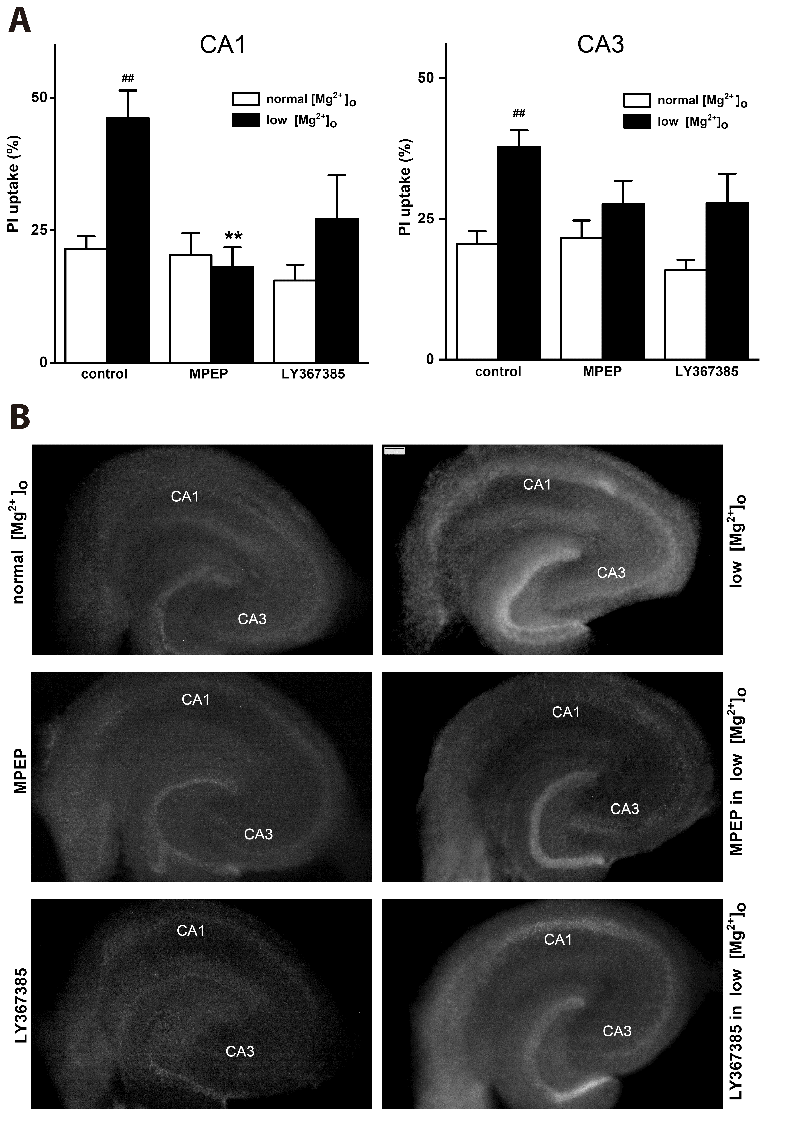
Low [Mg2+]o-induced epileptiform activity has been reported to induce neuronal cell death in the hippocampal slice [24]. We test whether mGluR 5 is involved in low [Mg2+]o-induced neuronal cell death in the CA1 region and CA3 regions of rat hippocampal slice (Fig. 7). Hippocampal slices were exposed to normal [Mg2+]o or low [Mg2+]o DMEM for 24 h in the absence or presence of mGluR antagonists such as MPEP (50 µM) and LY367385 (100 µM). PI staining was performed to observe the effects of mGluR antagonists on the cell death after the exposure. PI incorporation was shown as a percentage of ethanol-induced maximal PI uptake (See METHODS). Exposure to normal [Mg2+]o DMEM showed a basal PI incorporation (21.53 ± 2.33 relative to 70% ethanol-induced maximal PI uptake, n = 12) (Fig. 7A). Exposure to low [Mg2+]o DMEM significantly increased PI incorporation in the CA1 region (control = 46.11 ± 5.27, n = 14, p < 0.01) and in the CA3 region (control = 37.85 ± 2.90, n = 14, p < 0.01). MPEP significantly decreased the low [Mg2+]o DMEM-induced PI incorporation (MPEP = 18.14 ± 3.67, n = 6, p < 0.01), but LY367385 did not affect the PI incorporation (LY367385 = 27.17 ± 8.22, n = 6, p = 0.14). However, in the CA3 region, both MPEP and LY 367385 did not significantly affect the PI incorporation (MPEP = 27.60 ± 4.17, n = 6, p = 0.46; LY367385 = 27.80 ± 5.20, n = 6, p = 0.51). In addition, MPEP or LY367385 alone did not affect the PI corporation in the presence of normal [Mg2+]o DMEM. Photographs show effects of mGluR antagonists on the PI incorporation in rat hippocampal slices 24 h after exposure to normal [Mg2+]o or low [Mg2+]o DMEM solution in the absence or presence of mGluR antagonists in the CA1 and CA3 regions (Fig. 7B).
Present study shows that activation of mGluR 5 is involved in low [Mg2+]o-induced interictal epileptiform activity in the CA1 region of rat hippocampal slices by mobilization of Ca2+ from intracellular store via IP3 receptors and ryanodine receptors, and activation of PKC and TRPC channels. These effects of mGluR 5 on the epileptiform activity may be involved in low [Mg2+]o-induced neuronal cell death.
In this study, mGluR5 antagonist MPEP inhibited low [Mg2+]o- induced interictal epileptiform activity in the CA1 region of rat hippocampal slices, but mGluR1 antagonist LY367385 did not. Group 1 mGluR agonist DHPG increased the low Mg2+-induced interictal epileptiform activity. mGluR5 is expressed on principal cells in all rat hippocampal area and expressed highly on the hippocampal pyramidal cells in the CA1 regions, whereas mGluR1 is expressed on some pyramids in the CA3 regions and absent in the CA1 regions [12]. In our recent study, MPEP almost completely inhibited low Mg2+-induced [Ca2+]i spikes in cultured rat hippocampal neurons [19]. All these data suggest that mGluR 5 is involved in low [Mg2+]o-induced interictal epileptiform activity in the CA1 regions of rat hippocampal slices. However, since mGluR5 has also been suggested to be the principal subtype in initiating bursting activity in the rat hippocampus [25], we can not rule out a possibility that mGluR5 is also involved in epilepsy in the CA3 region. The detailed roles of mGluR5 in the CA3 region should be further studied.
Group 1 mGluRs, including mGluR5, bind to Gq/11 and activate PLC [8]. Activation of mGluRs induces mobilization of Ca2+ from intracellular stores via IP3 receptors through activation of PLC and Ca2+-induced Ca2+ release from the same stores by IP3 receptors and ryanodine receptors [26]. Activation of mGluRs and subsequent release of Ca2+ from intracellular stores further depolarize the postsynaptic membrane [27]. In this study, PLC inhibitor U73122 inhibited the low Mg2+-induced interictal epileptiform activity. ER Ca2+ ATPase antagonist thapsigargin inhibited the spike firing and amplitude of the low [Mg2+]o-induced epileptiform activity. In addition, both IP3 receptor antagonist 2-APB and ryanodine receptor antagonist dantrolene inhibited the spike firing of the epileptiform activity. The IP3 receptor antagonist 2-APB or the ryanodine receptor antagonist dantrolene inhibits Ca2+ release through IP3 receptors and ryanodine receptors, respectively, whereas thapsigargin depletes Ca2+ from intracellular stores [28], inhibiting Ca2+ release through both IP3 receptors and ryanodine receptors. This difference is thought to be why thapsigargin suppresses not only the spike firing but also the amplitude of spikes [29]. All these data suggest that mGluR 5 is involved in low [Mg2+]o-induced epileptiform activity by mobilization of Ca2+ from intracellular stores via IP3 receptors and ryanodine receptors through activation of PLC. These results are indirectly supported by other reports that Ca2+ release from intracellular stores participates in epileptogenesis in other hippocampal models of epilepsy [29,30].
Activation of PKC can induce phosphorylation of receptors, channels and/or other enzymes and change synaptic networks [20]. In this study, the PKC inhibitors such as chelerythrine and GF109203X, significantly increased the spike firing of the epileptiform activity. This result is supported by other quite similar result that the low [Mg2+]o condition induced by transient removal of extracellular Mg2+, elicits persistent suppression of LTP at hippocampal CA1 synapse via PKC activation [31,32]. All these findings suggest that the synaptic activity-induced activation of PKC suppresses synaptic strength in the low [Mg2+]o-induced epileptiform activity. These findings are also indirectly supported by the reports that PKC-induced phosphorylation may be beneficial for the prevention of mGluR-induced epileptogenesis [33] and reduction of status epilepticus [34].
Concomitant activation of mGluR and release of Ca2+ from intracellular stores activate TRPC channels that further depolarizes the post synaptic membrane, which is important for modulation of pyramidal neuron excitability by changing firing frequency in the CA1 region of the hippocampus [9]. Low [Mg2+]o has been reported to induce [Ca2+]i increase as well as seizure like events in the hippocampal slice [35]. TRPC1, TRPC 4 and TRPC 5 are expressed in rat CA1 pyramidal neurons [21,22]. Anti-TRPC1, anti-TRPC4 and anti-TRPC5 antibodies can block mGluR agonist DHPG-induced depolarization [9]. In the present study, the TRPC channel antagonist flufenamic acid, which inhibits more specifically TRPC 4 and TRPC 5 channels [36], inhibited the spike firing of the epileptiform activity, although TRPC channel antagonist SKF96365, which inhibits more specifically TRPC 3 and TRPC 6 channels [37,38], did not affect the epileptiform activity. In fact, TRPC1, TRPC 4 and TRPC 5 channels have been reported to play a critical role in the epileptiform burst firing [39]. In this study, therefore, concomitant activation of perisynaptic mGluR5 activates flufenamic acid-sensitive TRPC1, TRPC 4, TRPC 5 channels that further depolarize the postsynaptic membrane [9,14], which in turn, may be involved in low [Mg2+]o-induced epileptiform activity in the CA1 region of the hippocampus. All these data suggest that mGluR 5 is involved in low [Mg2+]o-induced interictal epileptiform activity in the CA1 region of rat hippocampal slice through activation of PLC, release of Ca2+ from intracellular stores and activation of PKC and TRPC channels, which is important for modulation of pyramidal neurons excitability by decreasing and increasing inter-ictal activity.
Activation of group 1 mGluRs promotes neuronal cell death [19,40] as well as epileptogenesis [10,11]. In the present study, mGluR5 antagonist MPEP inhibited neuronal cell death in the CA1 regions of the hippocampal slice as well as interictal epileptiform activity. However, MPEP did not affect low [Mg2+]o-induced neuronal cell death and epileptiform activity in the CA3 regions. These data suggest that mGluR5 is involved in epileptic activity and subsequent excitotoxicity in the CA1 region of rat hippocampal slice. These results are indirectly supported by our previous report [19] that mGluR5 is involved in low [Mg2+]o-induced-[Ca2+]i spikes and neuronal cell death in cultured rat hippocampal neurons.
This study presents that the mGluR 5 is involved in low [Mg2+]o- induced interictal epileptiform activity and neuronal cell death in the CA1 region of the rat hippocampal slices through PLC, release of Ca2+ from intracellular stores, activation of PKC and TRPC channels. Besides NMDA receptor, mGluR5 is involved in low [Mg2+]o-induced epileptiform activity in the CA1 regions of rat hippocampus. Hippocampal interictal activity has been suggest to control rather than promotes the ability of the entorhinal cortex to generate the seizure [41]. All these results suggest that mGluR5 in the CA1 regions of the rat hippocampus play an important role in temporal lobe epilepsy.
Notes
REFERENCES
1. Abele AE, Scholz KP, Scholz WK, Miller RJ. 1990; Excitotoxicity induced by enhanced excitatory neurotransmission in cultured hippocampal pyramidal neurons. Neuron. 4:413–419. DOI: 10.1016/0896-6273(90)90053-I. PMID: 1690567.
2. Shen M, Piser TM, Seysub VS, Thayer SA. 1996; Cannabinoid receptor agonists inhibit glutamatergic synaptic transmission in rat hippocampal cultures. J Neurosci. 16:4322–4334. DOI: 10.1523/JNEUROSCI.16-14-04322.1996. PMID: 8699243. PMCID: PMC6578864.
3. Mody I, Lambert JD, Heinemann U. 1987; Low extracellular magnesium induces epileptiform activity and spreading depression in rat hippocampal slices. J Neurophysiol. 57:869–888. DOI: 10.1152/jn.1987.57.3.869. PMID: 3031235.
4. Tancredi V, Hwa GG, Zona C, Brancati A, Avoli M. 1990; Low magnesium epileptogenesis in the rat hippocampal slice: electrophysiological and pharmacological features. Brain Res. 511:280–290. DOI: 10.1016/0006-8993(90)90173-9. PMID: 1970748.
5. Traub RD, Jefferys JG, Whittington MA. 1994; Enhanced NMDA conductance can account for epileptiform activity induced by low Mg2+ in the rat hippocampal slice. J Physiol. 478(Pt 3):379–393. DOI: 10.1113/jphysiol.1994.sp020259. PMID: 7965853. PMCID: PMC1155660.
6. Raimondo JV, Heinemann U, de Curtis M, Goodkin HP, Dulla CG, Janigro D, Ikeda A, Lin CK, Jiruska P, Galanopoulou AS, Bernard C. 2017; Methodological standards for in vitro models of epilepsy and epileptic seizures. A TASK1-WG4 report of the AES/ILAE Translational Task Force of the ILAE. Epilepsia. 58 Suppl 4(Suppl 4):40–52. DOI: 10.1111/epi.13901. PMID: 29105075. PMCID: PMC5679463.
7. Shigemoto R, Kinoshita A, Wada E, Nomura S, Ohishi H, Takada M, Flor PJ, Neki A, Abe T, Nakanishi S, Mizuno N. 1997; Differential presynaptic localization of metabotropic glutamate receptor subtypes in the rat hippocampus. J Neurosci. 17:7503–7522. DOI: 10.1523/JNEUROSCI.17-19-07503.1997. PMID: 9295396. PMCID: PMC6573434.
8. Niswender CM, Conn PJ. 2010; Metabotropic glutamate receptors: physiology, pharmacology, and disease. Annu Rev Pharmacol Toxicol. 50:295–322. DOI: 10.1146/annurev.pharmtox.011008.145533. PMID: 20055706. PMCID: PMC2904507.
9. El-Hassar L, Hagenston AM, D'Angelo LB, Yeckel MF. 2011; Metabotropic glutamate receptors regulate hippocampal CA1 pyramidal neuron excitability via Ca²+ wave-dependent activation of SK and TRPC channels. J Physiol. 589:3211–3229. DOI: 10.1113/jphysiol.2011.209783. PMID: 21576272. PMCID: PMC3145935.
10. Merlin LR, Wong RK. 1997; Role of group I metabotropic glutamate receptors in the patterning of epileptiform activities in vitro. J Neurophysiol. 78:539–544. DOI: 10.1152/jn.1997.78.1.539. PMID: 9242303.
11. Pan YZ, Rutecki PA. 2014; Enhanced excitatory synaptic network activity following transient group I metabotropic glutamate activation. Neuroscience. 275:22–32. DOI: 10.1016/j.neuroscience.2014.05.062. PMID: 24928353.
12. Lujan R, Nusser Z, Roberts JD, Shigemoto R, Somogyi P. 1996; Perisynaptic location of metabotropic glutamate receptors mGluR1 and mGluR5 on dendrites and dendritic spines in the rat hippocampus. Eur J Neurosci. 8:1488–1500. DOI: 10.1111/j.1460-9568.1996.tb01611.x. PMID: 8758956.
13. Lepannetier S, Gualdani R, Tempesta S, Schakman O, Seghers F, Kreis A, Yerna X, Slimi A, de Clippele M, Tajeddine N, Voets T, Bon RS, Beech DJ, Tissir F, Gailly P. 2018; Activation of TRPC1 channel by metabotropic glutamate receptor mGluR5 modulates synaptic plasticity and spatial working memory. Front Cell Neurosci. 12:318. DOI: 10.3389/fncel.2018.00318. PMID: 30271326. PMCID: PMC6149316.
14. Gualdani R, Gailly P. 2020; How TRPC channels modulate hippocampal function. Int J Mol Sci. 21:3915. DOI: 10.3390/ijms21113915. PMID: 32486187. PMCID: PMC7312571.
15. Nakatsuka H, Natsume K. 2014; Circadian rhythm modulates long-term potentiation induced at CA1 in rat hippocampal slices. Neurosci Res. 80:1–9. DOI: 10.1016/j.neures.2013.12.007. PMID: 24406747.
16. Avoli M, D'Antuono M, Louvel J, Köhling R, Biagini G, Pumain R, D'Arcangelo G, Tancredi V. 2002; Network and pharmacological mechanisms leading to epileptiform synchronization in the limbic system in vitro. Prog Neurobiol. 68:167–207. DOI: 10.1016/S0301-0082(02)00077-1. PMID: 12450487.
17. Stoppini L, Buchs PA, Muller D. 1991; A simple method for organotypic cultures of nervous tissue. J Neurosci Methods. 37:173–182. DOI: 10.1016/0165-0270(91)90128-M. PMID: 1715499.
18. Happ DF, Tasker RA. 2016; A method for objectively quantifying propidium iodide exclusion in organotypic hippocampal slice cultures. J Neurosci Methods. 269:1–5. DOI: 10.1016/j.jneumeth.2016.05.006. PMID: 27179931.
19. Yang JS, Jeon S, Jang HJ, Yoon SH. 2022; Group 1 metabotropic glutamate receptor 5 is involved in synaptically-induced Ca2+-spikes and cell death in cultured rat hippocampal neurons. Korean J Physiol Pharmacol. 26:531–540. DOI: 10.4196/kjpp.2022.26.6.531. PMID: 36302627. PMCID: PMC9614404.
20. Nishizuka Y, Shearman MS, Oda T, Berry N, Shinomura T, Asaoka Y, Ogita K, Koide H, Kikkawa U, Kishimoto A, Kose A, Saito N, Tanaka C. 1991; Protein kinase C family and nervous function. Prog Brain Res. 89:125–141. DOI: 10.1016/S0079-6123(08)61719-7. PMID: 1796138.
21. Strübing C, Krapivinsky G, Krapivinsky L, Clapham DE. 2001; TRPC1 and TRPC5 form a novel cation channel in mammalian brain. Neuron. 29:645–655. DOI: 10.1016/S0896-6273(01)00240-9. PMID: 11301024.
22. Chung YH, Sun Ahn H, Kim D, Hoon Shin D, Su Kim S, Yong Kim K, Bok Lee W, Ik Cha C. 2006; Immunohistochemical study on the distribution of TRPC channels in the rat hippocampus. Brain Res. 1085:132–137. DOI: 10.1016/j.brainres.2006.02.087. PMID: 16580647.
23. Rae MG, Hilton J, Sharkey J. 2012; Putative TRP channel antagonists, SKF 96365, flufenamic acid and 2-APB, are non-competitive antagonists at recombinant human α1β2γ2 GABA(A) receptors. Neurochem Int. 60:543–554. DOI: 10.1016/j.neuint.2012.02.014. PMID: 22369768.
24. Kovács R, Gutiérrez R, Kivi A, Schuchmann S, Gabriel S, Heinemann U. 1999; Acute cell damage after low Mg2+-induced epileptiform activity in organotypic hippocampal slice cultures. Neuroreport. 10:207–213. DOI: 10.1097/00001756-199902050-00002. PMID: 10203310.
25. Lanneau C, Harries MH, Ray AM, Cobb SR, Randall A, Davies CH. 2002; Complex interactions between mGluR1 and mGluR5 shape neuronal network activity in the rat hippocampus. Neuropharmacology. 43:131–140. DOI: 10.1016/S0028-3908(02)00086-2. PMID: 12213267.
26. Simpson PB, Challiss RA, Nahorski SR. 1995; Neuronal Ca2+ stores: activation and function. Trends Neurosci. 18:299–306. DOI: 10.1016/0166-2236(95)93919-O. PMID: 7571010.
27. Hagenston AM, Fitzpatrick JS, Yeckel MF. 2008; MGluR-mediated calcium waves that invade the soma regulate firing in layer V medial prefrontal cortical pyramidal neurons. Cereb Cortex. 18:407–423. DOI: 10.1093/cercor/bhm075. PMID: 17573372. PMCID: PMC3005283.
28. Thastrup O, Cullen PJ, Drøbak BK, Hanley MR, Dawson AP. 1990; Thapsigargin, a tumor promoter, discharges intracellular Ca2+ stores by specific inhibition of the endoplasmic reticulum Ca2(+)-ATPase. Proc Natl Acad Sci U S A. 87:2466–2470. DOI: 10.1073/pnas.87.7.2466. PMID: 2138778. PMCID: PMC53710.
29. Wülfert E, Margineanu DG. 1998; Thapsigargin inhibits bicuculline-induced epileptiform excitability in rat hippocampal slices. Neurosci Lett. 243:141–143. DOI: 10.1016/S0304-3940(98)00073-1. PMID: 9535133.
30. Rutecki PA, Sayin U, Yang Y, Hadar E. 2002; Determinants of ictal epileptiform patterns in the hippocampal slice. Epilepsia. 43 Suppl 5:179–183. DOI: 10.1046/j.1528-1157.43.s.5.34.x. PMID: 12121317.
31. Hsu KS, Ho WC, Huang CC, Tsai JJ. 2000; Transient removal of extracellular Mg(2+) elicits persistent suppression of LTP at hippocampal CA1 synapses via PKC activation. J Neurophysiol. 84:1279–1288. DOI: 10.1152/jn.2000.84.3.1279. PMID: 10980002.
32. Stanton PK. 1995; Transient protein kinase C activation primes long-term depression and suppresses long-term potentiation of synaptic transmission in hippocampus. Proc Natl Acad Sci U S A. 92:1724–1728. DOI: 10.1073/pnas.92.5.1724. PMID: 7878048. PMCID: PMC42592.
33. Cuellar JC, Griffith EL, Merlin LR. 2005; Contrasting roles of protein kinase C in induction versus suppression of group I mGluR-mediated epileptogenesis in vitro. J Neurophysiol. 94:3643–3647. DOI: 10.1152/jn.00548.2005. PMID: 16049142.
34. Terunuma M, Xu J, Vithlani M, Sieghart W, Kittler J, Pangalos M, Haydon PG, Coulter DA, Moss SJ. 2008; Deficits in phosphorylation of GABA(A) receptors by intimately associated protein kinase C activity underlie compromised synaptic inhibition during status epilepticus. J Neurosci. 28:376–384. DOI: 10.1523/JNEUROSCI.4346-07.2008. PMID: 18184780. PMCID: PMC2917223.
35. Kovács R, Szilágyi N, Barabás P, Heinemann U, Kardos J. 2000; Low-[Mg2+]-induced Ca2+ fluctuations in organotypic hippocampal slice cultures. Neuroreport. 11:2107–2111. DOI: 10.1097/00001756-200007140-00010. PMID: 10923653.
36. Jiang H, Zeng B, Chen GL, Bot D, Eastmond S, Elsenussi SE, Atkin SL, Boa AN, Xu SZ. 2012; Effect of non-steroidal anti-inflammatory drugs and new fenamate analogues on TRPC4 and TRPC5 channels. Biochem Pharmacol. 83:923–931. DOI: 10.1016/j.bcp.2012.01.014. PMID: 22285229.
37. Boulay G, Zhu X, Peyton M, Jiang M, Hurst R, Stefani E, Birnbaumer L. 1997; Cloning and expression of a novel mammalian homolog of Drosophila transient receptor potential (Trp) involved in calcium entry secondary to activation of receptors coupled by the Gq class of G protein. J Biol Chem. 272:29672–29680. DOI: 10.1074/jbc.272.47.29672. PMID: 9368034.
38. Shlykov SG, Yang M, Alcorn JL, Sanborn BM. 2003; Capacitative cation entry in human myometrial cells and augmentation by hTrpC3 overexpression. Biol Reprod. 69:647–655. DOI: 10.1095/biolreprod.103.015396. PMID: 12700192.
39. Phelan KD, Shwe UT, Abramowitz J, Wu H, Rhee SW, Howell MD, Gottschall PE, Freichel M, Flockerzi V, Birnbaumer L, Zheng F. 2013; Canonical transient receptor channel 5 (TRPC5) and TRPC1/4 contribute to seizure and excitotoxicity by distinct cellular mechanisms. Mol Pharmacol. 83:429–438. DOI: 10.1124/mol.112.082271. PMID: 23188715. PMCID: PMC3558807.
40. Strasser U, Lobner D, Behrens MM, Canzoniero LM, Choi DW. 1998; Antagonists for group I mGluRs attenuate excitotoxic neuronal death in cortical cultures. Eur J Neurosci. 10:2848–2855. DOI: 10.1111/j.1460-9568.1998.00291.x. PMID: 9758154.
41. Avoli M. 2001; Do interictal discharges promote or control seizures? Experimental evidence from an in vitro model of epileptiform discharge. Epilepsia. 42(Suppl 3):2–4. DOI: 10.1046/j.1528-1157.2001.042suppl.3002.x. PMID: 11520313.
Fig. 1
mGluR5 antagonist inhibits the spike firing of low [Mg2+]o-induced interictal epileptiform activity in the CA1 region of rat hippocampal slice.
Exposure to Mg2+-free ACSF induced interictal epileptiform activity in the CA1 (A1) and CA3 (B1) regions of rat hippocampal slices. The mGluR5 antagonist MPEP (50 μM) inhibited the spike firing of the low [Mg2+]o-induced interictal epileptiform activity, but the mGluR1 antagonist LY367385 (100 μM) did not affect the interictal epileptiform activity in the CA1 (A1). Graph summarizes the spike firing and amplitude of the low [Mg2+]o- induced epileptiform activity in non (control, n = 7)-, MPEP (n = 6)- and LY367385 (n = 6)-treated groups in the CA1 (A2) and in non (control, n = 6)-, MPEP (n = 7)- and LY367385 (n = 9)-treated groups CA3 (B2) region. Data are expressed as means ± SEM. mGluR, metabotropic glutamate receptor; [Mg2+]o, extracellular Mg2+ concentration; ACSF, artificial cerebrospinal fluid; MPEP, 6-Methyl-2-(phenylethynyl) pyridine. *p < 0.05 relative to respective control (one way ANOVA with Bonferroni’s test).
Fig. 2
DHPG enhances the spike firing of low [Mg2+]o-induced epileptiform activity in the CA1 region of rat hippocampal slice.
(A) Exposure to low [Mg2+]o induced epileptiform activity in the CA1 region of rat hippocampal slices. The group 1 mGluR agonist DHPG (10 µM) increased the spike firing of the epileptiform activity. (B) Graph summarizes the spike firing and amplitude of the low [Mg2+]o-induced epileptiform activity in non (control, n = 7)-, DHPG (n = 6)-treated groups in the CA1 region. Data are expressed as means ± SEM. DHPG, 3,5-dihydroxyphenylglycine; [Mg2+]o, extracellular Mg2+ concentration; mGluR, metabotropic glutamate receptor. **p < 0.01 relative to respective control (non-paired Student’s t-test).
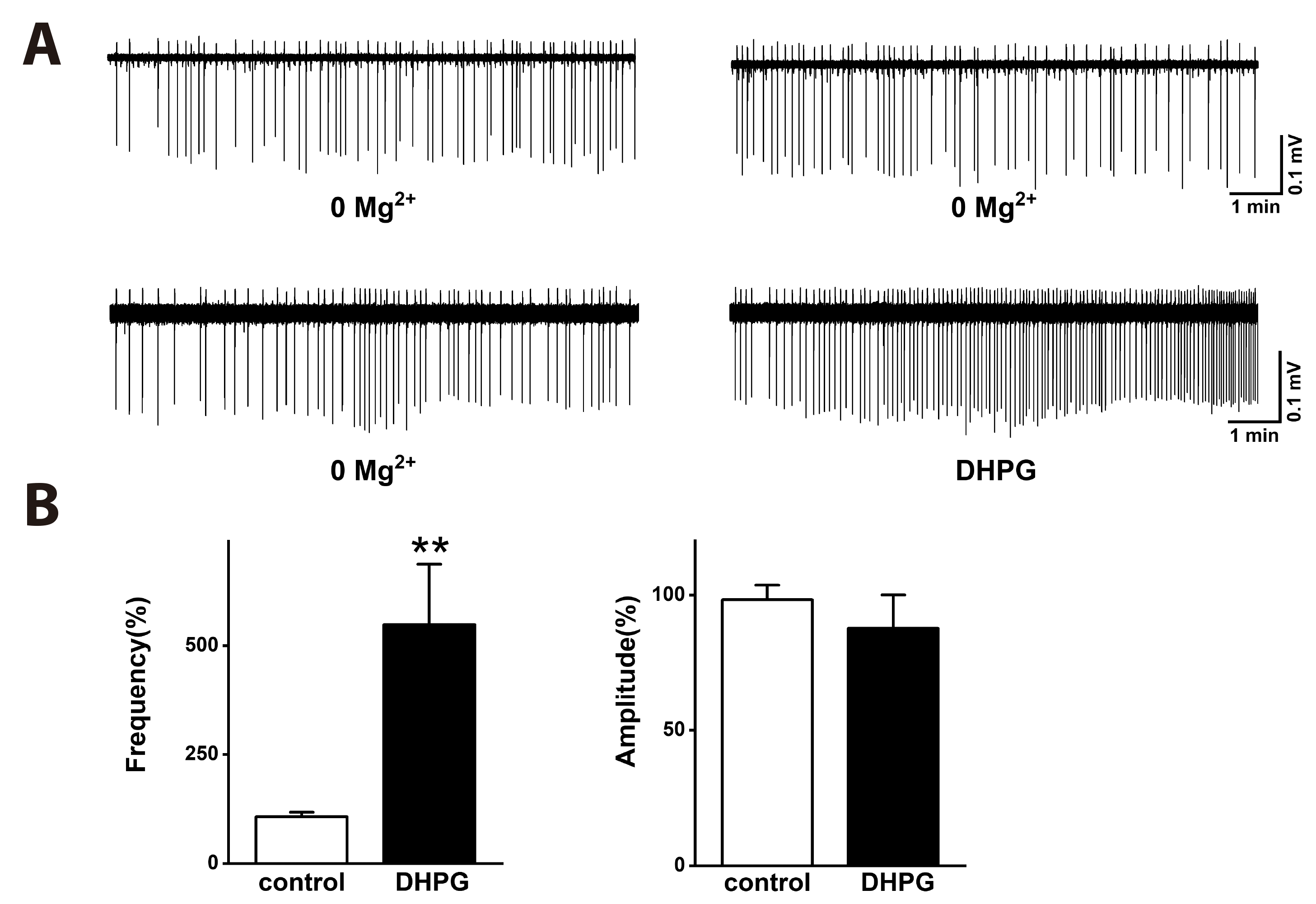
Fig. 3
The phospholipase C inhibitor U73122 inhibits the spike firing of low [Mg2+]o-induced epileptiform activity.
(A) Exposure to Mg2+-free ACSF induced epileptiform activity in the CA1 region. U73122 (10 μM) significantly inhibited the spike firing of low [Mg2+]o-induced epileptiform activity. (B) Graph summarizes the spike firing and amplitude of low [Mg2+]o-induced epileptiform activity in non (control, n = 6)- and U73122 (n = 7)-treated groups. Data are expressed as means ± SEM. [Mg2+]o, extracellular Mg2+ concentration; ACSF, artificial cerebrospinal fluid. **p < 0.01 relative to respective control (non-paired Student’s t-test).
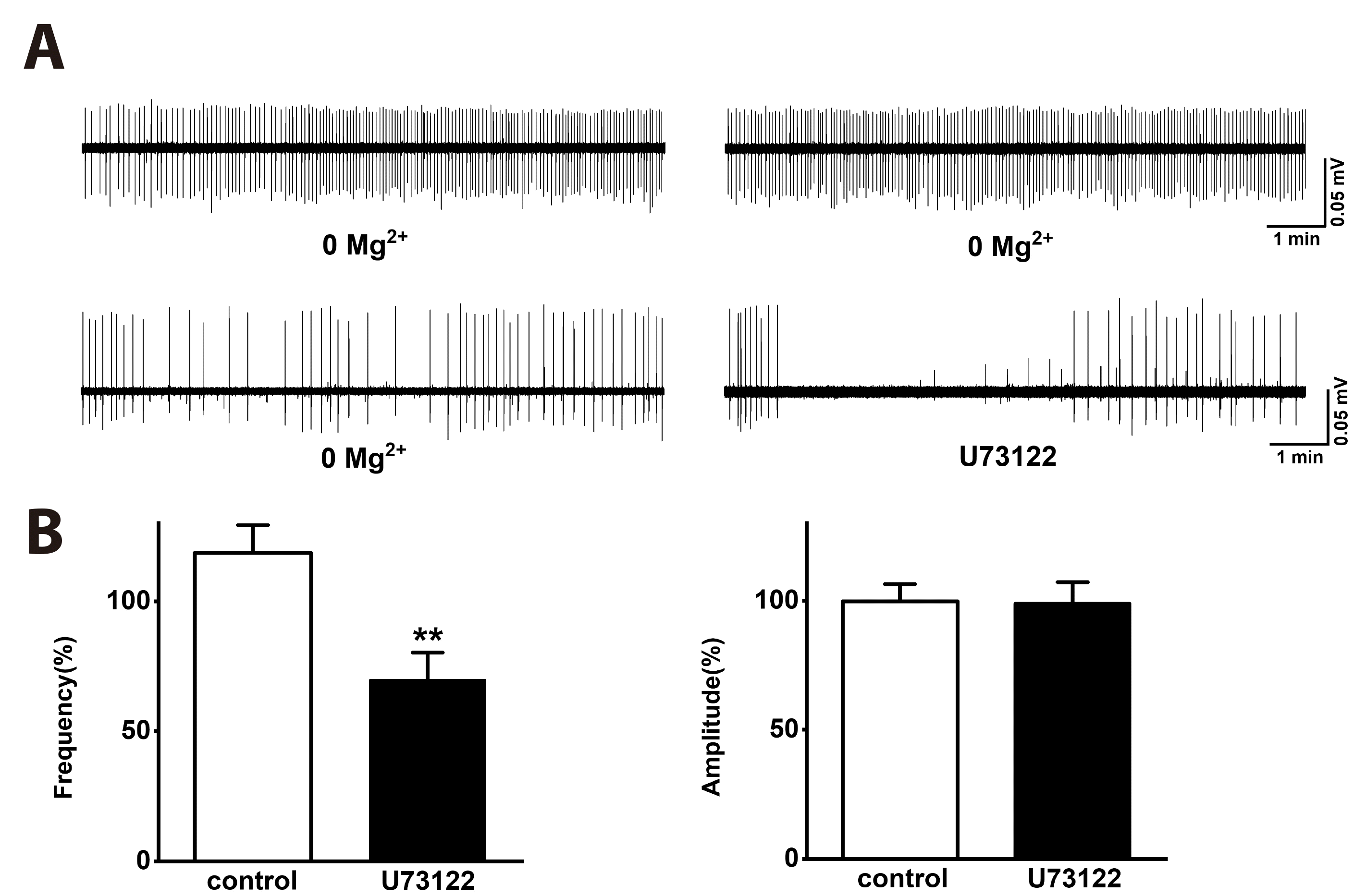
Fig. 4
Inhibitory effects of the ER Ca2+-ATPase inhibitor thapsigargin, the IP3 receptor antagonist 2-APB, and the ryanodine receptor antagonist dantrolene on low [Mg2+]o-induced epileptiform activity.
(A1) Exposure to Mg2+-free ACSF induced epileptiform activity in the CA1 region of acute hippocampal slices. Thapsigargin (10 μM), which induces depletion of ER Ca2+ stores, significantly inhibited the spike firing and amplitude of low [Mg2+]o-induced epileptiform activity. (A2) Graph summarizes the spike firing and amplitude of the epileptiform activity in non (control, n = 6)-, thapsigargin (n = 6)-treated groups. (B1) IP3 receptor antagonist 2-APB (30 μM) and ryanodine receptor antagonist dantrolene (30 μM) significantly inhibited the spike firing of low [Mg2+]o-induced epileptiform activity. (B2) Graph summarizes the spike firing and amplitude of the epileptiform activity in non (control, n = 8)-, 2-APB (n = 6)-, and dantrolene (n = 6)-treated groups. Data are expressed as means ± SEM. ER, endoplasmic reticulum; IP3, inositol-1,4,5-trisphosphate; 2-APB, 2-aminoethoxydiphenyl borate; [Mg2+]o, extracellular Mg2+ concentration; ACSF, artificial cerebrospinal fluid. *p < 0.05 relative to respective control (non-paired Student’s t-test), **p < 0.01 relative to respective control (non-paired Student’s t-test), #p < 0.05 relative to respective control (one way ANOVA with Bonferroni’s test).
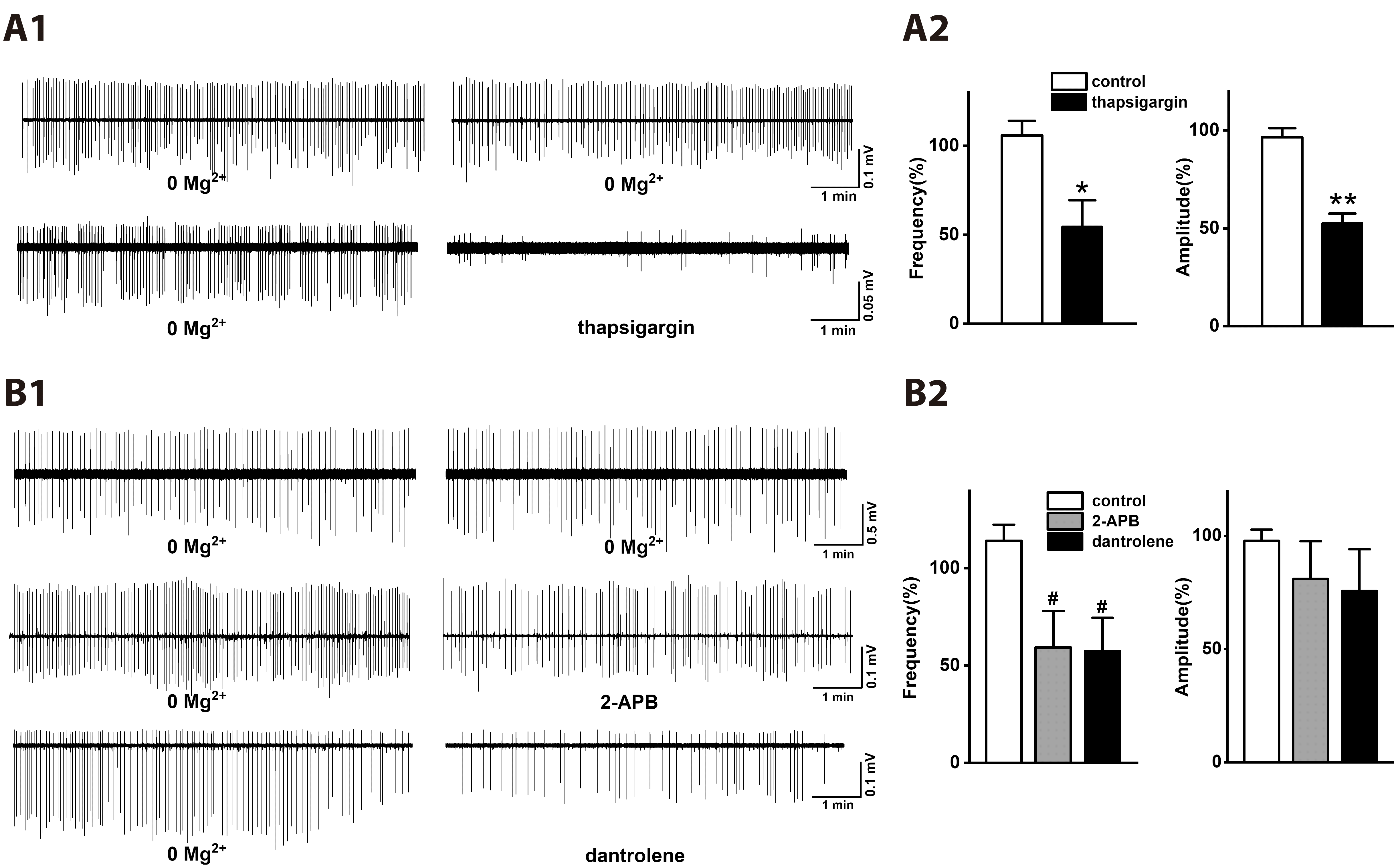
Fig. 5
The protein kinase C inhibitors GF109203X and chelerythrine increase the spike firing of low [Mg2+]o-induced epileptiform activity.
(A) GF109203X (1 μM) and chelerythrine (10 μM) significantly increased the spike firing of low [Mg2+]o-induced interictal epileptiform activity. (B) Graph summarizes the spike firing and amplitude of the interictal epileptiform activity in non (control, n = 8)-, GF109203X (n = 7)- and chelerythrine (n = 5)-treated groups. Data are expressed as means ± SEM. [Mg2+]o, extracellular Mg2+ concentration. *p < 0.05 relative to control, **p < 0.01 relative to control (one way ANOVA with Bonferroni’s test).
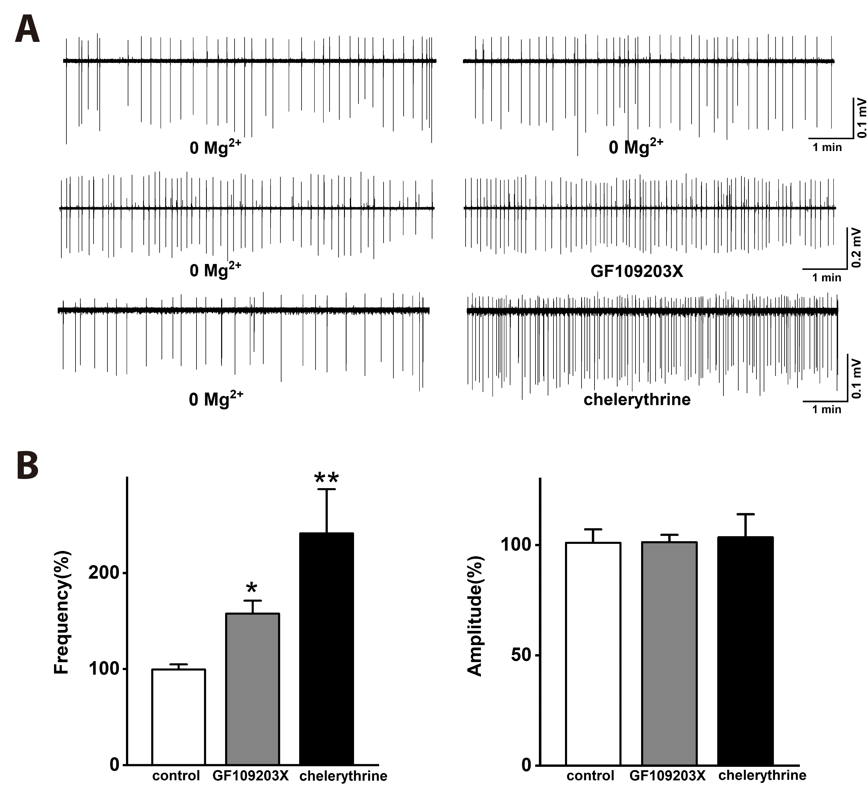
Fig. 6
The effects of the TRPC channel inhibitors such as flufenamic acid and SKF96365 on low [Mg2+]o-induced epileptiform activity.
(A) Flufenamic acid (100 μM) significantly inhibited the spike firing of low [Mg2+]o-induced epileptiform activity, but SKF96365 (10 μM) did not affect inhibited the spike firing of the epileptiform activity. (B) Graph summarizes the spike firing and amplitude of low [Mg2+]o-induced epileptiform activity in non (control, n = 7)-, flufenamic acid (n = 7)- and SKF96365 (n = 7)-treated groups. Data are expressed as means ± SEM. TRPC, transient receptor potential canonical; [Mg2+]o, extracellular Mg2+ concentration. *p < 0.05 relative to respective control (one way ANOVA with Bonferroni’s test).
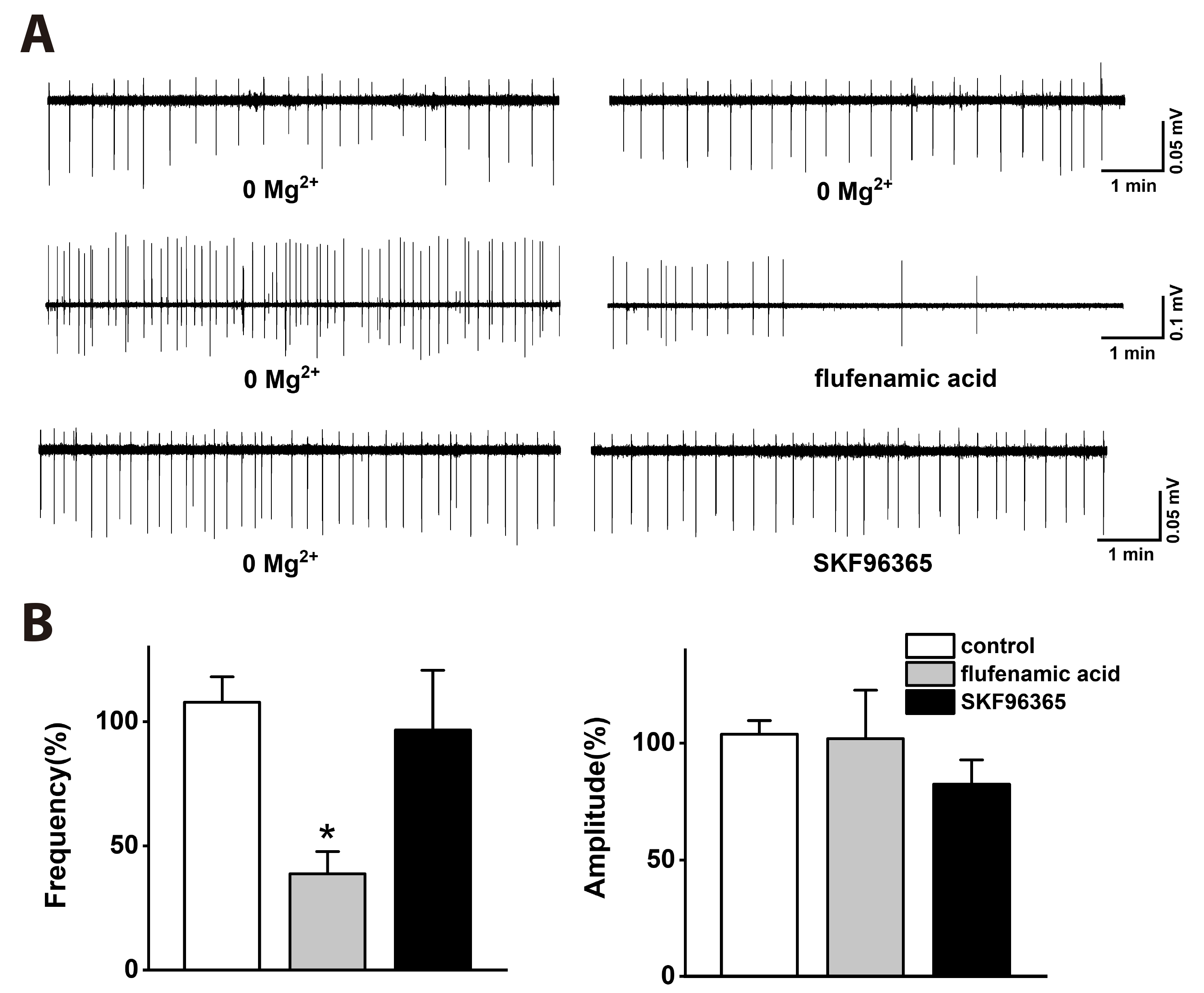
Fig. 7
The mGluR5 antagonist MPEP inhibits low [Mg2+]o-induced PI incorporation in the CA1 regions of rat hippocampal slices.
Hippocampal slices were exposed to normal [Mg2+]o or low [Mg2+]o DMEM solution (See METHODS) for 24 h in the absence or presence of mGluR antagonists such as MPEP (50 µM) and LY367385 (100 µM). The viability of neuronal cells was assessed by PI incorporation after 30 min exposure to the red fluorescent dye, PI. PI incorporation was shown as a percentage of 70% ethanol-induced maximal PI. (A) Bar graph shows PI incorporation in non (control: normal [Mg2+]o, n = 12; low [Mg2+]o, n = 14)-, MPEP (normal [Mg2+]o, n = 7; low [Mg2+]o, n = 6)- and LY367385 (normal [Mg2+]o, n = 6; low [Mg2+]o, n = 6)-treated groups in the CA1 and CA3 regions. Exposure to normal [Mg2+]o DMEM solution induced a basal PI incorporation. Exposure to low [Mg2+]o DMEM solution significantly increased PI incorporation in the CA1 and CA3 regions. MPEP significantly decreased the low [Mg2+]o-induced PI incorporation in the CA1 region. However, MPEP or LY367385 did not affect the low [Mg2+]o-induced PI incorporation in the CA3 region. (B) Representative phasecontrast photomicrographs showing rat hippocampal slices 24 h after exposure to normal [Mg2+]o or low [Mg2+]o DMEM solution in the absence or presence of mGluR antagonists. Data are expressed as means ± SEM. mGluR, metabotropic glutamate receptor; MPEP, 6-Methyl-2-(phenylethynyl) pyridine; [Mg2+]o, extracellular Mg2+ concentration; PI, propidium iodide; DMEM, Dulbecco’s modified eagle media. **p < 0.01 relative to respective control, ##p < 0.01 relative to normal [Mg2+]o. Scale bars: 100 μm.


 PDF
PDF Citation
 Print
Print


 XML Download