Abstract
During adolescence, traumas in the subscapularis muscle lead more often to osseous avulsion of the lesser tuberosity or subscapularis tendon, as opposed to often to intrasubstance tendinous injury in adults. This injury is commonly diagnosed as persistent shoulder pain, and it is difficult to diagnose immediately. This article is a case report of teenage patient who underwent open reduction and internal fixation for a displaced lesser tuberosity avulsion fracture. After experiencing a sports trauma 8 months ago, a 14-year-old male came to our clinic complaining of right shoulder pain and weakness. We used a cannulated screw to humeral lesser tuberosity and repaired infraspinatus additionally. We applied a progressive physical therapy regimen postoperatively. On postoperative 6 months, full range of motion of shoulder joint and strength was restored. We report our successful surgical treatment outcome of a fracture of the lesser tuberosity in an adolescent.
In the shoulder joint, lesser tuberosity avulsion fracture is a rare injury and consists of about 2% of proximal humerus fractures1. The first case report of an isolated lesser tuberosity avulsion fracture was published in 19852. Lesser tuberosity avulsion injuries in adolescents are even more uncommon but it was reported that those injuries have gradually increased in the recent literature3. With increased participation in athletic activity in younger patients, accidents and traumas have grown, especially at that age4.
The injury mechanism is acute forced shoulder joint external rotation with the abduction of arm, which has been reported in younger patients who are enjoying sports such as football, hockey, volleyball, wrestling, and skateboarding5. The avulsion injury occurs by sudden traction force on the lesser tuberosity caused by counter contraction of the subscapularis muscle to oppose the external rotation movement of the shoulder joint6. During adolescence, traumas in the subscapularis muscle lead more often to osseous avulsion of the lesser tuberosity or subscapularis tendon, as opposed to often to intrasubstance tendinous injury in adults7.
For acute and displaced injuries, treatment seems not debatable and results of surgical repair appear good. However, because of the variability of the mechanism and presentation of these injuries, the diagnosis is often missed initially8. Diagnosis of this injury is often challenging, leading to delays in treatment. Missing to diagnose these injuries may lead to nonunion of bony structures and eventually lead to chronic shoulder pain and muscle weakness9.
In this paper, we report the case of a 14-year-old boy who complained of persistent shoulder pain and muscle weakness after an injury 8 months ago.
This report was approved by the Institutional Review Board of Soonchunhyang University Bucheon Hospital (No. 2024-02-022). Written informed consent was obtained from the patient for the publication of this report including all clinical images.
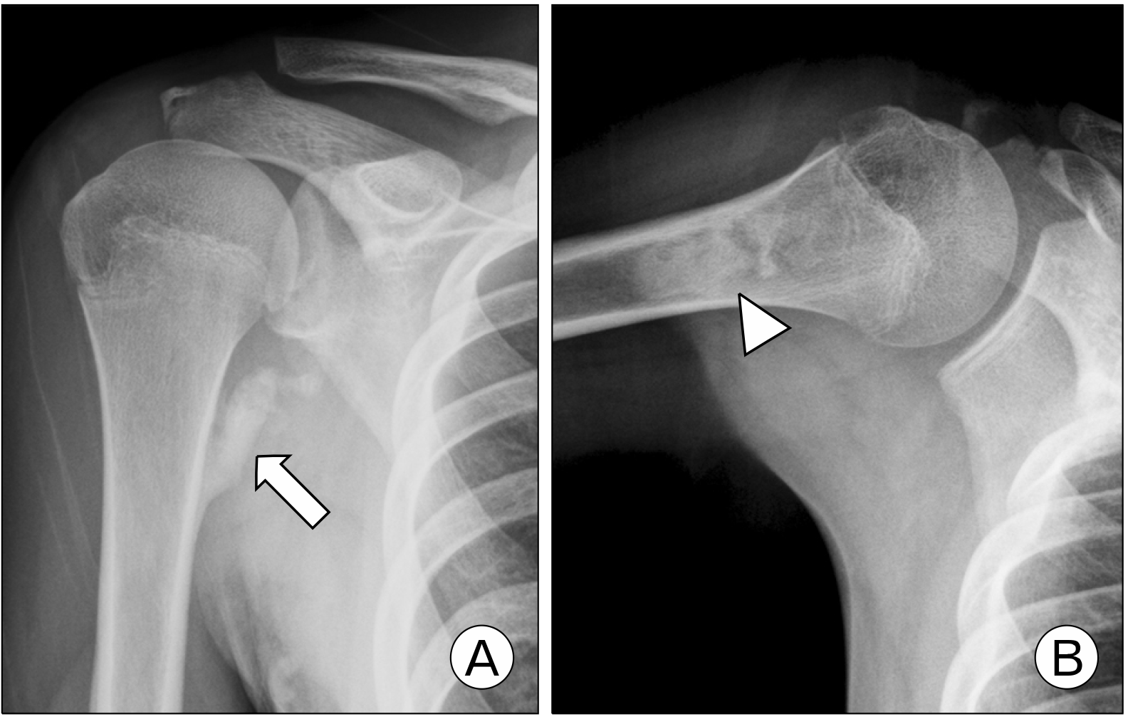
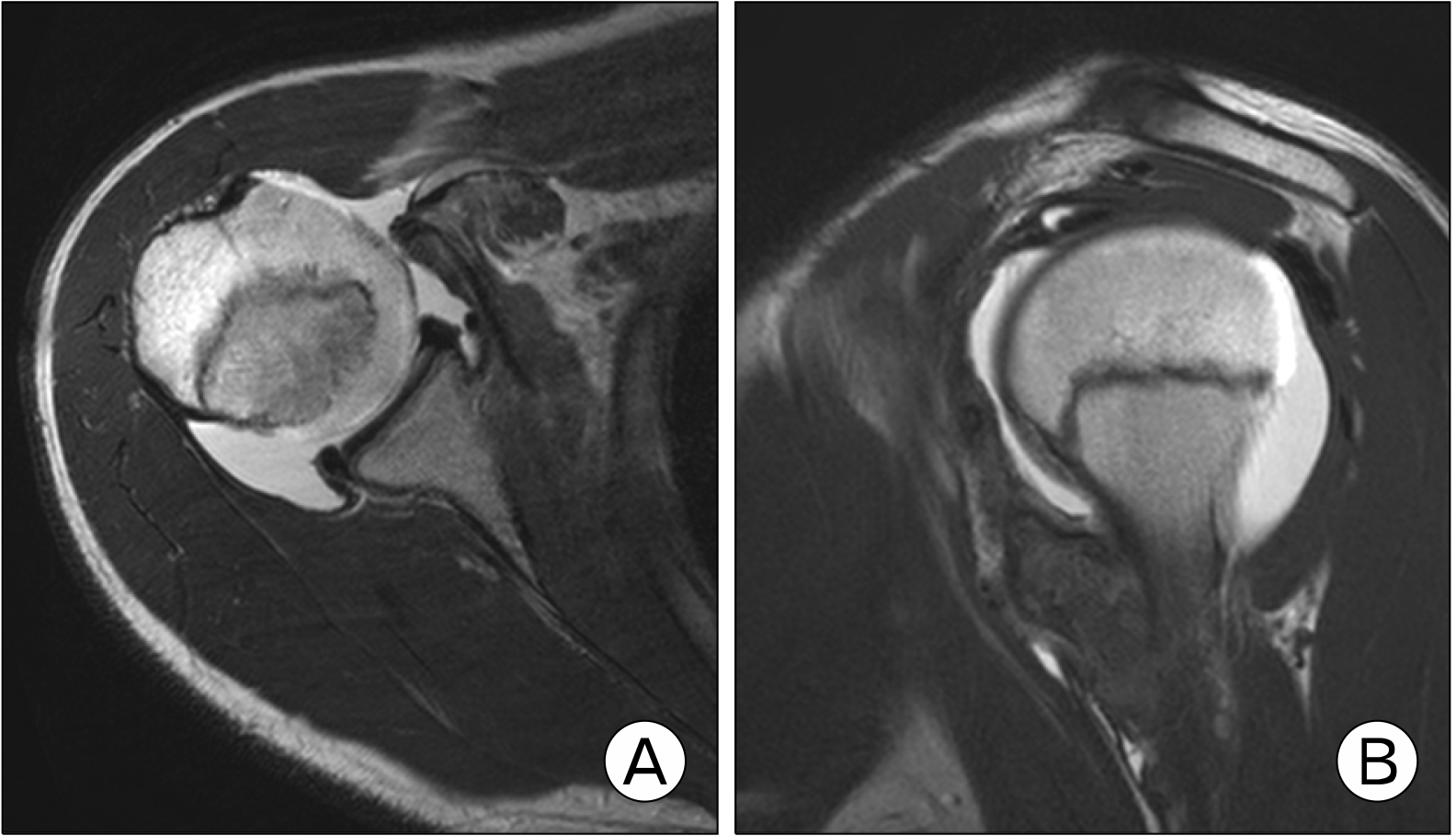
A 14-year-old male patient came to outpatient presenting with right shoulder joint pain and muscle weakness. Eight months ago, he had a collision on his right shoulder with another person’s elbow while playing soccer. Although he had no sign of dislocation and just had edema and severe bruise at that time, right shoulder joint pain and muscle weakness appeared continuously. At another local hospital, he heard that a benign bone tumor was suspected. On physical examination, frontal flexion and internal rotation showed reduced in both active and passive range of motion (ROM), and the subscapularis lift-off test and the belly-press test showed positive signs. On the right shoulder anteroposterior radiograph, about 55×10-mm size bone shadow was observed in the metaphysis of the proximal humerus; and on the lateral radiograph, a 40×13-mm size bone shadow was observed parallel with the diaphysis of the proximal humerus (Fig. 1). On magnetic resonance imaging (MRI), bone defect of the lesser tuberosity, complete rupture of subscapularis, and about 55×13×20-mm irregular shape of bony lesion were observed (Fig. 2).
The authors identified the condition as a missed isolated fracture of the lesser tuberosity of the proximal humerus in adolescents, and therefore planned surgery.
Under general anesthesia with beach chair position, surgery approached by axillary incision, between the deltoid muscle and pectoralis major muscle at the insertion site of the infraspinatus of the humerus lesser tuberosity. In the operative field, infraspinatus was not visible and the bone defect was observed at the medial aspect of the insertion site, as a 55×13-mm size bone fragment accompanying infraspinatus tendon with ossified tissue showed adhesion. Ossified tissue was removed from bone fragments by using a saw. After the bone fragment with infraspinatus was attached to humeral lesser tuberosity using a cannulated screw. Also, an additional suture of infraspinatus was done five times with Ethibond (Ethicon, Inc.). Finally, after meticulous coagulation, irrigation and skin suture were done.
On postoperative day 1, passive ROM of the shoulder joint was initiated except for external rotation. A brace was applied while gentle passive ROM exercises were performed for 4 weeks. After postoperative 7 weeks, active ROM of the shoulder joint started a using shoulder joint rehabilitation kit. At 3 months postoperatively, The University of California at Los Angeles (UCLA) shoulder score was 28 points and the constant score was 81 points. At 3 months postoperatively, full ROM of the shoulder joint and strength was restored, and UCLA score and constant score were 32 and 94 points, respectively.
The case of isolated fracture of the lesser tuberosity of the proximal humerus in children or adolescents is very rare worldwide. In Korea, Yum et al.10 reported one case. Compared to previous cases of lesser tuberosity avulsion fracture of the humerus in an adolescent, this patient had a much larger and displaced bony fragment. This injury is hard to diagnose early and about half of cases are presented as chronic shoulder joint pain. Missing early diagnosis of this could cause later clinical disorders.
The injury mechanism is as follows: avulsion of subscapularis when acute external rotation force was applied with proximal humerus externally rotated6. Diagnosis of isolated avulsion fracture of lesser tuberosity without dislocation of shoulder joint preferentially needs identification of injury mechanism. In addition, for patients complaining of shoulder pain, if there is no improvement in symptoms after sustained conservative treatment after injury, the disease should be suspected. Those patients may show mild anterior shoulder joint tenderness or mild limit of internal rotation of the shoulder joint2. In that case, a close review of a simple X-ray could be helpful, as specific appearance shows that contracted infraspinatus draws an avulsed bone fragment from infraspinatus’ insertion site to subarticular space11,12. Additional evaluation tools such as computed tomography (CT) and MRI can be considered. CT can accurately show bony defect area, degree of displacement of bony fragment, size of bone fragment, and fracture type. MRI is a helpful study showing the status of nearby soft tissue like infraspinatus and bone fragments, and soft tissue injury such as a tear of biceps brachii or glenoid labrum. After plain radiographs and magnetic resonance arthrography were performed, CT was not performed because we thought that sufficient data were obtained.
Standard surgical indication of humeral lesser tuberosity fracture is displaced fracture (fracture gap is more than 5 mm, fracture angulation is bigger than 45°), mechanical motion difficulty, failure of conservative treatment, and severe accompanied injury. But Ogawa and Takahashi6 conclude conservative treatment can obtain satisfying results in acute, nondisplaced humeral lesser tuberosity fracture, but regardless of size or displacement of a bony fragment, surgical treatment shows superior results. In complex comminuted fractures, by drilling a hole in the proximal humerus and then anchoring absorbable or nonabsorbable suture, fixation can be achieved causing minimal damage on the lesser tuberosity. After then fixation power can be increased by a screw with suture, in case of large size bone fragment fixation can be achieved by cortical screw13,14. However, usage of this screw can cause physeal injury to humeral lesser tuberosity and early closure of the growth plate; therefore, it should be implemented carefully.
We decided on surgical treatment considering clinical symptoms and MRI findings which show a complete rupture of the infraspinatus tendon and about 55×13-mm size irregular shape bony defect. Despite the possibility of physeal injury to firmly fix a large bone defect, the authors used a cannulated screw to humeral lesser tuberosity and repaired the infraspinatus additionally.
This study has several limitations. This patient was followed up for only 6 months. Later, continuous follow-up is needed about physeal injury and clinical course. Since this is a rare case, this paper consists of only one case. In the future, it is thought that systematic review will be needed by studying more cases at multiple institutions.
In adolescents, isolated avulsion fracture of humeral lesser tuberosity is far rare. This injury is hard to diagnose early and sometimes can appear as chronic shoulder joint pain. Missing early diagnosis of this injury case can cause important clinical disorders later. So early diagnosis and treatment open reduction and internal fixation to isolate fracture of the lesser tuberosity of the proximal humerus in adolescents if needed.
Acknowledgments
This work was supported by the Soonchunhyang University Research Fund (No. 2024-0020).
REFERENCES
1. Goeminne S, Debeer P. 2012; The natural evolution of neglected lesser tuberosity fractures in skeletally immature patients. J Shoulder Elbow Surg. 21:e6–11. DOI: 10.1016/j.jse.2012.01.017. PMID: 22572400.
2. White GM, Riley LH Jr. 1985; Isolated avulsion of the subscapularis insertion in a child: a case report. J Bone Joint Surg Am. 67:635–6. DOI: 10.2106/00004623-198567040-00021. PMID: 3980510.
3. Neogi DS, Bejjanki N, Ahrens PM. 2013; The consequences of delayed presentation of lesser tuberosity avulsion fractures in adolescents after repetitive injury. J Shoulder Elbow Surg. 22:e1–5. DOI: 10.1016/j.jse.2012.12.004. PMID: 23415817.
4. Vavken P, Bae DS, Waters PM, Flutie B, Kramer DE. 2016; Treating subscapularis and lesser tuberosity avulsion injuries in skeletally immature patients: a systematic review. Arthroscopy. 32:919–28. DOI: 10.1016/j.arthro.2015.10.022. PMID: 26786826.
5. Sugalski MT, Hyman JE, Ahmad CS. 2004; Avulsion fracture of the lesser tuberosity in an adolescent baseball pitcher: a case report. Am J Sports Med. 32:793–6. DOI: 10.1177/0095399703258620. PMID: 15090399.
6. Ogawa K, Takahashi M. 1997; Long-term outcome of isolated lesser tuberosity fractures of the humerus. J Trauma. 42:955–9. DOI: 10.1097/00005373-199705000-00029. PMID: 9191680.
7. Vezeridis PS, Bae DS, Kocher MS, Kramer DE, Yen YM, Waters PM. 2011; Surgical treatment for avulsion injuries of the humeral lesser tuberosity apophysis in adolescents. J Bone Joint Surg Am. 93:1882–8. DOI: 10.2106/JBJS.K.00450. PMID: 22012525.
8. Harper DK, Craig JG, van Holsbeeck MT. 2013; Apophyseal injuries of the lesser tuberosity in adolescents: a series of five cases. Emerg Radiol. 20:33–7. DOI: 10.1007/s10140-012-1064-x. PMID: 22895662.
9. Sikka RS, Neault M, Guanche CA. 2004; An avulsion of the subscapularis in a skeletally immature patient. Am J Sports Med. 32:246–9. DOI: 10.1177/0363546503258906. PMID: 14754752.
10. Yum JK, Chung HJ, Choi EO, Lee SL. 2006; Isolated avulsion of the lesser tuberosity of the humerus in an adolescent judo player: a case report. Clin Shoulder Elb. 9:119–23. DOI: 10.5397/CiSE.2006.9.1.119.
11. Coates MH, Breidahl W. 2001; Humeral avulsion of the anterior band of the inferior glenohumeral ligament with associated subscapularis bony avulsion in skeletally immature patients. Skeletal Radiol. 30:661–6. DOI: 10.1007/s002560100430. PMID: 11810162.
12. Shibuya S, Ogawa K. 1986; Isolated avulsion fracture of the lesser tuberosity of the humerus: a case report. Clin Orthop Relat Res. (211):215–8. DOI: 10.1097/00003086-198610000-00027. PMID: 3769259.
13. Levine B, Pereira D, Rosen J. 2005; Avulsion fractures of the lesser tuberosity of the humerus in adolescents: review of the literature and case report. J Orthop Trauma. 19:349–52. PMID: 15891546.
14. McAuliffe TB, Dowd GS. 1987; Avulsion of the subscapularis tendon: a case report. J Bone Joint Surg Am. 69:1454–5. DOI: 10.2106/00004623-198769090-00024. PMID: 3440808.


 PDF
PDF Citation
 Print
Print


 XML Download