Journal List > Korean J Sports Med > v.42(3) > 1516088232

무릎 손상 및 질환 환자에서 혈류 제한 운동의 적용: 최신 경향 분석

Abstract

In patients who have undergone knee joint surgery or suffer from osteoarthritis, weakened quadriceps muscle strength is often observed. This can lead to various pathological issues in the long term, such as abnormal gait and persistent knee pain. Due to the need to protect against pain or surgical site complications, high-intensity exercise is often restricted for most patients with muscle atrophy in the knee joint due to surgery or osteoarthritis or surgery. To overcome this challenge, various methods are being attempted, including exercise combined with neuromuscular electrical stimulation, blood flow restriction (BFR) exercise, and biofeedback exercise. Recently, BFR exercise has garnered attention in line with these strategic trends. Applying low-intensity BFR exercise to patients who have undergone anterior cruciate ligament reconstruction surgery or suffer from knee joint osteoarthritis, most studies report improvements in strength, muscle mass, and functional enhancement of the knee joint. Compared to non-BFR high-intensity exercise, it has been reported that increases in maximal strength and muscle mass are similar, but pain reduction is superior with BFR exercise. Engaging in low-intensity BFR exercise for a sufficient duration can minimize the risk of injury associated with high-intensity exercise while maximizing the exercise’s effectiveness, leading to symptom improvement and enhancement of knee joint function. Furthermore, when conducted according to specified manuals, the likelihood of cardiovascular imbalance, muscle damage, thrombosis, and embolism due to BFR is low, making it a safe rehabilitation method.

서 론

하지 근육 중에서 대퇴사두근은 다양한 일상적인 활동과 운동에서 필수적인 역할을 수행하는데, 이는 슬관절의 신전, 관절의 안정화, 신체의 자세 및 균형 유지 등의 기능을 하기 때문이다1. 슬관절 수술 환자들 혹은 골관절염 환자들에서 대퇴사두근력이 약화된 경우를 많이 보게 되는데, 이는 병적 보행, 지속적인 무릎 통증 등 여러 가지 병리학적 문제를 장기적으로 유발할 수 있다2. 슬관절 수술 후 대퇴사두근력의 저하가 지속되면 수술 실패로까지 이어질 수 있다. 실제로 전방십자인대 재건술을 받은 환자의 30%에서 대퇴사두근 근력 약화 및 근 위축이 보고되고 있고, 이는 수술 후 재파열 발생률 및 동요도 등의 지표와 연관성을 보이고 있다3. 이러한 대퇴사두근의 위축은 슬관절 수술 이후 단순히 환측에 국한되어 나타나는 것이 아니라 양측에서 동시에 발생하는 경우들도 많다4.
대퇴사두근 위축은 단순 통증에 의하기보다는 복잡한 기전으로 발생하는데, 대표적인 이론이 신경학적 억제에 의해 근 활성화가 제한된다는 관절기원성 근 억제(arthrogenic muscle inhibition, AMI) 이론이다. 이런 신경학적 억제를 방지하기 위해 저항성 운동이 대표적으로 사용되는데, 이때 운동 강도의 선택이 중요하다. 1회 최대 반복 중량의 20%–30%는 저강도, 60%는 중강도, 80%–90%는 고강도로 정의하는데, 근력을 향상하기 위해서는 1회 최대 반복 횟수(repeated maximum)의 60%–70% 이상, 근 비대를 위해서는 1회 최대 반복 중량의 70%–85% 수준으로 강도 높은 운동을 시행해야 한다. 하지만 근 위축을 보이는 대부분의 슬관절 골관절염 환자들 혹은 수술 후 환자들은 통증 혹은 수술 부위를 보호해야 한다는 이유로 이러한 고강도 운동 수행이 제한된다5-8. 또한, 고강도 운동으로 발생하는 기계적 스트레스가 관절이 감당할 수 있는 부하를 넘어서기에 관절 내 통증과 염증을 악화시킨다는 연구들도 있어 재활 프로그램 초기에 고강도 운동을 포함하기에는 제한점들이 많다. 그러므로 다양한 기전들로 인해 나타난 대퇴사두근의 위축은 일반적인 운동만으로는 회복하기 쉽지 않다. 이를 극복하기 위해 다양한 방법들이 시도되고 있는데, 대표적으로 근신경 전기자극을 병행한 운동, 혈류 제한(blood flow restriction) 운동, 바이오 피드백 운동 등이 있다9,10. 이런 운동들의 원리는 1회 최대 반복 중량의 60%보다 적은 저강도 운동을 하면서 고강도 운동을 할 때와 유사한 대사 스트레스 환경을 조성하여 근력 향상 및 근 비대 효과를 얻을 수 있다는 것이다11. 뿐만 아니라 고강도 운동 시 동반되는 통증 및 염증 반응을 감소시킬 수 있어 그 효용성이 점차 강조되고 있다12. 그러므로, 저강도에서 운동을 수행하면서 고강도 운동의 효과를 얻을 수 있는 운동 전략은 골관절염 및 수술 후 환자들의 기능 회복 프로그램에서 핵심이 될 것이다. 최근 혈류 제한 운동이 이러한 전략적 흐름에 따라 관심을 받고 있다.
혈류 제한 운동의 목적은 지속적인 통증, 수술 등 다양한 이유로 발생할 수 있는 근 위축을 방지하고 증상 발생 이전의 근력을 회복하는 것이다. 뇌경색, 척수 손상 환자 등 근 위약이 발생한 경우에 유용하게 사용되며, 근골격계 환자 중 특히 전방십자인대 재건술 환자 및 슬관절염 환자에서 사용되고 있다13. 전방십자인대 손상 및 슬관절염 환자들 모두 대퇴사두근의 위축이 발생할 가능성이 높아 적극적인 하지 근력 보존을 위한 근력 운동이 필수적이지만 상대적으로 고강도 훈련을 하기 어렵기 때문이다.
그러나, 아직 혈류 제한 운동의 적용 및 임상 결과에 대해 정리된 연구가 많지 않기에 본 종설에서는 (1) 혈류 제한 운동의 정의 및 원리, (2) 전방십자인대 손상 환자들의 혈류 제한 운동 적용 및 임상 결과, (3) 골관절염 환자들의 혈류 제한 운동 적용 및 임상 결과 순서로 최신의 문헌들을 정리해서 살펴보고자 한다.

본 론

1. 혈류 제한 운동의 정의 및 원리

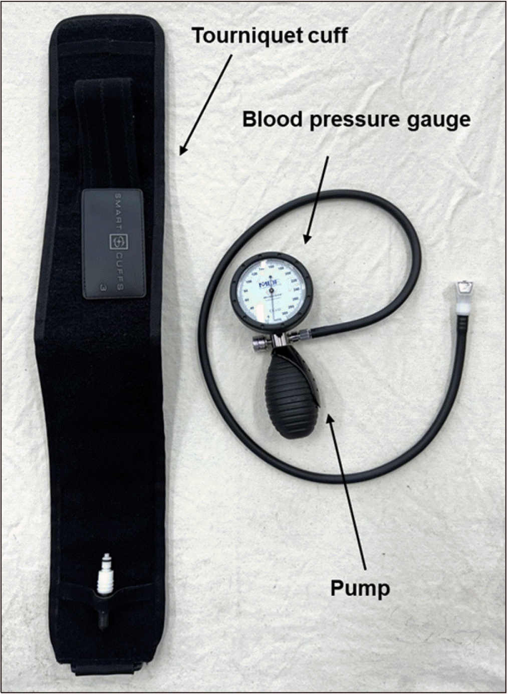
혈류 제한 운동은 가압 훈련(occlusion training) 또는 KAATSU 훈련이라고도 불리며, 혈압 커프 혹은 스트랩 등을 사지의 근위부에 적용하여 사지 원위부의 동맥 혈류를 부분적으로, 정맥 혈류를 완전히 폐쇄한 후 운동하는 것을 말한다. 이 운동은 2004년 일본에서 Sato 박사에 의해 개발되었다14,15. 혈류 제한 운동을 위한 장비는 토니켓 커프(tourniquet cuff), 혈압계(blood pressure gauge), 펌프(pump) 등으로 구성된다(Fig. 1)16.
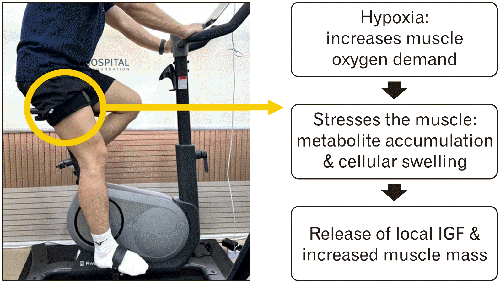
혈류 제한 기기의 원리는 토니켓 커프에 공기를 주입하여 근육의 혈류를 제한함으로써 조직 내 저산소(hypoxia) 환경을 만들어 수행하는 부위에 일시적인 허혈(ischemia) 상태를 만드는 것이다17. 근육 내 조성된 저산소성 환경은 대사 스트레스를 유발하여 혈류 제한에 의해 근 합성의 주요 신호전달 체계인 mammalian target of rapamycin (mTOR) 경로 발현 및 동화작용의 증가를 유도한다18,19. 이는 세포 증식에 관여하는 인슐린양 성장인자(insulin-like growth factor) 등 여러 성장인자(growth factors)의 생성을 유도하여 보다 많은 근섬유의 동원 및 근육의 비후화로 이어지게 된다(Fig. 2)20. 이는 고강도 훈련을 시행하였을 때와 비슷한 수준의 외측광근 근 합성 및 mTOR 경로의 발현 증가이다21. 또한 단백질 티로신 키나아제(protein tyrosine kinase) 같은 신호전달을 통해 미토콘드리아 투과성 전이 기공(mitochondrial permeability transition pore)을 억제하고 전체 활성산소(oxygen-free radical)의 농도를 줄여 손상을 최소화할 수 있으며, 근육의 대사 요구량(metabolism requirement)을 충족시켜 운동 수행 능력(exercise performance ability) 향상 효과를 기대해 볼 수 있다18.

2. 전방십자인대 손상 환자에서의 혈류 제한 운동 적용 및 임상 결과 분석

전방십자인대 재건술을 받은 환자의 30%가 대퇴사두근 근력 약화 및 근 위축을 보이면서 수술 후에도 상당 기간 유지될 수 있는데, 이는 수술 후 재파열 발생 및 잔존 동요도 등의 지표와 연관이 있는 것으로 알려져 있다3. 이를 극복하기 위한 방법으로 전방십자인대 재건술 후 혈류 제한 운동은 대부분의 연구에서 대퇴사두근과 더불어 햄스트링, 내전근 등의 근력 상승 및 근 비대 효과를 보여줬다5,22-25.
3개월 이상의 혈류 제한 적용에 대해서는 임상적으로 유효하다는 연구 결과들이 많다(Table 1)5,22-27. Ohta 등23의 연구에서는 전방십자인대 재건술을 받은 젊은 남녀 44명을 대상으로 3개월간 혈류 제한 저강도 운동 그룹과 비 혈류 제한 저강도 운동 그룹으로 나누어 최대 근력(슬관절 신전, 굴곡), 근 단면적(대퇴사두근, 햄스트링 및 내전근)을 비교한 결과, 혈류 제한 저강도 운동 그룹에서 비 혈류 제한 저강도 운동 그룹보다 유의미한 최대 근력의 상승과 근 단면적의 상승을 보였다고 하였다. 또한 그룹별로 8명의 환자에서 근 생검을 통한 type 1, 2 근섬유의 직경을 비교하였는데, 혈류 제한 저강도 운동 그룹에서 근섬유 직경의 증가를 관찰할 수 있었지만 그 차이가 유의미한 정도는 아니었다. 이 외에도 Table 1에 기술된 다른 연구들에서 골밀도 검사(dual-energy X-ray absorptiometry)를 통한 근육량, 근지구력(실패 지점까지의 최대 횟수), 근전도 활성화, 근육으로의 혈류 등이 혈류 제한 운동 그룹에서 유의미하게 높았다.
또한 Hughes 등5 은 전방십자인대 재건술을 받은 24명의 젊은 남녀를 대상으로 혈류 제한 저강도 운동 그룹, 비 혈류 제한 고강도 운동 그룹으로 나누어 근력, 근육량뿐만 아니라 슬관절 가동 범위, 신체기능 점수, 통증 정도를 비교하였다. 혈류 제한 저강도 운동 그룹에서 고강도 운동 그룹에 준하는 근력 및 근육량의 상승효과를 확인할 수 있었고, 오히려 고강도 운동 그룹에 비해 슬관절 가동 범위, 기능성 지표는 더 높고 통증 점수는 더 낮은 것을 확인할 수 있었다.

3. 슬관절 골관절염 환자에서의 혈류 제한 운동 적용 및 임상 결과 분석

슬관절 골관절염 환자에서 대퇴사두근의 근력 약화 및 근 위축이 관절염의 주요한 원인 및 악화 인자로 알려져 있다2. 특히, 영상학적인 관절염의 진행과 더불어 대퇴사두근의 근력이 일상생활 능력 및 기능 수행 능력과 밀접한 연관이 있어 그 중요성이 강조되고 있다28. 다수의 연구들에서 슬관절 골관절염 환자들에게 혈류 제한 운동을 적용한 결과, 근력 및 근육량 증가를 보였고 기능성 검사와 통증 정도 또한 개선되었다6-8.
슬관절 골관절염 환자들에게 혈류 제한 운동을 적용한 대표적인 연구들을 살펴보면 다음과 같다(Table 2)6-8,29,30. 슬관절 골관절염에서 단기간의 혈류 제한의 유용성에 대해서는 성별에 따른 상반된 연구 결과가 보고되었다. Segal 등29,30은 남성과 여성에 대해 각각 연구를 시행하였는데, 남성의 경우 45세 이상 골관절염이 있거나 골관절염 위험요인을 가진 41명을 대상으로 4주 동안 혈류 제한 저강도 운동 그룹과 비 혈류 제한 저강도 운동 그룹으로 나누어 최대 근력 및 통증 점수를 비교하였다. 최대 근력은 두 군 간 유의미한 차이가 없었으나, Knee Injury and Osteoarthritis Outcome Score (KOOS) 통증 점수는 혈류 제한 저강도 운동 그룹에서 더 높은 것을 확인할 수 있었다. 여성의 경우, 45–65세의 골관절염이 있거나 골관절염 위험요인을 가진 총 40명을 대상으로 4주 동안 혈류 제한 저강도 운동 그룹과 비 혈류 제한 저강도 운동 그룹으로 나누어 최대 근력, 근육량, 통증 점수를 비교하였는데, 최대 근력은 혈류 제한 저강도 운동 그룹에서 유의미하게 높았고, KOOS 통증 점수에서는 두 군 간 유의미한 차이가 없었다.
반면, 6주 이상의 혈류 제한을 다룬 연구들에서는 혈류 제한 운동이 임상적으로 유효하다는 연구 결과들이 많다. 대표적으로 Ferraz 등7의 연구에서 American College of Rheumatology 기준에 해당하는 Kellgren and Lawrence 등급 2, 3의 골관절염 여성 48명을 혈류 제한 저강도 운동 그룹, 비 혈류 제한 고강도 운동 그룹, 비 혈류 제한 저강도 운동 그룹으로 나누어 12주 동안 최대 근력, 근육량, 기능성 검사, 삶의 질 및 통증 점수 등을 비교하였다. 혈류 제한 저강도 운동 그룹에서 비 혈류 제한 고강도 운동 그룹에 준하는 최대 근력 및 근육량의 상승을 보였고, 비 혈류 제한 저강도 운동 그룹은 유의미한 변화가 없는 것을 확인할 수 있었다. 기능성 검사 중 timed stands test, Western Ontario and McMaster Universities Osteoarthritis Index (WOMAC) 기능성 지표에서 혈류 제한 저강도 운동 그룹과 비 혈류 제한 고강도 운동 그룹에서 유의미한 상승을 관찰하였고, WOMAC stiffness 지표는 혈류 제한 저강도 운동 그룹에서만 유의미한 개선을 보였으며, WOMAC 통증 지표는 혈류 제한 저강도 운동 그룹과 비 혈류 제한 저강도 운동 그룹에서 감소한 것을 확인할 수 있었다. 한편, 비 혈류 제한 고강도 운동 그룹에서만 25%의 환자들이 운동 중 통증으로 연구 도중 이탈되었다. 이외 타 연구들 또한 혈류 제한 저강도 운동 그룹이 비 혈류 제한 저강도 운동 그룹에 비해 근력 및 근육량의 상승, 기능성 지표의 개선을 확인할 수 있었다.

4. 혈류 제한 운동의 적용법

혈류 제한 운동 시, 혈류 제한 압력의 정도, 커프의 크기, 제한하는 시간, 운동 사이 휴식 등 설정해야 할 구체적인 방법은 연구마다 차이를 보인다. 본 종설에서 다룬 연구들에서 혈류 제한 압력은 130–200 mm Hg로 적용하였는데, 도플러를 통해 측정한 동맥 폐쇄 압력의 80% 등으로 혈류 제한을 적용하여 시행하였다. 커프의 크기 또한 6–20 cm까지 다양한 크기로 진행되었고 운동 사이 휴식은 30초–1분 등 다양하게 세팅하여 연구를 진행하였다. 혈류 제한 시간은 운동 수행 당시에만 적용하여 진행하거나 세션 내내 11분 이상 유지하는 연구자들도 있었다. 최근 혈류 제한 가이드라인을 구성하여 혈류 제한의 안정성과 효과를 극대화하기 위한 시도들이 있다. Patterson 등16은 혈류 제한 압력은 그 동맥의 폐쇄 정도가 개인마다 다르기 때문에 특정 수치로 하기보다는 개인의 동맥 폐쇄 압력의 40%–80% 정도로 하는 것이 가장 안전하다고 하였다. Mattocks 등31 은 혈류 제한 적용 환자들을 동맥 폐쇄 압력의 0%, 10%, 20%, 30%, 50%, 90%로 나누어 임상 시험을 실시하였는데, 혈류 제한 압력이 증가할수록 근육으로의 혈류 증가와 같은 심혈관적 반응을 강화할 수 있지만, 혈류 제한 압력이 너무 높으면 운동 강도가 상승하여 운동 자체의 총량이 감소하여 근 비대 효과는 떨어지는 것을 확인하였다. 반면, Hughes와 Patterson32은 낮은 혈류 제한(동맥 폐쇄 압력의 40%) 저강도 운동 그룹과 높은 혈류 제한(동맥 폐쇄 압력의 80%) 저강도 운동 그룹을 비교한 결과, 높은 혈류 제한 저강도 운동 그룹에서 통증 감소 효과가 뚜렷하였다고 하였다. Neto 등33은 혈류 제한 저강도 운동 그룹에서 지속적인 혈류 제한과 휴식 도중 혈류 제한을 풀어주는 간헐적인 혈류 제한으로 나누어 최대수의적 근력(등척성 근력 및 동적 근력), 근 비대, 근지구력 등을 비교하였는데, 근력, 근 비대는 두 그룹에서 차이가 없었고 근지구력의 경우는 지속적인 혈류 제한 그룹에서 더 높은 것을 확인하였다. Nielsen 등34은 혈류 제한 저강도 운동 그룹(100 mm Hg 압박)에서 고빈도, 수의적 실패 지점까지의 운동, 30초의 쉬는 시간, 지속적인 혈류 제한으로 세팅하고 적용하였을 때 근육 손상 발생은 없었다고 하였다. Mouser 등35은 커프의 너비는 동맥 폐쇄 압력의 분율로 혈류 제한을 할 경우 혈류 제한 운동 효과에 정도의 차이가 거의 없다고 하였다.

5. 혈류 제한 운동의 안정성

혈류 제한 운동의 적용법에 따른 안전성은 여러 연구들을 통해 입증되었다. Okuno 등36은 혈류 제한을 통한 1회 최대 반복 중량의 40% 수준의 저강도 운동은 1회 최대 반복 중량의 80% 수준의 고강도 운동에 비해 미주신경 조절(vagal regulation)의 빠른 회복을 가져올 수 있다고 하였다. Neto 등37은 지속적인(continuous) 혈류 제한 운동을 한 그룹과 간헐적인(intermittent) 혈류 제한 운동을 한 그룹 모두에서 근 손상이 일어나지 않았으며, 오히려 군인과 같이 규칙적인 운동과 훈련을 했던 그룹이 단관절 근육의 근지구력 향상을 보였고, 근 손상이 적었다고 하였다.
혈류 제한 운동 적용 시 발생할 수 있는 합병증 등으로 심혈관계 이상 반응, 하지 허혈, 혈전 생성 등이 있다38. Patterson과 Ferguson39은 혈류 제한 저강도 운동 후 심혈관계 합병증에 대해 조사한 결과, 말초 혈류 역학의 변동을 보인다고 하였다. 그러나, Clark 등40은 맥 경직도를 반영하는 맥파 속도(pulse wave velocity), 발목상완지수(ankle brachial index) 값에서 큰 변화가 없었다고 하였다. Takano 등41은 혈류 제한 저강도 운동이 고강도 운동에 비해 혈압, 심박수 등의 상승이 현저히 줄어든다고 하였고, 심박출량의 변화 또한 미비하여 상대적으로 안전하다고 하였다. Clark 등40은 젊은 성인 16명을 대상으로 4주 동안 혈류 제한 저강도 운동을 시행하였을 때 정맥 혈전 색전증의 대표적 지표인 D-dimer, 고감도 C-반응 단백질(high-sensitive C-reactive protein, hs-CRP), 프로트롬빈 시간과 같은 수치들이 상승하지 않았던 것을 확인하고, 정맥 혈전 색전증에 대한 안전성을 입증하였다. Tennent 등42도 18–65세에서 슬관절 관절경 수술을 받은 환자들 17명을 대상으로 6주 동안 운동을 시행하고, 혈류 제한 운동 그룹(동맥 폐쇄 압력의 80%)과 비 혈류 제한 운동 그룹으로 나누어 초음파 검사를 하였을 때, 혈전 발생은 관찰되지 않았다고 하였다. Madarame 등43은 항응고제 복용 중이 아닌 안정성 허혈성 심장병 환자 9명을 대상으로 혈류 제한 저강도 운동을 적용한 결과, 심박수와 노르아드레날린(noradrenaline) 수치는 수술 후 유의미한 상승을 보였고 D-dimer, 피브리노겐/피브린 분해 생성물(fibrinogen/fibrin degradation products), hsCRP 수치는 운동 전후로 유의미한 상승을 보이지 않아 안정성 허혈성 심장병 환자에게 상대적으로 안전하게 혈류 제한 저강도 운동을 적용해 볼 수 있다고 하였다. 단, 연구 결과로 볼 때 심장에 부하를 주는 것은 맞기에 신중하게 적용해야 하며 불안정성 허혈성 심장병의 경우 혈류 제한 운동이 위험하다고 주장했다. 본 저자들은 동맥 석회화(arterial calcification), 겸상 적혈구(sickle cell trait), 심한 고혈압, 심부전증, 허혈성 심질환 등 심혈관 계통이 좋지 않은 경우, 응고 위험을 증가시키는 약물 복용 등에서는 혈류 제한 운동을 시행하지 않고 있다. 혈류 제한 적용 후 국소적으로 나타날 수 있는 합병증으로는 피하출혈(subcutaneous hemorrhage), 말초 신경 압박에 의한 저림 등이 있다. 그러나, 이러한 증상들은 대부분 시간이 지나면서 해소되는 것으로 보고되었다44. 이러한 전신적 혹은 국소적 혈류 제한 운동 관련 합병증을 예방하기 위해서는 혈류 제한 운동 교육을 이수한 전문가들의 가이드 설정이 선행되어야 하고, 적용 전 충분한 검사를 통해 대상자를 선별하여 적용할 필요가 있다.

6. 혈류 제한 운동의 통증 완화 효과

혈류 제한 운동 후 통증 완화 효과에 대한 연구의 결과 또한 다양하다. 전방십자인대 재건술 환자에서 운동 시 통증 및 운동 강도 인식(rating of perceived exertion)의 경우, 혈류 제한 저강도 운동 그룹이 비 혈류 제한 고강도 운동 그룹과 비슷하거나 더 높게 나타난다는 연구들이 있다45,46. 이러한 통증에 대한 민감성은 혈류 제한을 통한 근육 내 저산소성 환경 및 젖산의 축적으로 인해 substance P, bradykinin, histamine, prostaglandin 등이 유도되어 증가하는 것으로 보인다. Martín-Hernández 등47은 혈류 제한 저강도 운동 그룹과 비 혈류 제한 고강도 운동 그룹을 비교하였을 때 초기 두 그룹 간 운동 강도 인식은 비슷한 수준이었으나, 세션이 진행될수록 저강도에 적응이 된 혈류 제한 저강도 운동 그룹에서 운동 강도 인식이 유의미하게 감소한다고 하였다. 반면 전방 무릎 통증은 혈류 제한 저강도 운동 그룹에서 적게 발생하였다. Hughes와 Patterson32은 저강도 운동 그룹, 고강도 운동 그룹, 낮은 혈류 제한 저강도 운동 그룹(동맥 폐쇄 압력의 40%, 1회 최대 반복 중량의 30%), 높은 혈류 제한 저강도 운동 그룹(동맥 폐쇄 압력의 80%, 1회 최대 반복 중량의 30%)으로 나누어 압력 통증 역치(pressure pain threshold) 및 운동 유발 통 각저하(exercised induced hypoalgia)를 비교한 결과, 낮은 혈류 제한 저강도 운동 그룹, 높은 혈류 제한 저강도 운동 그룹, 고강도 운동 그룹에서 압력 통증 역치가 증가했고, 높은 혈류 제한 저강도 운동 그룹에서 그 증가 폭이 가장 높았다고 하였다. 또한 높은 혈류 제한 저강도 운동 그룹, 낮은 혈류 제한 저강도 운동 그룹에서 24시간 후 운동 유발 통각이 저하된 상태로 유지되었다. Segal 등29,30이 슬관절 골관절염 환자들을 대상으로 한 연구에서는 12주 이상 운동 시 혈류 제한 저강도 운동 그룹이 비 혈류 제한 고강도 그룹에 비해 통증의 유의미한 감소를 보였다고 하였다.

7. 저자들의 혈류 제한 운동 적용 방식

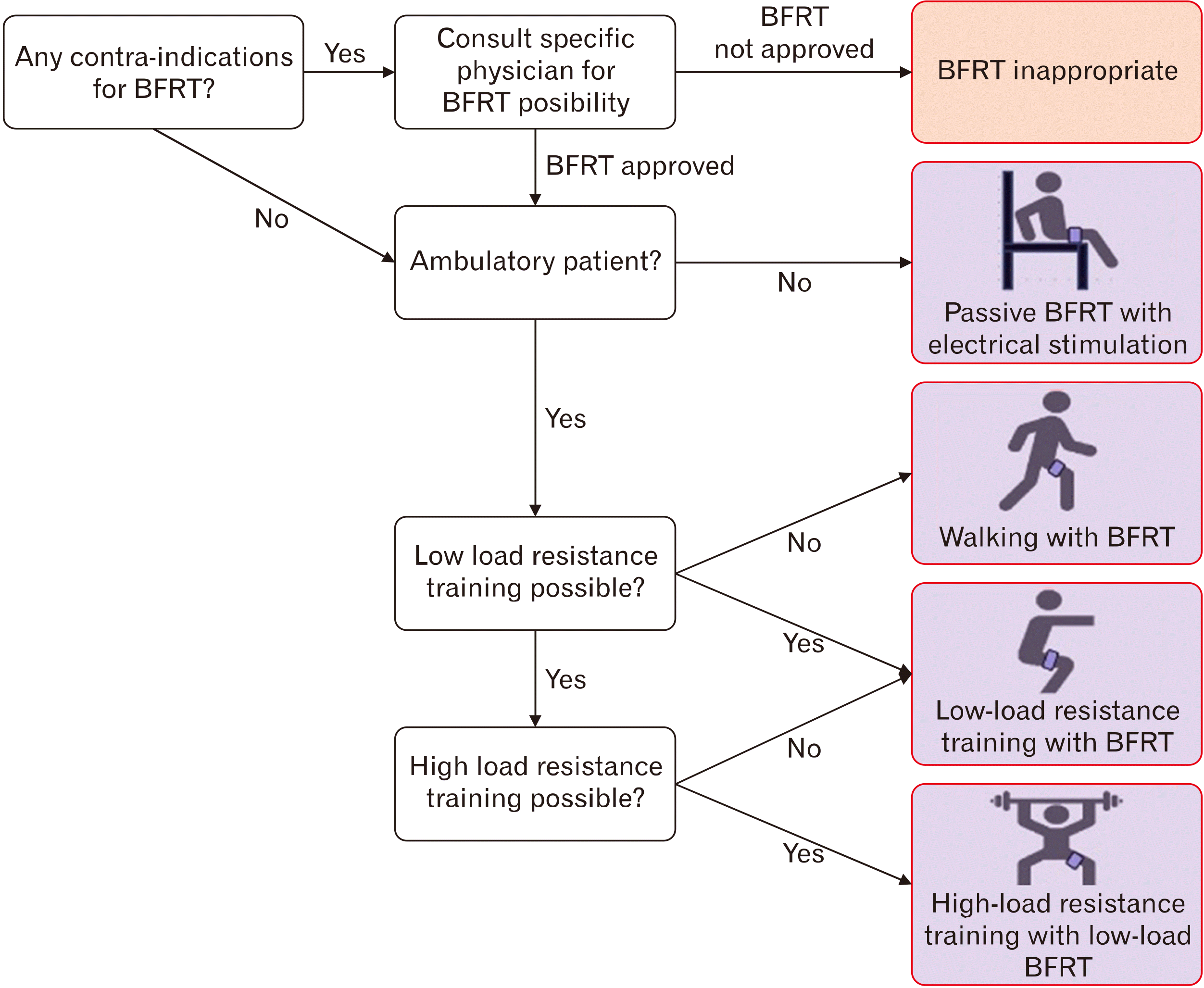
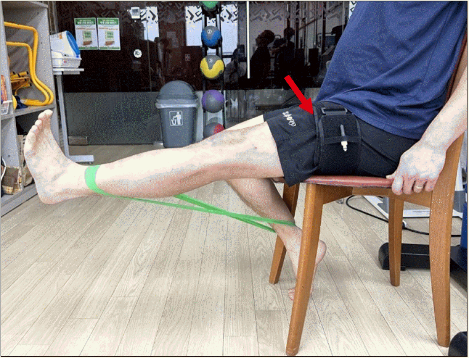
본 종설에서 다룬 전방 십자인대 손상 및 골관절염 환자들은 다음과 같이 보행 가능 여부 및 적응할 수 있는 운동 강도 등에 따라 수동적 혈류 제한, 혈류 제한 유산소 운동, 혈류 제한 근력 운동 순으로 진행을 한다(Fig. 348, Table 3). 수동적 혈류 제한은 체중 지지가 힘들거나 보행이 어려운 경우 적용한다(Table 3). 정상 보행이 가능한 경우 혈류 제한과 함께 주로 걷기나 자전거 운동과 같은 유산소 운동을 시행한다(Table 3, Fig. 2). 혈류 제한 근력 운동은 1회 최대 반복 중량의 20%–40%를 견딜 수 있는 수준이면 시행한다(Table 3). 이때 혈류 제한 저강도 근력운동의 효과가 일반 저강도 근력운동보다는 더 효과적이기 때문에 슬관절 수술 후 환자나 고령의 슬관절 관절염 환자에게도 적용할 수 있다(Fig. 4).

결 론

혈류 제한 운동은 고강도 운동이 어렵고 하지 근육의 위약이 발생할 가능성이 높은 슬관절 수술 후 재활이 필요한 환자들(특히, 전방십자인대 재건술 후 환자들) 혹은 슬관절 골관절염 환자들에서 유용하다고 결론내릴 수 있다. 충분한 기간 동안 혈류 제한 저강도 운동을 시행하면 고강도 운동 시 발생할 수 있는 부상 위험을 줄여주면서 운동 효과를 극대화하고, 증상 호전 및 슬관절 기능 개선 효과를 얻을 수 있다. 또한 정해진 매뉴얼대로 진행하면 혈류 제한에 따른 심혈관계의 불균형, 근육 손상, 혈전 및 색전증 발생 등의 가능성은 낮아 안전한 재활 방법이라고 볼 수 있다.

Notes

Conflict of Interest

No potential conflict of interest relevant to this article was reported.

Author Contributions

Conceptualization: DWL. Data curation: SIC. Formal analysis: SHK, JWL, SWK, BSP. Investigation: SWK. Resources: JWL, SIC. Supervision: DWL. Visualization: JWL, SIC, BSP. Writing–original draft: DWL, SHK. Writing–review & editing: DWL.

REFERENCES

1. Hassan BS, Mockett S, Doherty M. 2001; Static postural sway, proprioception, and maximal voluntary quadriceps contraction in patients with knee osteoarthritis and normal control subjects. Ann Rheum Dis. 60:612–8. DOI: 10.1136/ard.60.6.612. PMID: 11350851. PMCID: PMC1753664.
2. Segal NA, Glass NA, Felson DT, et al. 2010; Effect of quadriceps strength and proprioception on risk for knee osteoarthritis. Med Sci Sports Exerc. 42:2081–8. DOI: 10.1249/MSS.0b013e3181dd902e. PMID: 20351594. PMCID: PMC2921020.
3. Gupta R, Singhal A, Malhotra A, Soni A, Masih GD, Raghav M. 2020; Predictors for Anterior Cruciate Ligament (ACL) re-injury after successful primary ACL Reconstruction (ACLR). Malays Orthop J. 14:50–6. DOI: 10.5704/MOJ.2011.009. PMID: 33403062. PMCID: PMC7752004.
4. Thomas AC, Wojtys EM, Brandon C, Palmieri-Smith RM. 2016; Muscle atrophy contributes to quadriceps weakness after anterior cruciate ligament reconstruction. J Sci Med Sport. 19:7–11. DOI: 10.1016/j.jsams.2014.12.009. PMID: 25683732. PMCID: PMC4500756.
5. Hughes L, Rosenblatt B, Haddad F, et al. 2019; Comparing the effectiveness of blood flow restriction and traditional heavy load resistance training in the post-surgery rehabilitation of anterior cruciate ligament reconstruction patients: a UK national health service randomised controlled trial. Sports Med. 49:1787–805. DOI: 10.1007/s40279-019-01137-2. PMID: 31301034.
6. Harper SA, Roberts LM, Layne AS, et al. 2019; Blood-flow restriction resistance exercise for older adults with knee osteoarthritis: a pilot randomized clinical trial. J Clin Med. 8:265. DOI: 10.3390/jcm8020265. PMID: 30795545. PMCID: PMC6406824.
7. Ferraz RB, Gualano B, Rodrigues R, et al. 2018; Benefits of resistance training with blood flow restriction in knee osteoarthritis. Med Sci Sports Exerc. 50:897–905. DOI: 10.1249/MSS.0000000000001530. PMID: 29266093.
8. Bryk FF, Dos Reis AC, Fingerhut D, et al. 2016; Exercises with partial vascular occlusion in patients with knee osteoarthritis: a randomized clinical trial. Knee Surg Sports Traumatol Arthrosc. 24:1580–6. DOI: 10.1007/s00167-016-4064-7. PMID: 26971109.
9. Kong DH, Jung WS, Yang SJ, Kim JG, Park HY, Kim J. 2022; Effects of neuromuscular electrical stimulation and blood flow restriction in rehabilitation after anterior cruciate ligament reconstruction. Int J Environ Res Public Health. 19:15041. DOI: 10.3390/ijerph192215041. PMID: 36429760. PMCID: PMC9690111.
10. Gabler C, Kitzman PH, Mattacola CG. 2013; Targeting quadriceps inhibition with electromyographic biofeedback: a neuroplastic approach. Crit Rev Biomed Eng. 41:125–35. DOI: 10.1615/CritRevBiomedEng.2013008373. PMID: 24580566.
11. Okita K, Takada S, Morita N, et al. 2019; Resistance training with interval blood flow restriction effectively enhances intramuscular metabolic stress with less ischemic duration and discomfort. Appl Physiol Nutr Metab. 44:759–64. DOI: 10.1139/apnm-2018-0321. PMID: 30566362.
12. Asadi MB, Sharifi H, Abedi B, Fatolahi H. 2020; Acute inflammatory response to a single bout of resistance exercise with or without blood flow restriction. Int J Sport Stud Health. 3:e110594. DOI: 10.5812/intjssh.110594.
13. Bobes Álvarez C, Issa-Khozouz Santamaría P, Fernández-Matías R, et al. 2020; Comparison of blood flow restriction training versus non-occlusive training in patients with anterior cruciate ligament reconstruction or knee osteoarthritis: a systematic review. J Clin Med. 10:68. DOI: 10.3390/jcm10010068. PMID: 33375515. PMCID: PMC7796201.
14. Sato Y. 2005; The history and future of KAATSU training. Int J KAATSU Training Res. 1:1–5. DOI: 10.3806/ijktr.1.1.
15. Abe T, Kearns CF, Sato Y. 2006; Muscle size and strength are increased following walk training with restricted venous blood flow from the leg muscle, Kaatsu-walk training. J Appl Physiol (1985). 100:1460–6. DOI: 10.1152/japplphysiol.01267.2005. PMID: 16339340.
16. Patterson SD, Hughes L, Warmington S, et al. 2019; Blood flow restriction exercise: considerations of methodology, application, and safety. Front Physiol. 10:533. DOI: 10.3389/fphys.2019.00533. PMID: 31156448. PMCID: PMC6530612.
17. Hughes L, Paton B, Rosenblatt B, Gissane C, Patterson SD. 2017; Blood flow restriction training in clinical musculoskeletal rehabilitation: a systematic review and meta-analysis. Br J Sports Med. 51:1003–11. DOI: 10.1136/bjsports-2016-097071. PMID: 28259850.
18. Fry CS, Glynn EL, Drummond MJ, et al. 2010; Blood flow restriction exercise stimulates mTORC1 signaling and muscle protein synthesis in older men. J Appl Physiol (1985). 108:1199–209. DOI: 10.1152/japplphysiol.01266.2009. PMID: 20150565. PMCID: PMC2867530.
19. Fujita S, Abe T, Drummond MJ, et al. 2007; Blood flow restriction during low-intensity resistance exercise increases S6K1 phosphorylation and muscle protein synthesis. J Appl Physiol (1985). 103:903–10. DOI: 10.1152/japplphysiol.00195.2007. PMID: 17569770.
20. Hwang PS, Willoughby DS. 2019; Mechanisms behind blood flow-restricted training and its effect toward muscle growth. J Strength Cond Res. 33 Suppl 1:S167–79. DOI: 10.1519/JSC.0000000000002384. PMID: 30011262.
21. Dreyer HC, Fujita S, Cadenas JG, Chinkes DL, Volpi E, Rasmussen BB. 2006; Resistance exercise increases AMPK activity and reduces 4E-BP1 phosphorylation and protein synthesis in human skeletal muscle. J Physiol. 576(Pt 2):613–24. DOI: 10.1113/jphysiol.2006.113175. PMID: 16873412. PMCID: PMC1890364.
22. Lambert B, Hedt CA, Jack RA, et al. Blood flow restriction therapy preserves whole limb bone and muscle following ACL reconstruction. Orthop J Sports Med. 2019; 7(3_suppl2):DOI: 10.1177/2325967119S00196. PMCID: PMC6447983.
23. Ohta H, Kurosawa H, Ikeda H, Iwase Y, Satou N, Nakamura S. 2003; Low-load resistance muscular training with moderate restriction of blood flow after anterior cruciate ligament reconstruction. Acta Orthop Scand. 74:62–8. DOI: 10.1080/00016470310013680. PMID: 12635796.
24. Takarada Y, Takazawa H, Ishii N. 2000; Applications of vascular occlusion diminish disuse atrophy of knee extensor muscles. Med Sci Sports Exerc. 32:2035–9. DOI: 10.1097/00005768-200012000-00011. PMID: 11128848.
25. Žargi T, Drobnič M, Stražar K, Kacin A. 2018; Short-term preconditioning with blood flow restricted exercise preserves quadriceps muscle endurance in patients after anterior cruciate ligament reconstruction. Front Physiol. 9:1150. DOI: 10.3389/fphys.2018.01150. PMID: 30197599. PMCID: PMC6118218.
26. Karampampa C, Papasoulis E, Hatzimanouil D, Koutras G, Totlis T. 2023; The effects of exercise with blood flow restriction (BFR) in the post operative rehabilitation of anterior cruciate ligament (ACL) reconstruction patients: a case series. J Orthop Res Ther. 8:1314. DOI: 10.29011/2575-8241.001314.
27. Jack RA 2nd, Lambert BS, Hedt CA, Delgado D, Goble H, McCulloch PC. 2023; Blood flow restriction therapy preserves lower extremity bone and muscle mass after ACL reconstruction. Sports Health. 15:361–71. DOI: 10.1177/19417381221101006. PMID: 35762124. PMCID: PMC10170230.
28. McAlindon TE, Cooper C, Kirwan JR, Dieppe PA. 1993; Determinants of disability in osteoarthritis of the knee. Ann Rheum Dis. 52:258–62. DOI: 10.1136/ard.52.4.258. PMID: 8484690. PMCID: PMC1005622.
29. Segal NA, Williams GN, Davis MC, Wallace RB, Mikesky AE. 2015; Efficacy of blood flow-restricted, low-load resistance training in women with risk factors for symptomatic knee osteoarthritis. PM R. 7:376–84. DOI: 10.1016/j.pmrj.2014.09.014. PMID: 25289840. PMCID: PMC4385750.
30. Segal N, Davis MD, Mikesky AE. 2015; Efficacy of blood flow-restricted low-load resistance training for quadriceps strengthening in men at risk of symptomatic knee osteoarthritis. Geriatr Orthop Surg Rehabil. 6:160–7. DOI: 10.1177/2151458515583088. PMID: 26328230. PMCID: PMC4536503.
31. Mattocks KT, Jessee MB, Counts BR, et al. 2017; The effects of upper body exercise across different levels of blood flow restriction on arterial occlusion pressure and perceptual responses. Physiol Behav. 171:181–6. DOI: 10.1016/j.physbeh.2017.01.015. PMID: 28088558.
32. Hughes L, Patterson SD. 2020; The effect of blood flow restriction exercise on exercise-induced hypoalgesia and endogenous opioid and endocannabinoid mechanisms of pain modulation. J Appl Physiol (1985). 128:914–24. DOI: 10.1152/japplphysiol.00768.2019. PMID: 32105522.
33. Neto GR, Sousa MS, Costa e Silva GV, Gil AL, Salles BF, Novaes JS. 2016; Acute resistance exercise with blood flow restriction effects on heart rate, double product, oxygen saturation and perceived exertion. Clin Physiol Funct Imaging. 36:53–9. DOI: 10.1111/cpf.12193. PMID: 25257752.
34. Nielsen JL, Aagaard P, Prokhorova TA, et al. 2017; Blood flow restricted training leads to myocellular macrophage infiltration and upregulation of heat shock proteins, but no apparent muscle damage. J Physiol. 595:4857–73. DOI: 10.1113/JP273907. PMID: 28481416. PMCID: PMC5509865.
35. Mouser JG, Dankel SJ, Jessee MB, et al. 2017; A tale of three cuffs: the hemodynamics of blood flow restriction. Eur J Appl Physiol. 117:1493–9. DOI: 10.1007/s00421-017-3644-7. PMID: 28501908.
36. Okuno NM, Pedro RE, Leicht AS, de Paula Ramos S, Nakamura FY. 2014; Cardiac autonomic recovery after a single session of resistance exercise with and without vascular occlusion. J Strength Cond Res. 28:1143–50. DOI: 10.1519/JSC.0000000000000245. PMID: 24077384.
37. Neto GR, da Silva JC, Freitas L, et al. 2019; Effects of strength training with continuous or intermittent blood flow restriction on the hypertrophy, muscular strength and endurance of men. Acta Scientiarum Health Sci. 41:42273. DOI: 10.4025/actascihealthsci.v41i1.42273.
38. Manini TM, Clark BC. 2009; Blood flow restricted exercise and skeletal muscle health. Exerc Sport Sci Rev. 37:78–85. DOI: 10.1097/JES.0b013e31819c2e5c. PMID: 19305199.
39. Patterson SD, Ferguson RA. 2010; Increase in calf post-occlusive blood flow and strength following short-term resistance exercise training with blood flow restriction in young women. Eur J Appl Physiol. 108:1025–33. DOI: 10.1007/s00421-009-1309-x. PMID: 20012448.
40. Clark BC, Manini TM, Hoffman RL, et al. 2011; Relative safety of 4 weeks of blood flow-restricted resistance exercise in young, healthy adults. Scand J Med Sci Sports. 21:653–62. DOI: 10.1111/j.1600-0838.2010.01100.x. PMID: 21917016. PMCID: PMC6152804.
41. Takano H, Morita T, Iida H, et al. 2005; Hemodynamic and hormonal responses to a short-term low-intensity resistance exercise with the reduction of muscle blood flow. Eur J Appl Physiol. 95:65–73. DOI: 10.1007/s00421-005-1389-1. PMID: 15959798.
42. Tennent DJ, Hylden CM, Johnson AE, Burns TC, Wilken JM, Owens JG. 2017; Blood flow restriction training after knee arthroscopy: a randomized controlled pilot study. Clin J Sport Med. 27:245–52. DOI: 10.1097/JSM.0000000000000377. PMID: 27749358.
43. Madarame H, Kurano M, Fukumura K, Fukuda T, Nakajima T. 2013; Haemostatic and inflammatory responses to blood flow-restricted exercise in patients with ischaemic heart disease: a pilot study. Clin Physiol Funct Imaging. 33:11–7. DOI: 10.1111/j.1475-097X.2012.01158.x. PMID: 23216760.
44. de Queiros VS, Dantas M, Neto GR, et al. 2021; Application and side effects of blood flow restriction technique: a cross-sectional questionnaire survey of professionals. Medicine (Baltimore). 100:e25794. DOI: 10.1097/MD.0000000000025794. PMID: 33950976. PMCID: PMC8104249.
45. Hollander DB, Reeves GV, Clavier JD, Francois MR, Thomas C, Kraemer RR. 2010; Partial occlusion during resistance exercise alters effort sense and pain. J Strength Cond Res. 24:235–43. DOI: 10.1519/JSC.0b013e3181c7badf. PMID: 19935100.
46. Hughes L, Paton B, Haddad F, Rosenblatt B, Gissane C, Patterson SD. 2018; Comparison of the acute perceptual and blood pressure response to heavy load and light load blood flow restriction resistance exercise in anterior cruciate ligament reconstruction patients and non-injured populations. Phys Ther Sport. 33:54–61. DOI: 10.1016/j.ptsp.2018.07.002. PMID: 30014968.
47. Martín-Hernández J, Ruiz-Aguado J, Herrero AJ, et al. 2017; Adaptation of perceptual responses to low-load blood flow restriction training. J Strength Cond Res. 31:765–72. DOI: 10.1519/JSC.0000000000001478. PMID: 27191690.
48. Scott BR, Loenneke JP, Slattery KM, Dascombe BJ. 2015; Exercise with blood flow restriction: an updated evidence-based approach for enhanced muscular development. Sports Med. 45:313–25. DOI: 10.1007/s40279-014-0288-1. PMID: 25430600.

Fig. 1
Blood flow restriction instruments consist of tourniquet cuff, blood pressure gauge, and pump. Tourniquet cuff is used to partially restrict arterial inflow and fully restrict venous outflow during exercise. The blood pressure gauge is used to measure the pressure within the cuff. The pump is used to inflate the tourniquet cuff to the desired pressure.
kjsm-42-3-165-f1.tif
Fig. 2
Mechanism of blood flow restriction exercise. IGF; insulin like growth factor.
kjsm-42-3-165-f2.tif
Fig. 3
Algorithm for blood flow restriction training (BFRT) in a similar protocol as reported in the previous review article of Scott et al.48 (Sports Med 2015;45:313-25).
kjsm-42-3-165-f3.tif
Fig. 4
Blood flow restriction-voluntary resistance exercise.
kjsm-42-3-165-f4.tif
Table 1
Outcomes of blood flow restriction (BFR) exercise in anterior cruciate ligament reconstruction
Study Year Study design Method (cuff and pressure) Outcome
Ohta et al.23 2003 RCT
BFR (n=22): 180 mm Hg, low load
Control (n=22): BFR (−), low load
- Operative/contralateral knee extensor power: BFR>control at CC60, CC180, IM60
- CSA ratio of operative/contralateral knee extensor: BFR>control
Lambert et al.22 2019 RCT
BFR (n=7): 80% LOP 20% 1 RM
Control (n=7): BFR (−) 20% 1 RM
- Thigh lean muscle mass decreased more in the control group at week 6 and remained decreased in only the control group at week12
Zargi et al.25 2018 RCT
BFR (n=10): 150 mm Hg, 14 cm, 30% 1 RM
Control (n=10): 20 mm Hg, 14 cm, 30% 1 RM
- Muscle isometric endurance: Shortened significantly in control group at week 4 and returned to preoperative values at week 12, no change in BFR group at any time point
- EMG amplitude increased significantly at week 4 after surgery in BFR group only
- Muscle blood flow increased significantly in BFR group, and decreased in control group at week 4 after surgery
Hughes et al.5 2019 RCT
BFR (n=12): 80% LOP 11.5 cm, 30% 1 RM
Control (n=12): BFR (−) 70% 1 RM
- Isotonic strength: 10 RM strength significantly increased in both limbs with BFR-RT and HL-RT with no group differences
- Isokinetic strength: Significantly greater attenuation of knee extensor peak torque loss at 150°/sec and 300°/sec and knee flexor torque loss at all speeds with BFR-RT
- Muscle morphology: significant increases in muscle thickness with BFR-RT and HL-RT with no group differences
- Significantly greater increases in self-reported function, Y-balance performance and reductions in knee joint pain and effusion with BFR-RT compared to HL-RT
Karampampa et al.26 2023 RCT
BFR (n=8): 60%−80% LOP 10−20% 1 RM
Control (n=8): BFR (−) 10−20% 1 RM
Significant improvements in thigh circumference of the BFR group compared to the control group
Significantly greater attenuation of knee flexors strength deficit at 60°/sec in the BFR group
No statistically significant differences found neither in the strength deficit of the extensor muscles
Jack RA 2nd et al.27 2023 RCT
BFR (n=17): 80% LOP 20% 1 RM
Control (n=15): BFR (−) 20% 1 RM
Only the control group experienced decreases in LE-LM at week 6 and week 12
LE bone mass was decreased only in the control group at week 6 and week 12
Return to sports time was reduced in the BFR group compared with the control group

RCT: randomized control trial, SLR: straight leg raise, CC60: angular velocities of 60°/sec, CC180: angular velocities of 180°/sec, IM60: isometric contraction strength at 60° knee flexion, CSA: cross sectional area, LOP: limb occlusion pressure, RM: repeated maximum, RT: resistance training, HL: heavy load, LE: lower extremity, LM: lean mass.

Table 2
Outcomes of blood flow restriction (BFR) exercise in osteoarthritis patients
Study Year Study design Methods (cuff and pressure) Outcomes
Segal et al.30 2015 RCT
BFR (n=19): 30−200 mm Hg 30% 1 RM
Control (n=22): BFR (−) 30% 1 RM
Bilateral leg press 1 RM increased significantly in both the control and the BFR groups with no significant group differences
Significant improvements in isokinetic knee extensor strength in the control group, but not in the BFR group
Significant improvements in KOOS scores in the control group, but not in the BFR group
Segal et al.29 2015 RCT
BFR (n=19): 30−200 mm Hg 30% 1 RM
Control (n=21): BFR (−) 30% 1 RM
Bilateral leg press 1 RM increased significantly in the BFR group than in the control group
Isokinetic knee extensor strength increased significantly more in the BFR group than in the control group
Quadriceps volume with no intergroup difference
No worsening of knee pain in both group with no significant intergroup differences
Bryk et al.8 2016 RCT
BFR (n=17): 200 mm Hg 30% 1 RM
Control (n=17): pressure not described; 70% 1 RM
Higher level of quadriceps strength, function, less pain at 6-week with no inter-group difference
The BFR group with decreased anterior knee discomfort compared to the control group
Ferraz et al.7 2018 RCT
BFR (LI-RT, n=16): 70% LOP 20−30% 1 RM
1st Control (HI-RT, n=16): BFR (−): 50%−80% 1 RM
2nd Control (LI-RT, n=16): BFR (−) 20%−30% 1 RM
Significant 1 RM increase in HI-RT and BFRT but not in LI-RT, with no inter-group difference
Significant quadriceps CSA increase in HI-RT and BFRT but not in LI-RT with no inter-group difference
Significant improvement by TST in HI-RT and BFRT and no significant change by TUG
WOMAC physical function improved in HI-RT and BFRT
WOMAC pain improved in BFRT and LI-RT
Harper et al.6 2019 RCT
BFR (n=19): [pressure=0.5×SBP+2 (thigh circumference)+5] 20% 1 RM
Control MI-RT (n=16): BFR (−) 60% 1 RM
Isokinetic knee extension improvement in both groups of 60°/sec, 90°/sec, and 120°/sec with no significant difference
Function (400 m walk, SPPB, LLFDI) improvement in both groups with no significant difference
Pain (WOMAC and NPRS) improvement in both groups with no significant difference

RCT: randomized control trial, RM: repeated maximum, KOOS: Knee injury and Osteoarthritis Outcome Score, LOP: limb occlusion pressure, HI-RT: high-intensity resistance training, BFRT: blood flow restriction training, LI-RT: low-intensity resistance training, CSA: cross-sectional area, TST: timed stands test, TUG: timed up and go test, WOMAC: Western Ontario and McMaster Universities Osteoarthritis Index, SBP: systolic blood pressure, MI-RT: moderate-intensity resistance training, SPPB: short physical performance battery, LLFDI: late life function and disability instrument, NPRS: numeric pain rating scale.

Table 3
Protocol of blood flow restriction
Protocol Passive BFR Aerobic exercise Resistance training
Frequency 1−2 times/day 2−3 times/wk 2−3 times/wk
Cuff pressure 70%−100% arterial occlusive pressure 40%−80% arterial occlusive pressure 40%−80% arterial occlusive pressure
Restriction form Continuous Continuous or intervals Continuous or intermittent
Restriction time 5 min intervals 5−20 min intervals 5−10 min per exercise
Load - - 20%−40% 1 repeated maximum
Sets 3−5 - 4
Rest between sets 3−5 min - 30−45 sec
Repetitions - - 75 (30−15−15−15)
Execution speed - - 1−2 sec (concentric–eccentric)
Type of exercise - Walking or cycling Resistance training
TOOLS
Similar articles