Abstract
Lateral spinal artery (LSA) aneurysms are extremely rare lesions that can rupture and cause subarachnoid hemorrhage (SAH) even though the spinal arteries communicate directly with the subarachnoid space. To date, six cases of LSA aneurysms have been reported in the literature. Herein, three such cases are reported. All patients presented to the emergency department with headaches. The patients in the first two cases were confirmed to have SAH and LSA aneurysms on a brain computed tomography (CT) angiography performed at the hospital. Two patients had prior instances of cerebral infarction and coronary disease, respectively, and were undergoing antiplatelet therapy. The antiplatelet medication was stopped for 2 weeks and 1 week, respectively, while conservative care was provided. Subsequently, a suboccipital craniectomy was performed, followed by aneurysm clipping. Following the surgery, both patients were discharged without any significant neurological deficits. Regarding the third patient, no aneurysm was found on brain CT angiography, and cerebral angiography was performed during the patient’s hospital stay. She was hospitalized, where she received medication and conservative care, and was discharged with an improvement in bleeding without neurological symptoms. Subsequently, a LSA aneurysm was identified on a brain CT angiography performed at an outpatient clinic; however, the patient was transferred because she wanted to be treated at another hospital. LSA aneurysms are difficult to visualize using CT angiography; therefore, careful angiographic studies are required. Surgical clipping is the treatment of choice if the aneurysm is inaccessible by the endovascular treatment.
Intracranial subarachnoid hemorrhage (SAH) due to the rupture of spinal aneurysms accounts for approximately 1% of all SAHs [1,3,10]. Most of aneurysms involving the spinal artery are associated with arteriovenous malformations, spinal dural arteriovenous fistulas, or coarctation of the aorta. Thus, isolated saccular spinal artery aneurysms are extremely rare (Table 1) [1,4]. Herein, we describe three cases of intracranial SAH associated with lateral spinal artery (LSA) aneurysms. We also performed a brief review of the relevant literature regarding this clinical condition to elucidate methods for accurate diagnosis and appropriate management of patients with LSA aneurysm-related complications.
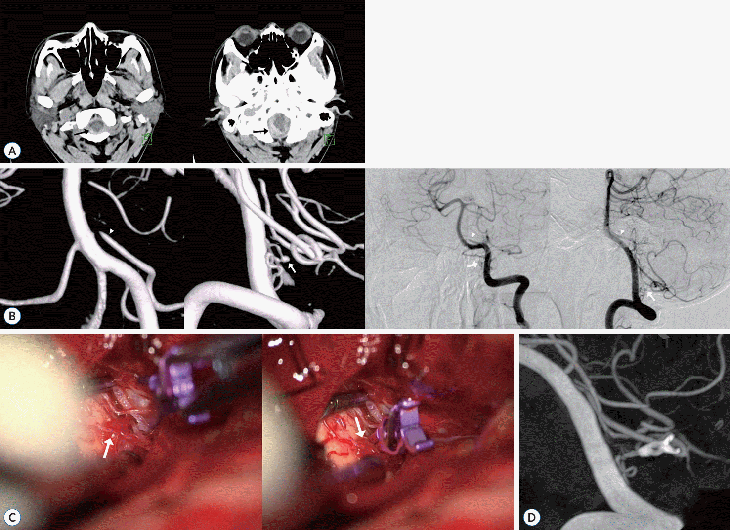
A 55-year-old male patient presented with sudden onset of severe headache and dizziness. Several months prior, he was diagnosed with the right basal ganglia infarction and had received dual antiplatelet medications for the prevention of secondary infarction. The patient had no definitive neurological deficits upon admission. The patient underwent brain computed tomography (CT) and was diagnosed with acute SAH of the posterior fossa, including the cerebellomedullary cistern, cisterna magna, and fourth ventricle (Fig. 1A). However, no visible cause of the SAH was detected, such as vascular malformation, on brain CT angiography. For further evaluation, cerebral angiography was performed, which detected a 2 mm-sized small aneurysm at the left LSA that anastomosed rostrally with branches of the posterior inferior cerebellar artery (PICA) at the tonsillo-medullary segment. We also found a fine, tortuous artery originating from the proximal V4 segment (Fig. 1B). Additionally, the ipsilateral proximal PICA showed severe stenosis.
Endovascular treatment was not possible because of the small diameters of the LSA and branch of the PICA. After 2 weeks of conservative treatment and the cessation of dual antiplatelet therapy, a follow-up cerebral angiography was performed to identify differences in previous angiographic findings. On follow-up cerebral angiography, the LSA aneurysm had exhibited a slight increase in size. Therefore, we decided to perform a surgical clipping. The patient was positioned prone and underwent a suboccipital craniectomy without C1 arch removal. A straight midline incision was then made. A suboccipital craniectomy was performed. The aneurysm was located on the posterolateral aspect of the medulla and changed during healing. After temporal clipping of the proximal LSA, indocyanine green (ICG) videoangiography revealed minimal flow into the aneurysm (Fig. 1C). Therefore, 5 mmsized straight “L-type” mini-clip was applied for managing the aneurysm. Post-clipping ICG videoangiography and Doppler ultrasonography (US) were performed. The aneurysm was completely occluded on the follow-up cerebral angiography on the 14th postoperative day (Fig. 1D). His clinical condition was satisfactory at discharge, and he is currently healthy without any neurologic deficits or recurrent aneurysms on follow-up imaging.
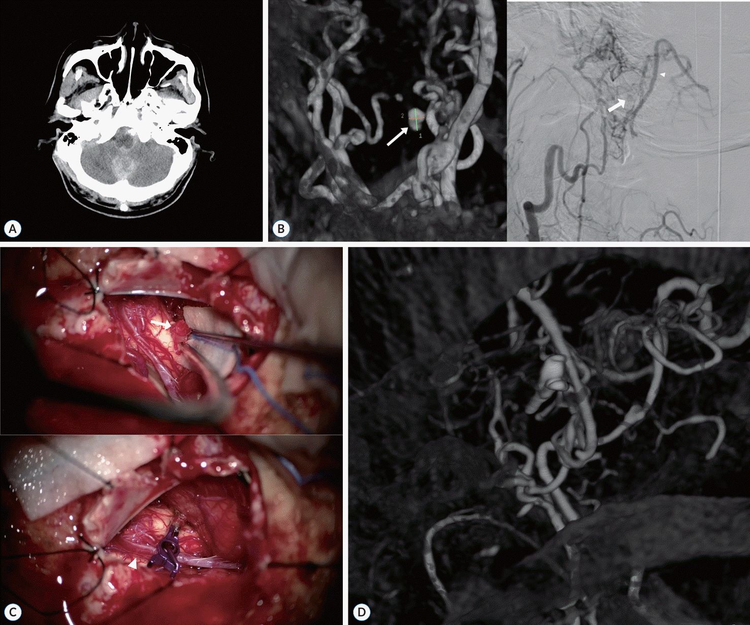
A 64-year-old male patient was admitted to our hospital with sudden onset of headache and vomiting. His past medical history included coronary disease, left ophthalmic artery occlusion, and hypertension. He was on dual antiplatelet medication and no neurological deficits on admission. He underwent a brain CT, and there was acute SAH in the cisterna magna and perimedullary cistern was observed (Fig. 2A). The CT angiography revealed severe stenosis of both vertebral arteries (VAs) and the right M1.
Cerebral angiography was then performed. The right VA was inaccessible because of the fine parent artery and the inlet of the left VA had severe stenosis. A 4-Fr angiographic catheter was then inserted into the left VA. Cerebral angiography revealed a faint contrast filling of a 2 mm sized-small aneurysm at the origin of the left LSA, forming an anastomosis with the left PICA (Fig. 2B). After conservative treatment for 1 week and cessation of dual antiplatelet therapy, cerebral angiography was repeated to identify differences from the previous angiography. On examination, the aneurysm had exhibited a slight increase in size. The patient had a suboccipital craniectomy, and underwent aneurysm clipping.
After remove the hematoma in the posterior fossa, a berry-type aneurysm with bleb formation was observed in the posterolateral aspect of the medulla. A 5 mm-sized straight “L-type” mini-clip was applied (Fig. 2C). Post-clipping ICG video-angiography and Doppler US were performed, which confirmed that the aneurysm was occluded. One month later, a follow-up cerebral angiography revealed an undetectable aneurysm (Fig. 2D). The patient was discharged without any neurological deficits.
A 73-year-old woman presented to the emergency room with a headache and vomiting. Her medical history included medications for high blood pressure and hyperlipidemia. The patient had no definitive neurological deficits upon admission. She underwent a brain CT angiography, and acute SAH in the prepontine and perimedullary cisterns were detected (Fig. 3A). However, brain CT angiography revealed no visible cause of the SAH, such as vascular malformations. A cerebral angiography was performed for further evaluation. However, there was no visible aneurysm (Fig. 3B). Therefore, perimesencephalic non-aneurysmal SAH (PNSAH) was diagnosed, and the patient was treated in the intensive care unit. Two days after the patient underwent brain CT angiography, no aneurysm was detected and the SAH resolved. On the 10th day of hospitalization, another cerebral angiography was performed and no visible aneurysm was identified. On the 13th day of hospitalization, the symptoms and SAH improved, and the patient was discharged with no neurological deficits. Brain CT angiography performed 19 days after discharge confirmed a 2 mm-sized small aneurysm in the right LSA of the right VA branch (Fig. 3C). However, the patient wanted to undergo treatment at another hospital and was transferred accordingly.
The LSA branches off from the intradural VA or PICA, runs along the medulla, and enters the posterolateral aspect of the cervical spine to connect to the posterior spinal artery (PSA) [1,5,7,8]. However, the definition of the LSA is unclear and still controversial because the LSA is sometimes considered the same blood vessel as the PSA, and the LSA stands for the rostral region of the PSA. Although the LSA is a part of the PSA, it has its own name because it travels toward the upper cervical spinal cord and medulla, forming an anastomosis with the PICA and VA, and supplies blood to the medulla [9,10]. In another definition, the LSA is a blood vessel branching from the intradural VA at the C1 level and travelling to the lateral side of the spinal cord, meeting and joining the PSA at the C4 and 5 levels. It then travels upward to form an anastomosis with the PICA and provides blood supply to the medulla [1,7,8]. In our daily practice, we define the LSA as a separate vessel from the PSA that branches from the VA to form the PICA. Thus, we describe this report based on this definition of the LSA. Fortunately, no disagreement exists regarding the function of the LSA, even though different opinions remain regarding the definition of the LSA.
The LSA is usually small and poorly visible on angiography; therefore, it is often not identified on brain CT angiography. In addition, it is rarely seen on conventional cerebral angiography, and superselection during cerebral angiography is required to confirm it [1,8].
LSA aneurysms are extremely rare, and SAH related to ruptured LSA aneurysms are equally rare, even though the spinal arteries also have direct communication with the subarachnoid space [3]. Cerebral aneurysms in the LSA occur because of hemodynamic stress related to events such as occlusion of the VA, PICA, or various variants of the VA and PICA [1,2,5,8]. This hemodynamic stress causes enlargement of the LSA itself and LSA aneurysm, leading to rupture of the aneurysm and SAH. However, LSA aneurysms rarely occur in the absence of hemodynamic stress [6]. In clinical practice, SAH related to LSA aneurysm is often misdiagnosed as PNSAH, and rebleeding may occur because LSA aneurysms can be clinically challenging owing to their small size and poor visualization on imaging modalities. Therefore, in patients suspected of having PNSAH, the presence of an aneurysm should be carefully evaluated when in case of excessive bleeding and a stenotic lesion, as in case 3. In case 3, we found a causative aneurysm on repeat cerebral angiography and brain CT angiography. Considering that the aneurysm was not initially detected in the third case, this does not suggest that the aneurysm grew in size over time. Rather, when the aneurysm ruptured, it was not detected because the thrombus filled the aneurysm and, resulting in a lack of contrast. Subsequently, the aneurysm may become visible as the thrombus dissolves.
In cases 1 and 2, we opted for delayed operation instead of immediate intervention because the patients’ susceptibility to risks associated with dual antiplatelet medication, the relatively small size of the aneurysm, and the uncertainty surrounding the likelihood of its rupture. Three treatment options are available for LSA aneurysms: endovascular coil embolization, surgical clipping, and conservative management [1,2,4,8]. In the case of conservative management, natural regression may occur as the aneurysm progresses; therefore, natural regression can be considered [4]. However, because of the risk of aneurysm re-rupture, active treatment is generally recommended. Conservative treatment can be considered when the patient’s general condition is too poor, and cannot tolerate surgery or endovascular treatment [4]. In cases of posterior circulation vessel aneurysms, the endovascular approach is often used. However, in cases such as cases 1 and 2, where intravascular access is challenging because of the lesion location in the fine vessel group, clipping can be performed using suboccipital craniectomy [1,3,6]. Whether through endovascular intervention or surgical clipping, preserving the parent artery is the ideal treatment [1,6]. However, serious consequences such as brain stem infarction, do not always occur even if the parent artery is not feasible. This is because numerous microscopic anastomoses develop between the VA and PICA [8], which averts infarction by providing collateral arterial support.
In our cases, we emphasize the challenge of identifying LSA aneurysms in imaging studies, such as CT angiography, magnetic resonance angiography, or cerebral angiography, owing to their fine vascular structure. Therefore, cerebral angiography should be performed while keeping spinal artery lesions, such as LSA aneurysms, in mind when clinicians encounter patients with posterior fossa SAH showing radiologically negative findings that can be easily considered as PNSAH [2,8]. After diagnosis, endovascular treatment can be considered first. However, when the vascular structure makes this impossible, clipping through suboccipital craniectomy remains a viable option.
Herein, we described three cases of LSA aneurysms presenting as posterior fossa SAH. In the clinical practice, LSA aneurysms can be challenging to diagnose because they are difficult to visualize using CT angiography. Therefore, clinical suspicion and careful subsequent angiographic studies are required for timely diagnosis and management to prevent unexpected morbidity and mortality related to LSA aneurysms. Surgical clipping is the treatment of choice when an aneurysm is inaccessible by endovascular treatment.
Notes
References
1. Chen CC, Bellon RJ, Ogilvy CS, Putman CM. Aneurysms of the lateral spinal artery: report of two cases. Neurosurgery. 48:949–954. discussion 953-954. 2001.

2. Chonan M, Nishimura S, Kimura N, Ezura M, Uenohara H, Tominaga T. A ruptured aneurysm arising at the leptomeningeal collateral circulation from the extracranial vertebral artery to the posterior inferior cerebellar artery associated with bilateral vertebral artery occlusion. J Stroke Cerebrovasc Dis. 23:e135–e139. 2014.
3. Germans MR, Kulcsar Z, Regli L, Bozinov O. Clipping of ruptured aneurysm of lateral spinal artery associated with anastomosis to distal posterior inferior cerebellar artery: a case report. World Neurosurg. 117:186–189. 2018.
4. Karakama J, Nakagawa K, Maehara T, Ohno K. Subarachnoid hemorrhage caused by a ruptured anterior spinal artery aneurysm. Neurol Med Chir (Tokyo). 50:1015–1019. 2010.
5. Kubota H, Suehiro E, Yoneda H, Nomura S, Kajiwara K, Fujii M, et al. Lateral spinal artery aneurysm associated with a posterior inferior cerebellar artery main trunk occlusion. Case illustration. J Neurosurg Spine. 4:347. 2006.

6. Kurita M, Endo M, Kitahara T, Fujii K. Subarachnoid haemorrhage due to a lateral spinal artery aneurysm misdiagnosed as a posterior inferior cerebellar artery aneurysm: case report and literature review. Acta Neurochir (Wien). 151:165–169. 2009.

7. Lasjaunias P, Vallee B, Person H, Ter Brugge K, Chiu M. The lateral spinal artery of the upper cervical spinal cord. Anatomy, normal variations, and angiographic aspects. J Neurosurg. 63:235–241. 1985.
8. Morigaki R, Satomi J, Shikata E, Nagahiro S. Aneurysm of the lateral spinal artery: a case report. Clin Neurol Neurosurg. 114:713–716. 2012.

Fig. 1.
A : Axial brain computer tomography images show acute subarachnoid hemorrhage in the posterior fossa, mainly perimedullary cistern and cisterna magna (black arrows). B : On the left vertebral 3-dimensional digital subtraction angiography and cerebral angiography, the left posterior inferior cerebellar artery (arrowheads) and anterior inferior cerebellar artery show severe stenosis at the proximal portion. Moreover, a small aneurysm is observed in the left lateral spinal artery (LSA) (arrows). C : Suboccipital craniectomy was performed to access the lateral spinal artery aneurysm (arrows) and clipping was performed. D : On the 14th postoperative day, cerebral angiography shows complete occlusion of treated LSA aneurysm.

Fig. 2.
A : Axial brain computer tomography image shows acute subarachnoid hemorrhage in the cisterna magna and perimedullary cistern. B : On the left vertebral 3-dimensional digital subtraction angiography and cerebral angiography, an aneurysm is observed in the left lateral spinal artery (LSA) (arrows), forming anastomosis with left posterior inferior cerebellar artery (PICA) (arrowhead). C : After suboccipital craniectomy, the left LSA aneurysm (arrow) is clipped. PICA (arrowhead). D : On the follow-up cerebral angiography, no aneurysm can be detected.

Fig. 3.
A : Axial computer tomography (CT) angiography images of the brain show acute subarachnoid hemorrhage in the prepontine and perimedullary cisterns. B : On right vertebral artery angiography, no visible aneurysm is observed. The posterior inferior cerebellar artery shows hypoplastic, and the anterior inferior cerebellar artery has dominancy. C : Axial maximum intensity projection image of brain CT angiography shows a small aneurysm in the right lateral spinal artery (arrow).

Table 1.
Overview of six reported cases of ruptured LSA aneurysms
| Study | Age (years)/sex | Symptom | Location of aneurysm | Other vascular disease | Treatment | Outcome |
|---|---|---|---|---|---|---|
| Morigaki et al. [8] (2012) | 78/M | Headache, mental decline, tetraplegia | Left LSA | Occlusion of both VAs | Coil embolization | Mental alertness, mild paraparesis |
| Chen et al. [1] (2001) | 72/F | Mental decline, bilateral 4th cranial nerve palsy, left leg weakness | Right LSA | Bilateral high-grade stenosis of VA | Coil embolization | Mental alertness, left hemiparesis |
| Chen et al. [1] (2001) | 69/F | Headache, nausea, photophobia, nonverbal | Left LSA | Narrowing of the distal left VA | Clipping and resection of aneurysm | Improvement in neurological status |
| Germans et al. [3] (2018) | 49/M | Headache, dizziness, nausea | Left LSA | Occlusion of left PICA | Clipping of aneurysm | No neurological deficits |
| Kubota et al. [5] (2006) | 59/F | Severe headache, consciousness change | Right LSA | Occlusion of right PICA | Resection of aneurysm | Improvement in consciousness |
| Kurita et al. [6] (2009) | 61/M | Headache | Right LSA | None | Clipping of aneurysm | No neurological deficits |


 PDF
PDF Citation
 Print
Print


 XML Download