Journal List > Acute Crit Care > v.39(3) > 1516088177

Aggarwal, Karki, Gaind, Matlani, and Muthukumar: Serum procalcitonin and C-reactive protein as indices of early sepsis and mortality in North Indian pediatric burn injuries: a prospective evaluation and literature review

Abstract

Background

Delays in diagnosing sepsis in children afflicted with thermal injuries can result in high morbidity and mortality. Our study evaluated the role of the biomarkers Procalcitonin (PCT) and C-reactive protein (CRP) as predictors of early sepsis and mortality, respectively, in this group of patients.

Methods

This was a prospective evaluation of 90 pediatric burn cases treated at a tertiary care burn center in Northern India. Patients, aged 1–16 years, presenting within 24 hours of being burned, with >10% body surface area of burn injury were included in the study. Levels of PCT and CRP were measured on days 1, 3, 5, and 7. Patients were followed until discharge, 30th post-burn day, or death, whichever occurred first.

Results

Sepsis was clinically present in 49 of 90 (54.4%) cases with a median 30% total body surface area (TBSA) of burns. Mortality was seen in 31 of 90 (34.4%) cases with a median of 35% TBSA burns. High PCT and CRP were seen in the sepsis group, particularly on days 3, 5, and 7. PCT was also significantly higher in the mortality group (days 1 and 3).

Conclusions

While PCT was a good early predictor of sepsis and mortality in children with burns, CRP was reliable as a predictor of sepsis only. Both markers, however, can serve as adjuncts to culture sensitivity reports for diagnosing early onset sepsis and initiation of antibiotic therapy in appropriate patients.

INTRODUCTION

The average global estimate of pediatric burns is approximately 0.79%, with Asia (0.8%) and Africa (1.08%) accounting for the highest rates [1]. The overall global pediatric burn mortality rate is roughly 2.5 per 100,000 cases [2]. Sepsis is an important contributor to this mortality. Diagnosis of sepsis is often delayed due to excessive reliance on blood culture sensitivity reports [3]. We believe identification of rapid and more sensitive biomarkers for diagnosing sepsis is needed to ensure earlier commencement of antibiotic therapy and improvement in overall outcomes.
Several parameters such as procalcitonin (PCT); C-reactive protein (CRP); complete blood count (CBC); and CBC-derived indices such as red cell distribution width (RDW), neutrophil to lymphocyte ratio (NLR), platelet to lymphocyte ratio (PLR), and platelet distribution width have been identified as markers of sepsis in critically ill patients. However, diagnosing infection early is always a challenge in burn patients, as they are in a state of chronic inflammation and hypermetabolism—a profile similar to Systemic Inflammatory Response Syndrome (SIRS) or sepsis—based on the conventional criteria [4,5]. Furthermore, the parameter’s ability to predict mortality in sepsis can vary significantly between adults and children.
While both PCT and CRP have been shown to be predictors of mortality in septic adult burn patients [6-8], there is a paucity of literature concerning the evaluation of these markers in the pediatric population [9-16]. Moreover, the studies that are available are plagued by poor design and small sample size, which makes it difficult to draw any strong inferences. In this study, we aimed to identify and evaluate the role of these markers as predictors of sepsis and mortality in pediatric patients afflicted with burn injuries and discuss their usefulness in comparison to other parameters.

MATERIALS AND METHODS

This was a single-center prospective study carried out from January 2019 to June 2020. We received approval from the Institutional Ethics Committee of Vardhman Mahavir Medical College and Safdarjung Hospital, New Delhi (No. IEC/VMMC/SJH/Thesis/November/2018/18). Written informed consent was obtained from the patients or their authorized representatives. Pediatric burn patients aged 1–16 years presenting within 24 hours of burn injury affecting >10% total body surface area (TBSA) were included in the study. Electric and chemical burns as well as patients with severe malnutrition/debilitating disease were excluded from the study.
The standard of care was as per departmental protocols. CBC was performed using an automated analyzer (Pentra ES 60, Horiba Medical). PCT levels were measured via quantitative enzyme linked immunosorbent assay (ELISA) using Human Procalcitonin ELISA (BioVendor). CRP was estimated using semi-quantitative latex agglutination Ozotex-CRP test. PCT and CRP levels were measured according to the manufacturer’s instructions. All parameters were measured on burn days 1, 3, 5, and 7. Sepsis was diagnosed when at least three of the following American Burn Association (ABA) sepsis criteria were met [3]. (1) Temperature: >39 ᵒC or <36.5 ᵒC. (2) Progressive tachycardia: >2 standard deviations (SDs) above the age-specific norm (85% age-adjusted maximum heart rate). (3) Progressive tachypnea: >2 SDs above the age-specific norm (85% age-adjusted maximum respiratory rate). (4) Thrombocytopenia (will not apply until 3 days after initial fluid resuscitation): <2 SDs below the age-specific norm. (5) Hyperglycemia (in the absence of preexisting diabetes mellitus): Untreated plasma glucose >200 mg/dl. (6) Inability to continue enteral feedings >24 hr: (a) abdominal distension, (b) enteral feeding intolerance (residual volume > 150 ml/hr), and (c) uncontrollable diarrhea (>400 ml/day).
In addition, an infection (defined below) was identified as (1) positive culture infection; (2) pathologic tissue source; and (3) clinical response to antimicrobials. All patients were followed until discharge, 30th burn day, or death, whichever was earlier.

Statistical Analysis

Quantitative data were expressed as median and interquartile range. Statistical analysis was conducted using SPSS software version 22.0 (IBM Corp.). Significance of quantitative variables between two groups (sepsis versus non-sepsis; survival versus death) was calculated using the Mann-Whitney test because data were not normally distributed. Correlations of baseline PCT and CRP with TBSA were evaluated using the Pearson correlation coefficient. The efficiency of these parameters was assessed by calculating indices and area under the curve based on optimum cut off points determined by the receiver operating characteristic (ROC) curve. Significance was set to P<0.05 with 95% CI.

RESULTS

A total of 90 cases was included in this study, comprising 55 male and 35 female patients with a median age of 3 years. The median percentage burn was 25% (20%–35%). Thermal burns and scalds accounted for 31.1% (28) and 68.9% (62) of the total proportion of cases, respectively. Sepsis was clinically present in 49 cases of pediatric burns (54.44%), with an infectious agent isolated in 24 patients. The most common microbe identified was Acinetobacter baumannii (10/24 or 41.7% cases), followed by Klebsiella pneumoniae (7/24 or 29.1% cases). Average time to diagnose sepsis from the day of admission was 5 days (range, 3–7 days). At 1-month follow up, 59 cases (65.55%) remained alive. The study groups (sepsis vs. nnon-sepsis, survival vs. non-survival) did not differ in sex distribution or age. However, the average duration of hospital stay was significantly higher in the sepsis group than the non-sepsis group (11 vs. 7 days, P=0.000).
The average TBSA was also significantly higher in both the sepsis (30% vs. 20%) and mortality groups (35% vs. 20%). No strong correlation of day 1 PCT (r=0.10, P=0.34) or CRP (r=0.16, P=0.11) was seen with TBSA burns. PCT levels were higher in the sepsis group in comparison to the non-sepsis group (P<0.05), except on day 1 (Table 1). The mean increase in PCT level from day 1 to day 3 in the sepsis group was 0.81 ng/ml compared to the mean 1.9 ng/ml decrease in the non-sepsis group (P=0.048). Overall, PCT level showed a decline from day 3 to day 7 (0.4 ng/ml) in the sepsis group compared to 2.8 ng/ml (P=0.013) for the non-sepsis group (Figure 1).
Higher PCT levels were observed in the dead patients compared to the survival group, with differences being statistically significant on days 1 (6.48±17.2 ng/ml vs. 1.80±2.14 ng/ml, P=0.009) and 3 (4.85±6.0 ng/ml vs. 2.08±2.8 ng/ml, P=0.009) (Table 2). Although Δ PCT showed a higher decreasing trend in patients who had died in comparison to the survival group, the difference was not statistically significant (Figure 1). CRP levels on days 3, 5, and 7 were higher in the sepsis group in comparison to the non-sepsis group (day 3, P=0.024; day 5, P<0.001; day 7, P<0.001) (Table 1). ∆CRP levels also showed an increasing trend in the sepsis group from day 1 to days 3, 5, and 7, while a decreasing trend was seen in the latter after an initial increase (P<0.05) (Figure 2).
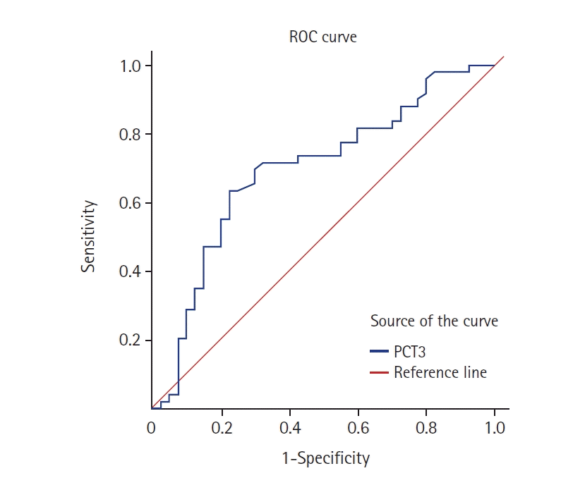
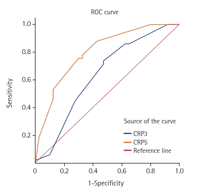
There was no significant difference in the CRP levels between the death and the survival groups, although higher levels were seen in the former. Moreover, in the survival group, an increasing trend in CRP level was seen from day 1 to day 5 followed by a decrease. In the mortality group, maximum increase was seen from day 1 to day 3, followed by a decreasing trend (Figure 2). ROC curve analysis of these markers as predictors of sepsis and survival in pediatric burns is shown in Figures. 3 and 4, respectively.

DISCUSSION

Burn injuries represent a major public health concern, especially in low-to-middle-income countries around the world. A major portion of disability-adjusted life years lost can be attributed to such injuries [17]. Burn victims are also more susceptible to infection due to a number of reasons such as loss of protective skin barrier, presence of necrotic tissue, humoral as well as cellular immunodepression, microbial dissemination and loss of airway clearance [17]. Sepsis can be defined as infection developing in the setting of SIRS [18]. A prompt diagnosis is key to ensuring reduced morbidity and improved overall survival. However, per the latest definition of sepsis in burn patients [3], this process may be inadvertently delayed. Prophylactic use of antibiotics, while beneficial, is not recommended due to lack of strong evidence in the literature [19]. Another issue with the routine usage of broad-spectrum empirical antibiotics is the emergence of anti-microbial resistance and increased frequency of nosocomial infections [20]. Early initiation of antibiotic therapy may lead to false negative reports on aerobic culture analysis. To preclude the overreliance on blood culture reports, novel biomarkers have been assessed for their ability to successfully predict sepsis in patients sustaining burn injuries. However, body physiology of children is quite different from those of adults. As a result, biomarkers like PCT and CRP, which have been shown to correlate with sepsis in adult burns, need to be validated before they can be used extensively in the pediatric age group [6-8].
The demographic parameters of our study population were similar to those reported in previous studies [21-24]. Male children and children aged 0–6 years were found to be at a higher risk than others [21-23]. This highlights the inquisitive nature of minors, which, if coupled with a lack of watchfulness by family members, can result in accidental burns at home. Moreover, a higher proportion of scald injuries (68.9%) occur in developing countries due to crowding, lack of access to electricity, etc. [21,25].
In our study, overall mortality was 34.4% (n=31), with a rate of 67.4% (n=21) in patients having sepsis. This was similar to a prospective evaluation by Dhopte et al. [26] involving 475 burn patients, which revealed a mortality rate of 31.3%, with 71.3% of subjects afflicted with sepsis. On the other hand, a recent analysis of pediatric burn mortality by Oncul et al. [22] showed infection in only 11 of 72 subjects (15.2%). However, the mean time to diagnosis of sepsis was identical to what was reported in our study, i.e., 5 days. Duration of hospital stay and median TBSA burns were significantly higher in the sepsis group compared to the non-sepsis group. Similar correlation was demonstrated by Hollén et al. [15]. This indicates the increasing risk of infection with increasing percentage of burns, which reiterates the importance of prompt initiation of antibiotic therapy.
PCT is usually detectable 3–4 hours after an inflammatory stimulus, peaking at 14 hours and remaining elevated for 24 hours, with a serum half-life of 22–35 hours. In contrast, an increase is seen 6–12 hours after initiation of bacterial infection, with a decrease noted only after control of infection [27]. This indicates PCT as an important marker to distinguish inflammation from infection, as in previous studies [28,29]. CRP is an acute phase protein that increases at 2–3 days after stimulus, reaching a peak at 50 hours, and then decreasing according to the rate of resolution of inflammation [6,30].
Our study clearly demonstrates that the levels of PCT are significantly higher in patients with sepsis, especially on days 3, 5, and 7 following burns. Previous studies support our findings [31-33]. Abdel-Hafez et al. [13], in their study of 42 cases of pediatric burns and Kundes and Kement [16] used PCT to predict sepsis in critically ill children with burn injuries. However, some authors did not find PCT to be a reliable indicator [34,35]. An earlier study by Rosanova et al. [12] showed no evidence of high PCT levels in relation to sepsis in burns. The same was observed by Neely et al. [14] in 20 pediatric burn cases. In 2018, a prospective evaluation of 27 patients by Hollén et al. [15] also found no correlation of CRP or PCT with sepsis. One reason for the conflicting results could be the small sample size in these studies. In 2016, Cabral et al. [17] published a meta-analysis of 14 studies (12 with adults and two with pediatric population) with 121 subjects. The authors opined that PCT had a strong diagnostic ability to discriminate septic from non-septic patients and could be used as a reliable determinant of sepsis.
Some studies have also shown PCT to be a more sensitive and specific marker for sepsis in children with burns than CRP [36-38]. A recent evaluation of 72 pediatric burn injuries by Oncul et al. [22] demonstrated a higher positive predictive value for PCT (64.3%) compared to CRP (47.6%) in determining sepsis. Likewise, Sharma and Sharma [39] and Jeschke et al. [11] reported no correlation of CRP with sepsis-associated mortality on evaluation of critically ill pediatric patients, a finding consistent with our study. While we demonstrated a significantly higher trend in the CRP levels on days 3, 5, and 7 in burn patients afflicted with sepsis, Pruchniewski et al. [9] noted that the biomarker peaked at 48 hours after burn. The reason for this discrepancy in findings can be attributed to the early wound excision and grafting performed in most burn injuries in this study. On the other hand, Neely et al. [14] found CRP to be a better marker for diagnosing early sepsis in children with burns, especially at day 2–3. A recent meta-analysis by Permatasari et al. [40] assessed the role of biomarkers in predicting infection in patients with thermal injuries and showed lower sensitivity (85.5% vs. 88%) and specificity (57.5% vs. 89%) with CRP compared to PCT.
These parameters require specific enzyme-backed immunosorbent assays for estimation. To address this problem, researchers have proposed utilizing indices such as RDW, NLR, and PLR, which can be easily derived from the CBC value, a test readily available everywhere [41-43]. While these parameters have demonstrated prognostic utility in community acquired pneumonia, autoimmune disorders, and critically ill patients with sepsis [44-48], their reliability as determinants of pediatric burn mortality is yet to be validated [49,50]. A summary of studies analyzing the roles of PCT and CRP in predicting sepsis/mortality and how they compare with our method is given in Table 3 [9,11-16,22].
There were certain limitations in our study. As it was a single-center study, results are not generally applicable and need further assessment. Second, there are confounding factors that can affect the significance and power of our study design. Also, we used the ABA criteria for burn-related sepsis in pediatric patients, instead of the more commonly used Pediatric Risk of Mortality (PRISM) and Pediatric Index of Mortality scores. The reason was that these scores are mainly used for predicting pediatric outcome in general and are not specific to burn injuries. Finally, while the study parameters (PCT and CRP) are continuous variables, we observed only their cross-sectional values on alternate days in the first week after burn injury.
Our study also offers some key highlights. First, it is a prospective evaluation and is thus free from selection bias. Second, our study cohort was homogeneous in nature, and sub-groups were similar demographically. Ours is also one of the largest studies prospectively analyzing the role of biomarkers in prognosticating pediatric burn sepsis.
PCT was a good early predictor of sepsis and mortality in pediatric burns, whereas CRP was reliable as a predictor of sepsis only. Both markers can serve as reliable adjuncts to blood culture sensitivity analysis for initiation of antibiotic therapy in children suffering from burn injuries. Future trends call for larger multi-center trials to draw a definite conclusion.

KEY MESSAGES

▪ Sepsis is an important cause of mortality in pediatric burns.
▪ Overreliance on blood culture may lead to delays in initiation of antibiotic therapy.
▪ Biomarkers can be used for faster diagnosis of sepsis in burns; however, there are limited data on these parameters in the pediatric age group.
▪ Procalcitonin and C-reactive protein are two such markers shown to be reliable in serving as determinants of sepsis and mortality in children afflicted with burn injuries.

Notes

CONFLICT OF INTEREST

No potential conflict of interest relevant to this article was reported.

FUNDING

None.

AUTHOR CONTRIBUTIONS

Conceptualization: DK. Methodology: RG, MM. Formal analysis: VM. Data curation: VM. Project administration: DK. Writing - original draft: NA. Writing - review & editing: DK. All authors read and agreed to the published version of the manuscript.

ACKNOWLEDGMENTS

None.

REFERENCES

1. Burd A, Yuen C. A global study of hospitalized paediatric burn patients. Burns. 2005; 31:432–8.
2. Sengoelge M, El-Khatib Z, Laflamme L. The global burden of child burn injuries in light of country level economic development and income inequality. Prev Med Rep. 2017; 6:115–20.
3. Greenhalgh DG, Saffle JR, Holmes JH, Gamelli RL, Palmieri TL, Horton JW, et al. American Burn Association consensus conference to define sepsis and infection in burns. J Burn Care Res. 2007; 28:776–90.
4. Greenhalgh DG. Sepsis in the burn patient: a different problem than sepsis in the general population. Burns Trauma. 2017; 5:23.
5. Martinez R, Rode H. Role of procalcitonin in paediatric burn wound sepsis. S Afr Med J. 2018; 108:793–4.
6. Barati M, Alinejad F, Bahar MA, Tabrisi MS, Shamshiri AR, Bodouhi NO, et al. Comparison of WBC, ESR, CRP and PCT serum levels in septic and non-septic burn cases. Burns. 2008; 34:770–4.
7. Mann EA, Wood GL, Wade CE. Use of procalcitonin for the detection of sepsis in the critically ill burn patient: a systematic review of the literature. Burns. 2011; 37:549–58.
8. Lavrentieva A, Papadopoulou S, Kioumis J, Kaimakamis E, Bitzani M. PCT as a diagnostic and prognostic tool in burn patients: whether time course has a role in monitoring sepsis treatment. Burns. 2012; 38:356–63.
9. Pruchniewski D, Pawlowski T, Morkowski J, Mackiewicz S. C-reactive protein in management of children's burns. Ann Clin Res. 1987; 19:334–8.
10. Neely AN, Smith WL, Warden GD. Efficacy of a rise in C-reactive protein serum levels as an early indicator of sepsis in burned children. J Burn Care Rehabil. 1998; 19:102–5.
11. Jeschke MG, Finnerty CC, Kulp GA, Kraft R, Herndon DN. Can we use C-reactive protein levels to predict severe infection or sepsis in severely burned patients? Int J Burns Trauma. 2013; 3:137–43.
12. Rosanova MT, Tramonti N, Taicz M, Martiren S, Basílico H, Signorelli C, et al. Assessment of C-reactive protein and procalcitonin levels to predict infection and mortality in burn children. Arch Argent Pediatr. 2015; 113:36–41.
13. Abdel-Hafez NM, Saleh Hassan Y, El-Metwally TH. A study on biomarkers, cytokines, and growth factors in children with burn injuries. Ann Burns Fire Disasters. 2007; 20:89–100.
14. Neely AN, Fowler LA, Kagan RJ, Warden GD. Procalcitonin in pediatric burn patients: an early indicator of sepsis? J Burn Care Rehabil. 2004; 25:76–80.
15. Hollén L, Hughes R, Dodds N, Coy K, Marlow K, Pullan N, et al. Use of procalcitonin as a biomarker for sepsis in moderate to major paediatric burns. Trauma. 2019; 21:192–200.
16. Kundes MF, Kement M. Value of procalcitonin levels as a predictive biomarker for sepsis in pediatric patients with burn injuries. Niger J Clin Pract. 2019; 22:881–4.
17. Cabral L, Afreixo V, Almeida L, Paiva JA. The use of procalcitonin (PCT) for diagnosis of sepsis in burn patients: a meta-analysis. PLoS One. 2016; 11:e0168475.
18. Mokline A, Garsallah L, Rahmani I, Jerbi K, Oueslati H, Tlaili S, et al. Procalcitonin: a diagnostic and prognostic biomarker of sepsis in burned patients. Ann Burns Fire Disasters. 2015; 28:116–20.
19. Avni T, Levcovich A, Ad-El DD, Leibovici L, Paul M. Prophylactic antibiotics for burns patients: systematic review and meta-analysis. BMJ. 2010; 340:c241.
20. Wibbenmeyer L, Danks R, Faucher L, Amelon M, Latenser B, Kealey GP, et al. Prospective analysis of nosocomial infection rates, antibiotic use, and patterns of resistance in a burn population. J Burn Care Res. 2006; 27:152–60.
21. Mukerji G, Chamania S, Patidar GP, Gupta S. Epidemiology of paediatric burns in Indore, India. Burns. 2001; 27:33–8.
22. Oncul U, Dalgıç N, Demir M, Karadeniz P, Karadağ ÇA. Use of procalcitonin as a biomarker for sepsis in pediatric burns. Eur J Pediatr. 2023; 182:1561–7.
23. Duke J, Wood F, Semmens J, Edgar DW, Spilsbury K, Hendrie D, et al. A study of burn hospitalizations for children younger than 5 years of age: 1983-2008. Pediatrics. 2011; 127:e971–7.
24. Ozkan GY, Yucel T. Burn, introduction, epidemiology, etiology. Turkiye Klin J Surg Med Sci. 2007; 3:1–3.
25. Peddi M, Segu S, Ramesha KT. The persistent paradigm of pediatric burns in India: an epidemiological review. Indian J Burn. 2014; 22:93–7.
26. Dhopte A, Bamal R, Tiwari VK. A prospective analysis of risk factors for pediatric burn mortality at a tertiary burn center in North India. Burns Trauma. 2017; 5:30.
27. Reinhart K, Karzai W, Meisner M. Procalcitonin as a marker of the systemic inflammatory response to infection. Intensive Care Med. 2000; 26:1193–200.
28. Arora S, Singh P, Singh PM, Trikha A. Procalcitonin levels in survivors and nonsurvivors of sepsis: systematic review and meta-analysis. Shock. 2015; 43:212–21.
29. Bouadma L, Luyt CE, Tubach F, Cracco C, Alvarez A, Schwebel C, et al. Use of procalcitonin to reduce patients' exposure to antibiotics in intensive care units (PRORATA trial): a multicentre randomised controlled trial. Lancet. 2010; 375:463–74.
30. Póvoa P. C-reactive protein: a valuable marker of sepsis. Intensive Care Med. 2002; 28:235–43.
31. Deepashree G, Rao C, Rau AT. Procalcitonin as marker of the severity of sepsis in critically ill children. Indian J Child Health. 2016; 3:102–5.
32. Solé-Ribalta A, Bobillo-Pérez S, Valls A, Girona-Alarcón M, Launes C, Cambra FJ, et al. Diagnostic and prognostic value of procalcitonin and mid-regional pro-adrenomedullin in septic paediatric patients. Eur J Pediatr. 2020; 179:1089–96.
33. Sachse C, Machens HG, Felmerer G, Berger A, Henkel E. Procalcitonin as a marker for the early diagnosis of severe infection after thermal injury. J Burn Care Rehabil. 1999; 20:354–60.
34. Lubis M, Lunis AD, Nasution BB. The usefulness of C-reactive protein, procalcitonin, and PELOD-2 score as a predictive factor of mortality in sepsis. Indones Biomed J. 2020; 12:85–188.
35. Siddiqui I, Jafri L, Abbas Q, Raheem A, Haque AU. Relationship of serum procalcitonin, C-reactive protein, and lactic acid to organ failure and outcome in critically ill pediatric population. Indian J Crit Care Med. 2018; 22:91–5.
36. Malluvalasa S, Arigela V, Lanka S, Padala S. Comparative study of procalcitonin versus c-reactive protein in the diagnosis of sepsis in children below 16 years-a single centre observational study. J Pediatr Res. 2017; 4:68–75.
37. Rey C, Los Arcos M, Concha A, Medina A, Prieto S, Martinez P, et al. Procalcitonin and C-reactive protein as markers of systemic inflammatory response syndrome severity in critically ill children. Intensive Care Med. 2007; 33:477–84.
38. Prat C, Domínguez J, Rodrigo C, Giménez M, Azuara M, Blanco S, et al. Use of quantitative and semiquantitative procalcitonin measurements to identify children with sepsis and meningitis. Eur J Clin Microbiol Infect Dis. 2004; 23:136–8.
39. Sharma R, Sharma J. Role of C reactive protein in pediatric sepsis. Int J Healthc Biomed Res. 2018; 6:57–63.
40. Permatasari AA, Sanjaya IG, Widiana IG, Niryana IW, Asmarajaya AA, Hamid AR, et al. Role of procalcitonin and C-reactive protein as marker of sepsis in major burn patients: a systematic review and meta-analysis. Open Access Maced J Med Sci. 2021; 9:197–203.
41. Angulo M, Moreno L, Aramendi I, Dos Santos G, Cabrera J, Burghi G. Complete blood count and derived indices: evolution pattern and prognostic value in adult burned patients. J Burn Care Res. 2020; 41:1260–6.
42. Qiu L, Jin X, Wang JJ, Tang XD, Fang X, Li SJ, et al. Plasma neutrophil-to-lymphocyte ratio on the third day postburn is associated with 90-day mortality among patients with burns over 30% of total body surface area in two Chinese burns centers. J Inflamm Res. 2021; 14:519–26.
43. Lin JC, Wu GH, Zheng JJ, Chen ZH, Chen XD. Prognostic values of platelet distribution width and platelet distribution width-to-platelet ratio in severe burns. Shock. 2022; 57:494–500.
44. Rajagopal P, Ramamoorthy S, Jeslin AG. Utility of haemogram parameters in mortality risk prediction of critically ill patients. J Evol Med Dent Sci. 2018; 7:1024–9.
45. Mısırlıoğlu M, Bekdas M, Kabakus N. Platelet-lymphocyte ratio in predicting mortality of patients in pediatric intensive care unit. J Clin Anal Med. 2018; 9:488–92.
46. Can E, Hamilcikan Ş, Can C. The value of neutrophil to lymphocyte ratio and platelet to lymphocyte ratio for detecting early-onset neonatal sepsis. J Pediatr Hematol Oncol. 2018; 40:e229–32.
47. Dursun A, Ozsoylu S, Akyildiz BN. Neutrophil-to-lymphocyte ratio and mean platelet volume can be useful markers to predict sepsis in children. Pak J Med Sci. 2018; 34:918–22.
48. Mathews S, Rajan A, Soans ST. Prognostic value of rise in neutrophil to lymphocyte ratio (NLR) and platelet to lymphocyte ratio (PLR) in predicting the mortality in paediatric intensive care. Int J Contemp Pediatr. 2019; 6:1052–8.
49. Fuss J, Voloboyeva A, Poliovyj V. Prognostic value of using neutrophil-lymphocyte ratio in patients with burn injury for the diagnosis of sepsis and bacteraemia. Pol Przegl Chir. 2018; 90:13–6.
50. Ciftci A, Esen O, Yazicioglu MB, Haksal MC, Tiryaki C, Gunes A, et al. Could neutrophil-to-lymphocyte ratio be a new mortality predictor value in severe burns? J Surg Surg Res. 2019; 5:26–8.
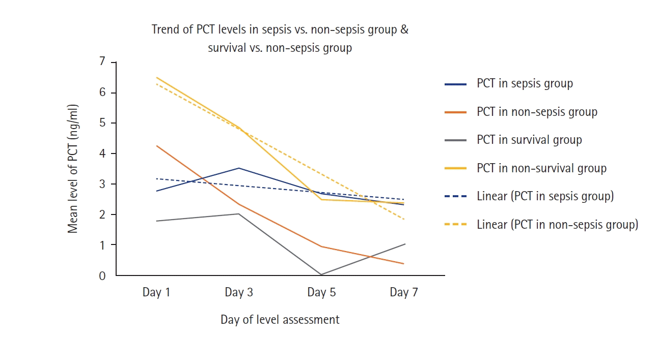
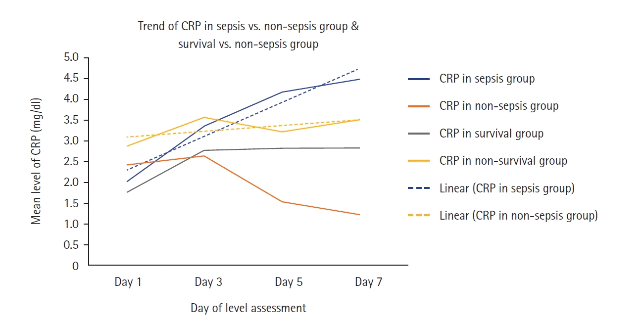
Figure 1.
Graphical representation of variation in procalcitonin (PCT) levels in the sepsis/non-sepsis and survival/non-survival groups on days 1, 3, 5, and 7 of assessment.
acc-2023-00759f1.tif
Figure 2.
Graphical representation of variation in C-reactive protein (CRP) levels in the sepsis/non-sepsis and survival/non-survival groups on days 1, 3, 5, and 7 of assessment.
acc-2023-00759f2.tif
Figure 3.
Receiver operating characteristic (ROC) curve of day 3 procalcitonin (PCT3) levels in the sepsis group. According to the ROC curve, measurement of PCT levels on day 3 after burn for correlation with sepsis has an AUC of 0.694. In addition, PCT levels above 1.4 ng/ml on day 3 after burn helps predict sepsis, with a sensitivity of 69.4% and specificity of 70%.
acc-2023-00759f3.tif
Figure 4.
Receiver operating characteristic (ROC) curve of day 3 and 5 C-reactive protein CRP3 and CRP5 levels in the sepsis group. On the ROC curve, area under the curve values for correlations of day 3 and 5 CRP levels with sepsis in pediatric burns were 0.636 and 0.801, respectively. Day 3 and 5 CRP levels above 1.3 mg/dl and 2 mg/dl in pediatric burns help predict sepsis, with a sensitivity of 75.5% and specificity of 50% and 70%, respectively.
acc-2023-00759f4.tif
Table 1.
Comparison of mean PCT and CRP levels between sepsis and non-sepsis group
Day Sepsis
Non-sepsis
P-value
PCT (ng/ml) CRP (mg/dl) PCT (ng/ml) CRP (mg/dl) PCT (ng/ml) CRP (mg/dl)
1 2.7±2.5 2.0±2.4 4.2±15.2 2.2±2.5 0.040 0.890
3 3.6±3.3 3.7±2.2 2.4±5.3 2.6±2.8 0.002 0.024
5 2.69±2.8 4.16±2.0 0.89±1.7 1.52±1.9 0.000 0.000
7 2.3±2.6 4.5±3.7 0.4±0.8 1.2±1.3 0.000 0.000

Values are presented as mean±standard deviation.

PCT: procalcitonin; CRP: C-reactive protein.

Table 2.
Comparison of mean PCT and CRP levels between non-survival and survival group
Day Non-survival
Survival
P-value
PCT (ng/ml) CRP (mg/dl) PCT (ng/ml) CRP (mg/dl) PCT (ng/ml) CRP (mg/dl)
1 6.5±17.2 2.9±3.0 1.8±2.14 1.8±2.0 0.009 0.065
3 4.9±6.0 3.6±2.7 2.1±2.8 2.8±2.0 0.009 0.165
5 2.5±2.9 3.2±2.1 1.6±2.9 2.8±2.7 0.719 1.000
7 2.3±2.8 3.5±3.1 1.0±1.8 2.8±3.3 0.303 0.451

Values are presented as mean±standard deviation.

PCT: procalcitonin; CRP: C-reactive protein.

Table 3.
A summary of studies evaluating the role of biomarkers in pediatric burns
Study Population Biomarker Study design Sample size Conclusion
Pruchniewski et al. (1987) [9] Pediatric CRP Prospective 48 CRP estimation is helpful.
Neely et al. (2004) [14] Pediatric PCT, CRP Prospective 20 CRP is better than PCT for sepsis.
Abdel-Hafez et al. (2007) [13] Pediatric CBC, CRP, IL-6, TNF, PCT, leptin, bFGF, TGF Prospective 42 Cases, 26 controls PCT can be used to predict sepsis along with IL-6.
Jeschke et al. (2013) [11] Pediatric CRP Prospective 918 No role
Rosanova et al. (2015) [12] Pediatric PCT, CRP Prospective 48 No role
Hollén et al. (2019) [15] Pediatric PCT Prospective 27 No role
Kundes and Kement (2019) [16] Pediatric PCT Retrospective 76 PCT may be predictor of sepsis.
Oncul et al. (2023) [22] Pediatric PCT, CRP Prospective 72 PCT perform better than CRP in predicting sepsis.
This study (2023) Pediatric PCT, CRP Prospective 90 PCT is a reliable predictor of both sepsis and mortality whereas CRP is a marker of only sepsis.

CRP: C-reactive protein; PCT: procalcitonin; CBC: complete blood count; IL: interleukin; TNF: tumor necrosis factor; bFGF: basic fibroblast growth factor; TGF: transforming growth factor.

TOOLS
Similar articles