Abstract
While primarily observed in adults, this case contributes valuable insights into the manifestation and management of this benign salivary gland tumor within the pediatric population. This paper reports the first documented case of sinonasal pleomorphic adenoma in pediatric otolaryngology, presenting a unique perspective on this rare nasal tumor in a 9-year-old boy. The patient presented with progressive nasal obstruction and epistaxis and underwent a smooth endoscopic resection of a 2-cm pleomorphic adenoma on the right anterior nasal septum. The subsequent discussion covered background, histology, imaging, and management strategies. Surgical removal with clear margins, particularly through the endoscopic approach, emerged as the primary and successful treatment method, minimizing morbidity and reducing recurrence risk. Long-term clinical surveillance is recommended due to an estimated 8.8% recurrence rate. In conclusion, this paper explains the challenges and solutions in diagnosing and treating sinonasal pleomorphic adenomas in children. It emphasizes the critical importance of early diagnosis, precise surgical intervention, and continuous monitoring, which are essential for achieving optimal patient outcomes.
Pleomorphic adenomas are the most common benign tumors of salivary glands, most commonly found in major salivary glands such as the parotid and submandibular glands [1].
About 28.6% of cases occur in minor salivary glands, which can be located anywhere in the upper aerodigestive tract. The hard and/or soft palate are most commonly affected [2]. It is important to note that sinonasal pleomorphic adenoma (PA) is an exceedingly rare phenomenon [3], particularly in children.
Existing literature on extra-salivary PAs is primarily limited to case reports. While these studies offer valuable insights, they often encompass non-sinonasal tract PAs or a small number of cases, leading to a lack of comprehensive information on the disease [4].
The etiology of extra-salivary PAs is unclear, but prevailing theories suggest they originate from vomeronasal organ residues or mature salivary gland tissue. Histologically, sinonasal and nasopharyngeal PAs are mainly epithelial with less mesenchymal stroma and lack capsules, unlike mixed tumors of the major salivary glands.
Clinically, sinonasal PAs present as well-defined, homogeneous soft tissue masses causing expansive bony changes, with common symptoms including unilateral nasal congestion and epistaxis, and additional symptoms such as mucopurulent rhinorrhoea, nasal swelling, and external deformities [5].
They have primarily been documented in adults, making our case an exceptional and informative contribution to the understanding of this benign salivary gland tumor in the pediatric population. We present the first reported case of this unusual nasal tumor in a previously healthy 9-year-old boy, adding a unique perspective to the limited literature on this condition.
A 9-year-old boy presented with a 5-month history of progressive unilateral nasal obstruction and epistaxis. He was otherwise well and had no medical history. The patient’s mother had also noted a mass in the right nostril. Clinical exam revealed a smooth mass arising from the right anterior nasal septum. The postnasal space and left nasal cavity had no abnormalities. Eye movements and neck examination were normal. The patient did not report any diplopia or changes in sense of smell. There was concern about etiology of the mass, with rhabdomyosarcoma being a significant differential diagnosis. Unfortunately, no clinical photographs were taken.
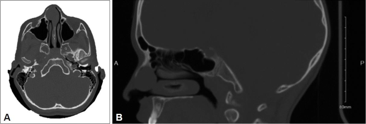
This patient underwent further investigations. A noncontrast computed tomography (CT) scan showed a 2-cm wellcircumscribed soft tissue density on the right septum without internal calcifications. There was no evidence of bony erosion or paranasal sinus abnormalities (Fig. 1).
A multidisciplinary decision was made regarding the management of this presentation. Due to the location and size of this mass, an excisional biopsy was considered to be the most effective approach. The parents were counseled about the potential for positive margins and the need for a repeat excision. The mass was successfully removed through endoscopic enbloc resection with no complications. Unfortunately, no images were taken. Mucosal and mucocartilaginous margins were taken to ensure nasal integrity in a young child.
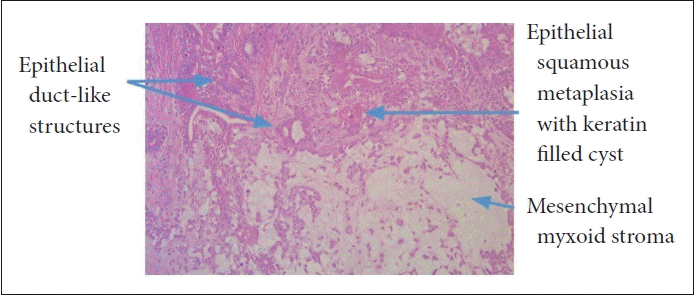
Histology showed a biphasic tumor composed of epithelial and mesenchymal components. The epithelial component is in the form of sheets, cords, and duct-like structures of small bland cells with eccentric eosinophilic cytoplasm and no significant cytological atypia or evidence of malignancy. The epithelial elements also include squamous cysts with abundant keratinous debris reflecting squamous metaplasia. There are well-formed duct-like structures with multiple layers. The mesenchymal component shows overt chondroid differentiation and elsewhere shows myxoid and hyalinised stroma. The lesion is partially covered by a fibrous capsule; which is consistent with a PA (Fig. 2).
Due to the uncommon nature of this presentation, the patient will undergo annual follow-ups for the next 5 years to watch for any signs of recurrence.
Pediatric head and neck tumors account for 3%–5% of all tumors. Pediatric sinonasal tumors have distinct epidemiology, pathology, and prognosis. They most commonly arise from the nasal cavity mucosa and its cartilaginous structures [6].
PA is the most common benign tumor of the salivary glands, predominantly occurring in the major salivary glands such as the parotid and submandibular glands [1]. PA can also occur in minor salivary glands throughout the upper aerodigestive tract. It represents a rare tumor of the sinonasal tract and has been reported in adults [3], but to our knowledge, this is the first reported case in a child.
In children, sinus and nasal masses are typically treated similarly, regardless of the prognosis. The primary objective is to make an accurate diagnosis and provide suitable treatment without causing serious complications. To establish a definite diagnosis, it is crucial to assess all patient characteristics along with clinical examination findings. This process is further improved by conducting investigations and procedures such as imaging, biopsy, and laboratory tests [6].
A multidisciplinary decision was made with regard to the management of this patient. Due to the location and size of this mass, an excisional biopsy was thought to be most fruitful. Parents were counseled with regard to the possibility of positive margins and the need for repeat excision. Mucosal and mucocartilaginous margins were taken, ensuring nasal integrity remains in a young child.
Histologically this tumor is made up of three components: epithelial, myoepithelial, and mesenchymal. These components are enclosed by a fibrous capsule, separating them from the surrounding tissues.
Identifying salivary gland tumors poses difficulties because of their infrequency and varied presentations. Diagnostic imaging methods such as ultrasound, CT scans, and magnetic resonance imaging (MRI) reveal specific characteristics of PAs. CT scans display PAs as smooth or lobulated masses with consistent soft tissue density, while MRI aids in pinpointing their location and size [7].
Given PA of the sinonasal tract is a rare entity, our knowledge comes from case reports and series reported in adults. Patients tend to report unilateral nasal obstruction, epistaxis, or pain. In advanced cases, they may present with an external nasal defect or deviation. On physical examination, nasal PAs appear smooth, soft, and polypoid. The diagnosis is made with tissue biopsy in conjunction with imaging [3].
Differential diagnoses for nasal masses in children include benign and malignant causes. The most worrisome would be rhabdomyosarcoma, which can be found throughout different sinonasal sites [6].
The primary treatment for nasal PAs involves extensive surgical removal with clear margins, which is crucial to prevent the potential development of malignancy referred to as “carcinoma ex pleomorphic adenoma,” a risk that may be present in up to 4% of cases. Various surgical approaches are available, including lateral rhinotomy, midface degloving, transpalatal surgery, and endoscopic procedures. Among these options, the endoscopic approach has proven to be a successful method for benign nasal cavity tumor removal. It results in reduced patient morbidity, minimized blood loss, shorter hospital stays, and avoids external scarring [8].
The endoscope provides improved visualization of the tumor and its boundaries. This reduces the need for unnecessary incisions and minimizes the risk of recurrence or malignant transformation due to incomplete resection. Recurrent disease can be more aggressive than PA in the major salivary glands. Precise tumor sampling and excision are crucial due to the potential for malignant transformation. In cases where tumors are unresectable, endoscopic debulking can improve patient symptoms and quality of life [9].
Chemoradiation is not commonly used because tumors do not respond well to this treatment. However, there is limited data on the role of chemoradiation in nasal PAs, primarily because most reported cases involve the parotid gland rather than the nasal region [10].
Research studies suggest that the likelihood of cancer recurrence is associated with factors such as the location of the tumor, the patient’s age at the time of diagnosis, and the completeness of surgical margin clearance. For instance, nasal PA has an estimated recurrence rate of about 8.8% after surgery. Therefore, it is recommended to establish a long-term clinical surveillance schedule with follow-up appointments every 4 to 6 months. Additionally, imaging evaluations such as CT or MRI should be conducted around one year after surgery. This systematic follow-up approach helps in the early detection of any potential local recurrence [3,5].
In summary, we reported a rare case of a 9-year-old boy with a nasal PA, a condition seldom seen in children. This case highlights the challenges in diagnosing and treating benign salivary gland tumors in unusual locations.
Diagnosing nasal PAs can be complex, as they may present with symptoms such as nasal obstruction and epistaxis. The primary treatment is surgical removal with clear margins, particularly using the endoscopic approach, to minimize complications. While chemoradiation is not typically recommended, more research is needed in this area. Long-term follow-up is crucial for detecting potential recurrences.
In conclusion, although rare, nasal PAs should be considered in diagnosing nasal masses in children. Early diagnosis, precise surgical management, and continuous monitoring are essential for optimal outcomes.
Notes
Availability of Data and Material
Data sharing not applicable to this article as no datasets were generated or analyzed during the study.
Author Contributions
Conceptualization: Michael Colreavy. Data curation: Raghad Alshammasi. Formal analysis: Holly Jones, Michael Walsh. Investigation: Nicholas Kruseman, Michael McDermott, Ian Robinson. Methodology: Michael Colreavy. Resources: Raghad Alshammasi. Supervision: Michael Colreavy. Writing—original draft: Raghad Alshammasi. Writing—review & editing: Holly Jones, Michael Walsh.
References
1. Sigdel B, Pokhrel A, Ranabhat S, KC S. Endoscopic management of huge pleomorphic adenoma of the nasal septum; a case report. Ann Med Surg (Lond). 2022; 77:103697.
2. Ritwik P, Brannon RB. A clinical analysis of nine new pediatric and adolescent cases of benign minor salivary gland neoplasms and a review of the literature. J Med Case Rep. 2012; 6:287.

3. Mackle T, Zahirovic A, Walsh M. Pleomorphic adenoma of the nasal septum. Ann Otol Rhinol Laryngol. 2004; 113(3 Pt 1):210–1.

4. Rha MS, Jeong S, Cho HJ, Yoon JH, Kim CH. Sinonasal pleomorphic adenoma: a single institution case series combined with a comprehensive review of literatures. Auris Nasus Larynx. 2019; 46(2):223–9.

5. Konsulov S, Milkov D, Markov D, Poryazova EG. Diagnostic challenges of sinonasal pleomorphic adenoma. Cureus. 2024; 16(2):e54010.

6. Lazim NM, Abdullah B. Multidisciplinary approach to children with sinonasal tumors: a review. Pediatr Investig. 2019; 3(3):173–9.
7. Kalwaniya DS, Meena R, Kumar D, Tolat A, Arya SV. A review of the current literature on pleomorphic adenoma. Cureus. 2023; 15(7):e42311.

8. Karakus MF, Ozcan KM, Dere H. Endoscopic resection of pleomorphic adenoma of the nasal septum. Tumori. 2007; 93(3):300–1.


 PDF
PDF Citation
 Print
Print


 XML Download