Journal List > Brain Tumor Res Treat > v.12(3) > 1516088069

Gwak, Yang, Yoo, Kim, Lee, and Kim: A Rare Case of Intracranial Growing Teratoma Syndrome in a Young Adult

Abstract

Intracranial growing teratoma syndrome (iGTS) is a rare phenomenon in patients with non-germinomatous germ cell tumor (NGGCT) after chemotherapy or radiotherapy. It manifests as paradoxical growth of teratomatous components, with multiple cystic lesions on cranial imaging despite normalized tumor markers. This paper presents a 22-year-old male with iGTS, diagnosed one month after chemotherapy against NGGCT. Initially diagnosed with presumptive pineal NGGCT causing obstructive hydrocephalus, the patient underwent endoscopic third ventriculostomy and extraventricular drainage with tumor biopsy followed by two chemotherapy cycles. Despite normalization of tumor markers, follow-up MRI showed increased tumor size with honeycomb-like cystic patterns. The patient underwent suboccipital craniotomy for tumor removal via combined telovelar and infratentorial supracerebellar approaches. The final pathology confirmed mature teratoma. However, postoperative bleeding and left thalamic infarction occurred, resulting in severe neurological deficits. Despite challenges, the patient eventually regained the ability to follow simple commands. To understand iGTS pathophysiology, several hypotheses, including the differentiation of immature components and the uninhibited growth of mature components induced by chemotherapy or radiotherapy, were explored. Surgical intervention remains as an ideal treatment, while clinical trials investigate chemotherapy options. Frequent imaging follow-ups are crucial for early detection in iGTS for NGGCT patients.

INTRODUCTION

Intracranial germ cell tumors (iGCTs) are rare, heterogeneous brain tumors that primarily arise in the pineal and suprasellar regions [12]. They account for about 10% of pediatric primary brain tumors in far East Asian countries [3]. iGCTs are germinomas and non-germinomatous germ cell tumors (NGGCTs), with the latter comprising teratomas categorized into mature and immature types. Frappaz et al. [4] established diagnostic criteria for iGCT based on clinical signs, radiology, markers in serum and cerebrospinal fluid, and histology.
Patients with NGGCTs show paradoxical growth of mature teratoma components after chemotherapy despite normalized tumor marker levels. Logothetis et al. [5] defined this phenomenon as growing teratoma syndrome (GTS). According to preceding studies, three criteria are required to diagnose: 1) normalization of tumor markers, such as alpha-fetoprotein (AFP) and/or beta-human chorionic gonadotropin (β-hCG), 2) rapid increase in tumor size during or after chemotherapy, and 3) absence of any NGGCT component in histopathology except mature teratoma [3]. Some researchers proposed that both chemotherapy and radiotherapy have to be included in definition of GTS [267]. GTS typically occurs in the gonad, retroperitoneum, or mediastinum [8]; however, it can manifest as intracranial GTS (iGTS). Michaiel et al. [6] reported that the incidence of iGTS is about 5% of all GCT patients due to a literature review from 2000 to 2017.
Here we report a rare case of iGTS one month after chemotherapy for pineal NGGCT.

CASE REPORT

A 22-year-old male patient complained of aggravating headaches and vomiting over the past few months. However, there was no specific illness in his past medical history. The initial contrast-enhanced brain MRI revealed a heterogeneously enhanced mass extending from the pineal gland to the fourth ventricle, causing obstructive hydrocephalus (Fig. 1A and B). A floating fatty droplet was also observed in the right lateral ventricle. The serum AFP level was elevated to 55.50 ng/mL, whereas the serum β-hCG level was within the normal range.
We performed an endoscopic third ventriculostomy (ETV) with tumor biopsy and extraventricular drainage (EVD). There was no definitive sign of drop metastasis on the whole spine MRI. At first, the tumor was presumed to be an NGGCT; however, the biopsy specimen includes only an ependymal cyst-like component (Fig. 2A). According to the follow-up tumor marker results, the AFP level further increased to 76.52 ng/mL, and the β-hCG level rose to 9.35 ng/mL. Based on these findings, the tumor was diagnosed as a mixed germ cell tumor. Consequently, he was transferred to the oncology department to initiate adjuvant chemotherapy. The regimen included etoposide, cisplatin, and bleomycin. Serum AFP and β-hCG levels normalized after two cycles of chemotherapy. Meanwhile, he complained of aggravating symptoms, such as tinnitus, hearing loss, and diplopia. Subsequent brain MRI for response evaluation showed an increase in mass size with honeycomb-like cystic patterns (Fig. 1C and D). Therefore, we performed suboccipital craniotomy with subtotal tumor removal by infratentorial supracerebellar and telovelar approaches. The final pathologic findings showed different types of mature tissues such as chondroid tissue with columnar epithelium and cartilaginous tissue with respiratory epithelium, confirming the diagnosis of mature cystic teratoma (Fig. 2B and C).
A postoperative hematoma was observed on the brain CT at the site where the mass had been located. While in an intensive care unit, the patient experienced drowsiness and quadriparesis, leading to a tracheostomy. Subsequent brain MRI revealed a left thalamic infarction. After several weeks of intensive care, the patient was successfully weaned off the ventilator and transferred to the general ward and then to the rehabilitation department. Currently, the patient can follow simple commands but is hospitalized for rehabilitation therapy and remains bedridden.

DISCUSSION

The patient exhibited normalization of tumor marker after two cycles of chemotherapy, whether the tumor rapidly increased in size with a pathognomonic honeycomb-like cystic lesion and final pathologic diagnosis corresponding to mature teratoma. According to Tasaka et al. [9], the onset of iGTS usually occurs at a median time of 12.8 months, with a range from 3 to 56 months. Additionally, Michaiel et al. [6] report a median onset of 2 months, with a range of 0.5 to 32 months. In our case, it took only a month to confirm the diagnosis of iGTS. Therefore, while the onset of the current case is not exceptionally short, it is still relatively rapid compared to the ranges reported in the literature.
There was no definite consensus about the pathogenesis of iGTS, but a few hypotheses have been proposed. First, chemotherapy and/or radiotherapy may induce the immature proportion of tumor differentiation to a mature phenotype. Second, the selective destruction by chemo-radiation therapy, excluding mature teratoma, may lead to uninhibited growth of teratomatous element [1011]. Satake et al. [12] claimed that DMRT1 loss and 12p gain contribute to teratomagenesis in GTS by copy number variation analysis. In addition to genetic factors, certain microenvironments, such as unlimited benign cell growth, and an immune-compromised state due to chemotherapy may lead to the rapid growth of mature teratoma [3].
Referring to the pathological findings of our case, the rapid increase in the mass size is attributed to the formation of multiple cysts with exudate, not the growth of tumor cells themselves, which is consistent with previous studies. According to recent research, multiple cyst-like lesions usually comprise fatty components rather than pure cystic content, and the fatty component is more important [913].
Thus, the treatment of iGCT depends on tumor classification and clinical symptoms. The primary treatment option for NGGCT is neoadjuvant or adjuvant chemotherapy. Recently, radiotherapy has become significant; however, it is not recommended for teratomas. For germinoma, chemo-radiotherapy approaches are essential. If obstructive hydrocephalus is present at the initial diagnosis, ETV with optional biopsy or at least EVD is preferred. If residual mass persists after chemotherapy, a second-look operation is highly recommended for maximal safe resection [414]. Considering iGTS as an extension of the iGCT treatment, the ideal treatment for iGTS requires surgical approaches with gross total resection, if possible [361516]. For unresectable tumors due to their location, a clinical trial of palbociclib as adjuvant chemotherapy was undertaken [17].
Although iGCT patients showed favorable overall survival, early detection and early surgical intervention can mitigate deterioration. This is important since immature teratoma show faster growth even during chemotherapy and have a significantly higher incidence of iGTS, for example, 40%, as indicated in related literature [3]. In our case, the tumor was considered a mixed germ cell tumor containing an epidermal cyst-like component. The growing teratoma exhibited very rapid growth, causing symptoms that necessitated earlier imaging with a second-look operation.
iGTS rarely occurs during or after chemotherapy for iGCTs. iGTS presents with paradoxical growth of tumor mass containing mature teratomatous components, despite normalized tumor markers. The pathogenesis of iGTS is associated with the differentiation or destruction of immature components. Hence, several aspects require clarification. Surgery is a common and preferred treatment for iGTS, while ongoing clinical trials are exploring additional options. To improve prognosis, practitioners should remain vigilant for the possibility of iGTS and frequent imaging follow-ups.

Acknowledgments

None

Notes

Ethics Statement: This case report, based solely on medical records, was reviewed by the Interstitial Review Board (IRB) and received an exemption from formal ethical approval (IRB No. VC24ZISI0148).

Author Contributions:

  • Conceptualization: Seung Ho Yang.

  • Data curation: Yujin Lee, Young Il Kim.

  • Investigation: Young Gook Gwak.

  • Methodology: Young Il Kim.

  • Project administration: Seung Ho Yang.

  • Resources: Yeun Ji Yoo, Hyun Ho Kim, Yujin Lee.

  • Supervision: Seung Ho Yang.

  • Validation: Yeun Ji Yoo.

  • Visualization: Young Gook Gwak, Yujin Lee.

  • Writing—original draft: Young Gook Gwak.

  • Writing—review & editing: Young Il Kim.

Conflicts of Interest: The authors have no potential conflicts of interest to disclose.

Funding Statement: None

Availability of Data and Material

Data sharing not applicable to this article as no datasets were generated or analyzed during the study.

References

1. Yanagisawa S, Okamoto K, Yamaguchi S, Tamai Y, Fujitani M, Inoue M, et al. Intracranial growing teratoma syndrome observed at 44 months after initial treatment; a case presentation and literature review. Childs Nerv Syst. 2020; 36:865–868. PMID: 31853895.
2. Khoo B, Ramakonar HH, Robbins P, Honeybul S. Intracranial monodermal teratoma presenting with growing teratoma syndrome. J Surg Case Rep. 2017; 2017:rjx038. PMID: 28560020.
3. Kim CY, Choi JW, Lee JY, Kim SK, Wang KC, Park SH, et al. Intracranial growing teratoma syndrome: clinical characteristics and treatment strategy. J Neurooncol. 2011; 101:109–115. PMID: 20532955.
4. Frappaz D, Dhall G, Murray MJ, Goldman S, Faure Conter C, Allen J, et al. EANO, SNO and Euracan consensus review on the current management and future development of intracranial germ cell tumors in adolescents and young adults. Neuro Oncol. 2022; 24:516–527. PMID: 34724065.
5. Logothetis CJ, Samuels ML, Trindade A, Johnson DE. The growing teratoma syndrome. Cancer. 1982; 50:1629–1635. PMID: 6288220.
6. Michaiel G, Strother D, Gottardo N, Bartels U, Coltin H, Hukin J, et al. Intracranial growing teratoma syndrome (iGTS): an international case series and review of the literature. J Neurooncol. 2020; 147:721–730. PMID: 32297094.
7. Moiyadi A, Jalali R, Kane SV. Intracranial growing teratoma syndrome following radiotherapy--an unusually fulminant course. Acta Neurochir (Wien). 2010; 152:137–142. PMID: 19404574.
8. Iwata H, Mori Y, Takagi H, Shirahashi K, Shinoda J, Shimokawa K, et al. Mediastinal growing teratoma syndrome after cisplatin-based chemotherapy and radiotherapy for intracranial germinoma. J Thorac Cardiovasc Surg. 2004; 127:291–293. PMID: 14752454.
9. Tasaka K, Umeda K, Kamitori T, Ogata H, Mikami T, Saida S, et al. Intracranial growing teratoma syndrome with intraventricular lipid accumulation. J Pediatr Hematol Oncol. 2021; 43:e505–e507. PMID: 32769571.
10. Oya S, Saito A, Okano A, Arai E, Yanai K, Matsui T. The pathogenesis of intracranial growing teratoma syndrome: proliferation of tumor cells or formation of multiple expanding cysts? Two case reports and review of the literature. Childs Nerv Syst. 2014; 30:1455–1461. PMID: 24633581.
11. Kong DS, Nam DH, Lee JI, Park K, Kim JH, Shin HJ. Intracranial growing teratoma syndrome mimicking tumor relapse: a diagnostic dilemma. J Neurosurg Pediatr. 2009; 3:392–396. PMID: 19409018.
12. Satake D, Natsumeda M, Satomi K, Tada M, Sato T, Okubo N, et al. Successful multimodal treatment of intracranial growing teratoma syndrome with malignant features. Curr Oncol. 2024; 31:1831–1838. PMID: 38668041.
13. Han NY, Sung DJ, Park BJ, Kim MJ, Cho SB, Kim KA, et al. Imaging features of growing teratoma syndrome following a malignant ovarian germ cell tumor. J Comput Assist Tomogr. 2014; 38:551–557. PMID: 24681864.
14. Kajiwara S, Nakamura H, Sakata K, Komaki S, Negoto T, Morioka M. Endoscopic aqueductal membrane fenestration was effective for intractable hydrocephalus after removal of a nongerminomatous germ cell tumor exhibiting growing teratoma syndrome: a case report. BMC Pediatr. 2022; 22:683. PMID: 36443673.
15. Kanamori M, Kumabe T, Watanabe M, Chonan M, Saito R, Yamashita Y, et al. Indications for salvage surgery during treatment for intracranial germ cell tumors. J Neurooncol. 2018; 138:601–607. PMID: 29582270.
16. Yagi K, Kageji T, Nagahiro S, Horiguchi H. Growing teratoma syndrome in a patient with a non-germinomatous germ cell tumor in the neurohypophysis--case report. Neurol Med Chir (Tokyo). 2004; 44:33–37. PMID: 14959935.
17. Schultz KA, Petronio J, Bendel A, Patterson R, Vaughn DJ. PD0332991 (palbociclib) for treatment of pediatric intracranial growing teratoma syndrome. Pediatr Blood Cancer. 2015; 62:1072–1074. PMID: 25417786.
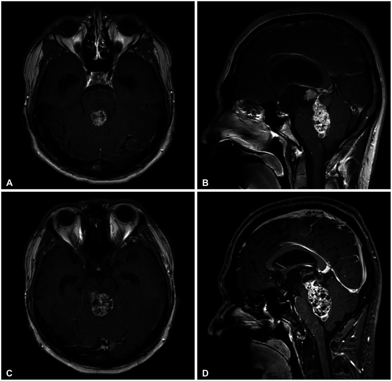
Fig. 1

Gadolinium-enhanced magnetic resonance (MR) brain images of intracranial growing teratoma syndrome (iGTS) patients. A and B: Initial MR images of germ cell tumor show heterogeneously enhanced mass obstructing the fourth ventricle. C and D: MR images after two cycles of chemotherapy show increased size of mass with honeycomb-like cystic patterns corresponding to iGTS.

btrt-12-200-g001
Fig. 2

Hematoxylin and eosin staining of the resected tumor. A: Tissue from ETV with biopsy shows only squamous epithelium containing keratin debris. B and C: Tissue from subtotal tumor resection shows typical mature cystic teratoma histopathology. It consists of different types of mature tissue components such as chondroid tissue (*) with columnar epithelium (**), cartilaginous tissue (black arrow), and respiratory epithelium (white arrow).

btrt-12-200-g002
TOOLS
Similar articles