Journal List > Korean Circ J > v.54(8) > 1516088057

Lee, Ahn, Jeon, Lee, Youn, Zhang, Hu, Wang, Lee, Hahn, Nam, Doh, Lee, Kim, Huang, Jiang, Zhou, Chen, Tang, Jiang, Chen, He, Yoon, Tahk, Kim, Ki, Shin, Hwang, Kang, Kim, and Koo: Discordance Between Angiographic Assessment and Fractional Flow Reserve or Intravascular Ultrasound in Intermediate Coronary Lesions: A Post-hoc Analysis of the FLAVOUR Trial

Author's summary

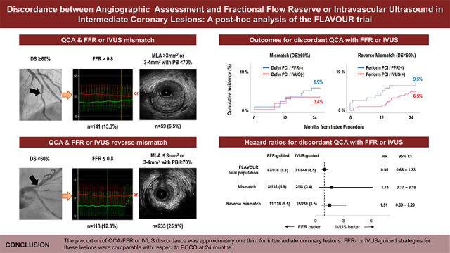
Assessment of coronary artery stenosis by quantitative coronary angiography (QCA) often disagrees with fractional flow reserve (FFR) or intravascular ultrasound (IVUS). We investigated the incidence of discrepancy between QCA and FFR or IVUS in intermediate coronary lesions and the impact of FFR- or IVUS-guided revascularization strategies for these discordant lesions. The proportion of lesions discordant with FFR or IVUS was 28.2% and 32.4%, respectively. Cumulative 2-year clinical outcomes were comparable between FFR- and IVUS-guided revascularization strategies for discordant coronary lesions. This provides valuable information for clinicians in deciding the optimal revascularization strategy for intermediate coronary lesions discordant with FFR or IVUS assessment.

Abstract

Background and Objectives

Angiographic assessment of coronary stenosis severity using quantitative coronary angiography (QCA) is often inconsistent with that based on fractional flow reserve (FFR) or intravascular ultrasound (IVUS). We investigated the incidence of discrepancies between QCA and FFR or IVUS, and the outcomes of FFR- and IVUS-guided strategies in discordant coronary lesions.

Methods

This study was a post-hoc analysis of the FLAVOUR study. We used a QCA-derived diameter stenosis (DS) of 60% or greater, the highest tertile, to classify coronary lesions as concordant or discordant with FFR or IVUS criteria for percutaneous coronary intervention (PCI). The patient-oriented composite outcome (POCO) was defined as a composite of death, myocardial infarction, or revascularization at 24 months.

Results

The discordance rate between QCA and FFR or IVUS was 30.2% (n=551). The QCA-FFR discordance rate was numerically lower than the QCA-IVUS discordance rate (28.2% vs. 32.4%, p=0.050). In 200 patients with ≥60% DS, PCI was deferred according to negative FFR (n=141) and negative IVUS (n=59) (15.3% vs. 6.5%, p<0.001). The POCO incidence was comparable between the FFR- and IVUS-guided deferral strategies (5.9% vs. 3.4%, p=0.479). Conversely, 351 patients with DS <60% underwent PCI according to positive FFR (n=118) and positive IVUS (n=233) (12.8% vs. 25.9%, p<0.001). FFR- and IVUS-guided PCI did not differ in the incidence of POCO (9.5% vs. 6.5%, p=0.294).

Conclusions

The proportion of QCA-FFR or IVUS discordance was approximately one third for intermediate coronary lesions. FFR- or IVUS-guided strategies for these lesions were comparable with respect to POCO at 24 months.

Trial Registration

ClinicalTrials.gov Identifier: NCT02673424

Graphical Abstract

kcj-54-485-abf001.jpg

INTRODUCTION

Angiographic assessment by quantitative coronary angiography (QCA) plays a pivotal role in guiding percutaneous coronary intervention (PCI) in patients with coronary artery disease (CAD). However, angiographic evaluation alone has limitations, such as over- or underestimation of stenosis severity and intra- or interobserver variability, specifically in intermediate coronary lesions.1) Thus, adjunctive physiological and intracoronary imaging examinations are often required for revascularization decision-making for intermediate coronary lesions. The fractional flow reserve (FFR) is used to enhance the diagnostic capability of coronary angiography for identifying coronary lesions that induce myocardial ischemia.2) Compared with angiography-guided PCI, FFR-guided PCI improves clinical outcomes, especially in patients with multivessel CAD.3)4) Intravascular ultrasound (IVUS) is another supplementary imaging tool for angiographic assessment as it outperforms QCA by providing detailed lesion information such as length and diameter, plaque characterization, or vascular remodeling.5)6) Several large-scale randomized clinical trials have demonstrated that IVUS-guided PCI is associated with a lower risk of major cardiovascular outcomes compared to angiography guidance alone.7)8)9) In particular, the Fractional Flow Reserve and Intravascular Ultrasound-Guided Intervention Strategy for Clinical Outcomes in Patients with Intermediate Stenosis (FLAVOUR) trial performed a head-to-head comparison between FFR and IVUS for the treatment of intermediate stenosis.10)
Coronary lesions with discrepancies between QCA-derived diameter stenosis (DS) and FFR, visual-functional mismatch,11)12) or IVUS13) are commonly encountered in clinical practice. However, the frequency of discrepancies between angiographic assessment and FFR or IVUS and the optimal revascularization strategy for these lesions has not been determined. Using data from the FLAVOUR trial, the present study investigated the incidence of discrepancy between angiographic assessment by QCA-derived DS and FFR or IVUS assessment in intermediate coronary lesions. We also evaluated the clinical outcomes of physiology- or IVUS-guided revascularization strategies in patients with discordant intermediate lesions between QCA and FFR or IVUS.

METHODS

Ethical statement

This study adhered to the International Council for Harmonization Guidelines for Good Clinical Practice and the principles of the 2013 version of the Declaration of Helsinki. The institutional review board of each participating site approved the study protocol, and written informed consent was obtained from all patients (Wonju Severance Christian Hospital, IRB: CR317104).

Study population

The FLAVOUR trial was an investigator-initiated, prospective, randomized, open-label, multinational trial conducted at 18 sites in Korea and China. A detailed explanation of the study protocol has been provided in previous studies.10)14) In summary, the FLAVOUR trial included patients with a de novo intermediate degree of stenosis in a target vessel size ≥2.5 mm eligible for stent implantation. A total of 1,682 patients who met the inclusion criteria were enrolled in this trial and randomly assigned in a 1:1 ratio to the FFR (838 patients) or IVUS arm (844 patients). Our study is a post-hoc analysis of the FLAVOUR trial, which included patients with de novo intermediate stenosis ranging from 40% to 70% by visual estimation. The distribution of QCA-derived DS among lesions included in the FLAVOUR trial is shown in Supplementary Figure 1. In this analysis, patients were categorized based on severe angiographic stenosis, defined as a QCA-derived DS of 60% or greater, the highest tertile of DS (inter-tertile range, 52–60%).

Procedure and quantitative coronary angiography analysis

Invasive coronary angiography and PCI were performed using current guidelines and conventional techniques. The angiographic core laboratory at Seoul National University Hospital, Seoul, South Korea, quantitatively analyzed the baseline and procedural coronary angiograms using a validated software program. Reference diameter, minimal lumen diameter, %DS, and lesion length were measured using QCA. The calculation of %DS was performed as follows: (reference vessel diameter - minimum lumen diameter)/reference vessel diameter × 100. After selecting the best projection showing the most significant stenosis, minimum lumen diameter, reference vessel diameter, lesion length, and end-diastolic %DS were measured.

Fractional flow reserve and intravascular ultrasound measurements

The FFR measurements were performed by conventional methods after maximum hyperemia was induced by intravenous infusion of adenosine (140 μg/kg/min), or by intracoronary injection of nicorandil (2 mg).15) IVUS measurements included minimal lumen area (MLA), external elastic membrane area, and plaque burden, calculated as a percentage of (1 − MLA/external elastic membrane area). In the FFR group, revascularization was performed if the FFR value showed 0.80 or less and deferred PCI according to FFR >0.8.16) In the IVUS group, revascularization was performed in lesions with an MLA of 3 mm2 or less or an MLA of 3–4 mm2 with a plaque burden of more than 70%.6)17)18) Raw data from FFR and IVUS were assessed and analyzed by core laboratories that were independent and unaware of the clinical and procedural characteristics.

Study endpoints and definitions

The primary endpoint of this study was to evaluate the rate of mismatch (severe angiographic stenosis with negative FFR or negative IVUS) and reverse mismatch (non-severe angiographic stenosis with positive FFR or positive IVUS criteria) in intermediate coronary lesions. Secondary endpoints included the incidence of patient-oriented composite outcomes (POCOs), a composite of all-cause death, myocardial infarction, and any revascularization 24 months after randomization according to revascularization strategy by FFR or IVUS for discordant lesions. The individual components of the endpoint, cardiac death, components of myocardial infarction, and target vessel failure were also matched. Each clinical endpoint was defined according to the Academic Research Consortium consensus.19) All clinical events were adjudicated in a blinded fashion by an independent clinical event adjudication committee.

Statistical analysis

Data are expressed as numbers (%), mean ± standard deviation, or median (interquartile range). Continuous variables were compared using Student’s t-test, and categorical data were compared using χ2 or Fisher’s exact tests. Correlation and linear regression analyses were used to compare the %DS, FFR values, and MLA. Event-free survival was analyzed using Kaplan-Meier survival curves, and differences between event-free survival curves were compared using the log-rank test. Statistical analyses were performed using SPSS version 25.0 (IBM, Armonk, NY, USA) and R Statistical Software version 3.0.1 (R Foundation for Statistical Computing, Vienna, Austria). Statistical significance was set at p<0.05.

RESULTS

Proportion of discordance between angiography and fractional flow reserve or intravascular ultrasound

Figure 1 outlines the selection process for the study population. Among the entire study population with 1,820 vessels, 680 lesions (37.4%) had DS ≥60%, while 1,140 lesions had DS <60% on QCA. Among the 680 lesions with DS ≥60%, 480 (70.6%) were treated with PCI according to FFR or IVUS positive results, and PCI was deferred in 200 lesions (29.4%) according to negative FFR (n=141, “FFR mismatch”) or negative IVUS criteria (n=59, “IVUS mismatch”). In contrast, among the 1,140 lesions with DS <60%, PCI was deferred in 789 lesions (69.2%) according to FFR or IVUS negative results, and 351 lesions (30.8%) underwent PCI according to positive FFR (n=118, “FFR reverse mismatch”) or positive IVUS (n=233, “IVUS reverse mismatch”).
Figure 1

Study flow. The highest tertile (60%) DS by QCA was used to categorize lesions discordant with the FFR or IVUS criteria. PCI was deferred in 200 patients with ≥60% DS (141 with FFR and 59 with IVUS), whereas 351 patients with DS <60% underwent PCI (118 with FFR and 233 with IVUS).

DS = diameter stenosis; FFR = fractional flow reserve; IVUS = intravascular ultrasound; QCA = quantitative coronary angiography.
kcj-54-485-g001
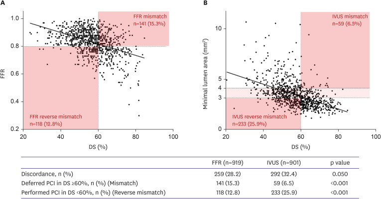
In the FLAVOUR trial, 919 were randomly assigned to the FFR group and 901 to the IVUS group. Figure 2 shows the inverse correlations between %DS and FFR (Figure 2A) and MLA on IVUS (Figure 2B). In the FFR group, 141 vessels (15.3%) had lesions with ≥60% DS and negative FFR (“FFR mismatch”), and 118 vessels (12.8%) showed DS <60% with positive FFR (“FFR reverse mismatch”). In the IVUS group, 59 vessels (6.5%) showed negative IVUS among the lesions with ≥60% DS (“IVUS mismatch”), and 233 vessels (25.9%) showed positive IVUS among the lesions with DS <60% (“IVUS reverse mismatch”). The proportion of discordant lesions between QCA-derived DS and IVUS was numerically higher than that of FFR (32.4% vs. 28.2%, respectively, p=0.050). Among the discordant lesions, the QCA-FFR mismatch rate was higher in the than the QCA-IVUS mismatch rate (15.3% vs. 6.5%, p<0.001), and the reverse mismatch rate was higher in the IVUS group than in the FFR group (25.9% vs. 12.8%, p<0.001).
Figure 2

Correlation between DS and FFR or IVUS minimum lumen area. There is an inverse correlation between %DS and FFR (A) or the minimum lumen area on IVUS (B). The proportions of discordance between QCA and FFR or IVUS were 28.2% and 32.4%, respectively (p=0.050). In the FFR group, 141 vessels (15.3%) had lesions with DS ≥60% but were FFR negative, and 118 vessels (12.8%) showed DS <60% but were FFR positive. In the IVUS group, 59 vessels (6.5%) showed IVUS negativity among lesions with DS ≥60%, and 233 vessels (25.9%) showed IVUS positivity among lesions with DS <60%.

DS = diameter stenosis; FFR = fractional flow reserve; IVUS = intravascular ultrasound; QCA = quantitative coronary angiography.
kcj-54-485-g002

Baseline and angiographic characteristics

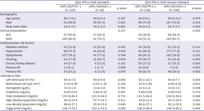
The baseline characteristics of the study population are summarized in Table 1. Among patients with a QCA-FFR or IVUS mismatch, there were no differences between the FFR and IVUS arms regarding demographic characteristics, clinical presentation, risk factors, and laboratory findings. In patients with QCA-FFR or IVUS reverse mismatch, baseline characteristics showed no significant differences between the FFR and IVUS groups, except for a history of myocardial infarction and hemoglobin level.
Table 1

Baseline characteristics of patients with discordance between QCA and FFR or IVUS

kcj-54-485-i001
QCA-FFR or IVUS mismatch QCA-FFR or IVUS reverse mismatch
≥60% DS/FFR (−) (n=135) ≥60% DS/IVUS (−) (n=58) p value <60% DS/FFR (+) (n=116) <60% DS/IVUS (+) (n=230) p value
Demographic
Age (years) 66.7±9.5 64.8±8.4 0.187 64.9±9.1 64.9±10.3 0.979
Male 93 (68.9) 39 (67.2) 0.821 88 (75.9) 167 (72.6) 0.516
Body mass index 24.4±3.5 24.5±3.0 0.901 24.9±3.0 24.7±3.5 0.605
Clinical presentation 0.157 0.240
ACS 27 (20.0) 17 (29.3) 52 (44.8) 88 (38.3)
SIHD 108 (80.0) 41 (70.7) 64 (55.2) 142 (61.7)
Cardiovascular risk factors
Diabetes mellitus 43 (31.9) 15 (25.9) 0.405 45 (38.8) 81 (35.2) 0.514
Hypertension 99 (73.3) 34 (58.6) 0.043 81 (69.8) 177 (77.0) 0.151
Dyslipidemia 107 (79.3) 45 (77.6) 0.794 99 (85.3) 183 (79.6) 0.191
Smoking 24 (17.8) 12 (20.7) 0.634 24 (20.7) 45 (19.6) 0.805
Chronic kidney disease* 24 (17.8) 6 (10.3) 0.191 20 (17.2) 51 (22.2) 0.283
Previous MI 11 (8.1) 1 (1.7) 0.090 10 (8.6) 7 (3.0) 0.023
Previous PCI 34 (25.2) 8 (13.8) 0.079 26 (22.4) 46 (20.0) 0.602
Laboratory data
Left ventricular EF (%) 64.4±7.0 64.6±9.8 0.842 62.1±10.1 64.4±7.7 0.058
White blood cell (per mm3) 6.55±2.06 6.07±1.30 0.104 6.53±1.92 6.64±2.01 0.631
Hemoglobin (g/dL) 13.5±1.9 13.6±2.0 0.761 13.4±1.6 13.8±1.6 0.036
Creatinine (mg/dL) 0.85±0.21 0.83±0.18 0.467 0.84±0.20 0.93±0.67 0.170
Total cholesterol (mg/dL) 155.9±46.4 153.2±44.9 0.715 155.5±44.8 150.3±38.6 0.268
High-density lipoprotein (mg/dL) 46.2±10.6 47.7±14.7 0.411 44.6±11.4 45.3±10.6 0.581
Low-density lipoprotein (mg/dL) 86.6±37.1 83.9±35.0 0.640 88.8±37.1 83.1±32.8 0.146
Triglyceride (mg/dL) 142.7±70.3 123.5±73.0 0.092 138.3±89.8 140.2±88.2 0.849
Values are mean ± standard deviations or number (%).
ACS = acute coronary syndrome; DS = diameter stenosis; EF = ejection fraction; FFR = fractional flow reserve; IVUS = intravascular ultrasound; MI = myocardial infarction; PCI = percutaneous coronary intervention; QCA = quantitative coronary angiography; SIHD = stable ischemic heart disease.
*Chronic kidney disease was defined as a history of chronic kidney disease or an estimated glomerular filtration rate of less than 60 mL per minute per 1.73 m2 of body-surface area.
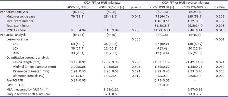
The angiographic and procedural characteristics of the study population are presented in Table 2. Among patients with a QCA-FFR or IVUS mismatch, lesion severity assessed using the SYNTAX score or QCA parameters was similar between the 2 groups. In contrast, among patients with a QCA-FFR or IVUS reverse mismatch, the SYNTAX scores were higher in the FFR group than in the IVUS group. An FFR reverse mismatch was more frequently observed in the left anterior descending artery (LAD) than an IVUS reserves mismatch (82.2% vs. 59.2%; p<0.001). QCA revealed that lesions in the FFR reverse mismatch were longer and smaller than those in the IVUS mismatch.
Table 2

Angiographic and procedural characteristics of patients with discordance between QCA and FFR or IVUS

kcj-54-485-i002
QCA-FFR or IVUS mismatch QCA-FFR or IVUS reverse mismatch
≥60% DS/FFR (−) ≥60% DS/IVUS (−) p value <60% DS/FFR (+) <60% DS/IVUS (+) p value
Per patient analysis (n=135) (n=58) (n=116) (n=230)
Multi-vessel disease 79 (58.5) 25 (43.1) 0.049 75 (64.7) 129 (56.1) 0.126
Total stent number - - 1.16±0.51 1.12±0.38 0.387
Total stent length - - 31.8±16.5 29.5±14.2 0.203
SYNTAX score 8.36±4.94 8.16±3.94 0.786 11.23±6.21 9.46±6.41 0.015
Per vessel analysis (n=141) (n=59) (n=118) (n=233)
Lesion location 0.385 <0.001
LAD 69 (48.9) 35 (59.3) 97 (82.2) 138 (59.2)
LCX 39 (27.7) 12 (20.3) 4 (3.4) 30 (12.9)
RCA 33 (23.4) 12 (20.3) 17 (14.4) 65 (27.9)
Quantitative coronary analysis
Lesion length (mm) 18.19±8.92 17.82±9.38 0.793 24.53±11.93 21.42±11.90 0.021
Minimal lumen diameter (mm) 1.02±0.25 1.03±0.26 0.805 1.32±0.22 1.38±0.25 0.039
Reference diameter (mm) 2.91±0.53 2.96±0.58 0.594 2.92±0.46 2.93±0.40 0.858
Diameter stenosis (%) 65.1±4.7 65.2±4.4 0.812 54.5±5.3 52.9±5.3 0.009
Pre-PCI FFR 0.87±0.05 - 0.75±0.05 -
Post-PCI FFR - - 0.87±0.05 -
MLA measured by IVUS (mm2) - 3.96±1.22 - 2.97±0.66
Plaque burden at MLA site (%) - 67.4±8.5 - 73.3±7.7
Values are mean ± standard deviations or number (%).
DS = diameter stenosis; FFR = fractional flow reserve; IVUS = intravascular ultrasound; LAD = left anterior descending artery; LCX = left circumflex artery; MLA = minimal lumen area; PCI = percutaneous coronary intervention; QCA = quantitative coronary angiography; RCA = right coronary artery.

Clinical outcomes in mismatched and reverse mismatched lesions

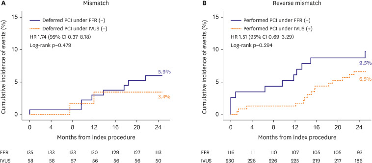
The 2-year clinical outcomes of the study population are summarized in Table 3. There was no difference in the incidence of POCO at 2 years between FFR and IVUS mismatches. Likewise, the incidence of POCO was similar between FFR and IVUS reverse mismatches. Figure 3 shows the incidence of POCO of deferred PCI in lesions with ≥60% DS and PCI with DS <60% according to the FFR- or IVUS-guided decision. Among the individual components of POCO, all-cause death and revascularization were similarly observed in both the FFR/IVUS mismatch and FFR/IVUS reverse mismatch. However, the rate of myocardial infarction was significantly higher in the FFR reverse mismatch group than in the IVUS reverse mismatch group (4.4% vs. 0.4%, p=0.026), mainly due to periprocedural myocardial infarction (2.6% vs. 0.0%, p=0.014). No significant effect was observed for the interaction between angiographic stenosis severity, QCA-FFR or IVUS concordance, and both revascularization strategies on the incidence of POCO (Supplementary Figure 2).
Table 3

Two-year clinical outcomes of patients with discordance between QCA and FFR or IVUS

kcj-54-485-i003
QCA-FFR or IVUS mismatch QCA-FFR or IVUS reverse mismatch
≥60% DS/FFR (−) (n=135) ≥60% DS/IVUS (−) (n=58) p value <60% DS/FFR (+) (n=116) <60% DS/IVUS (+) (n=230) p value
POCO* 8 (5.9) 2 (3.4) 0.479 11 (9.5) 15 (6.5) 0.294
All-cause death 3 (2.2) 0 (0.0) 0.250 1 (0.9) 5 (2.2) 0.384
Myocardial infarction 1 (0.7) 0 (0.0) 0.512 4 (4.4) 1 (0.4) 0.026
Any revascularization 4 (3.0) 2 (3.4) 0.865 7 (6.0) 10 (4.3) 0.483
Cardiac death 1 (0.7) 0 (0.0) 0.506 1 (0.9) 2 (0.9) >0.999
Myocardial infarction
Any 1 (0.7) 0 (0.0) 0.512 4 (4.4) 1 (0.4) 0.026
Periprocedural 1 (0.7) 0 (0.0) 0.512 3 (2.6) 0 (0.0) 0.014
Spontaneous 0 (0.0) 0 (0.0) >0.999 1 (0.9) 1 (0.4) 0.618
Target vessel 0 (0.0) 0 (0.0) >0.999 1 (0.9) 1 (0.4) 0.618
Target vessel failure 2 (1.5) 2 (3.4) 0.384 2 (1.7) 1 (0.4) 0.218
Values are number (%). The listed percentages were estimated with the use of the Kaplan-Meier method, so values may not calculate mathematically.
DS = diameter stenosis; FFR = fractional flow reserve; IVUS = intravascular ultrasound; POCO = patient-oriented composite outcome; QCA = quantitative coronary angiography.
*POCO is a composite of all-cause death, myocardial infarction, and any revascularization.
Figure 3

Cumulative 2-year clinical outcomes based on FFR- or IVUS-guided decision making. The 2-year cumulative event POCO was matched between the FFR and IVUS arms in deferred lesions with DS ≥60% but FFR- or IVUS-negative (A), and (B) in PCI lesions with DS <60% but FFR- or IVUS-positive. POCO was defined as a composite of all-cause death, myocardial infarction, and revascularization.

CI = confidence interval; DS = diameter stenosis; FFR = fractional flow reserve; HR = hazard ratio; IVUS = intravascular ultrasound; PCI = percutaneous coronary intervention; POCO = patient-oriented composite outcome; QCA = quantitative coronary angiography.
kcj-54-485-g003

DISCUSSION

In the present study, 30.2% of the FLAVOUR cohort showed discordance between angiographic assessment by QCA and FFR (28.2%) or IVUS (32.4%). The QCA-FFR mismatch (DS ≥60% but negative FFR) rate was higher than the QCA-IVUS mismatch (DS ≥60% but negative IVUS) rate, whereas the QCA-FFR reverse mismatch (<60% DS but positive FFR) rate was lower than the QCA-IVUS reverse mismatch rate (<60% DS but positive IVUS). The 2-year outcomes of FFR- or IVUS-guided deferral did not differ for mismatched intermediate coronary lesions. Similarly, the outcomes of the FFR- or IVUS-guided revascularization strategy for reverse mismatched lesions were comparable with respect to the incidence of POCO.
The coronary pressure-derived FFR is negatively correlated with the degree of DS and is recommended as the gold standard for the functional assessment of intermediate-grade stenosis.20)21) However, discordance between angiographic DS and FFR values is frequently observed in clinical practice. Indeed, only 35% were FFR-positive in lesions with 50–70% DS, and almost 20% were FFR-negative in lesions with 71–90% DS in the Fractional Flow Reserve Versus Angiography in Multivessel Evaluation (FAME) study.11) The FAME 2 trial showed that the incidence of discordance between %DS and the FFR value was 33.3%,22) which corresponds well with the present analysis in which the proportion of discordant coronary lesions with FFR was 28.2%. Several clinical and angiographic factors, such as age, lesion location, plaque morphology, and microvascular function, can induce discordance between %DS and FFR values.12)23) However, FFR value is a more critical determinant of clinical outcomes than angiographic %DS.22)24) In our study, lesions located on the LAD and longer lesion length were more frequently observed in FFR reverse mismatches than in IVUS reverse mismatches. Because lesion location in the LAD and diffuseness of coronary stenosis are known determinants of low FFR in intermediate coronary lesions,12) an additive physiological assessment is required for revascularization decision making, especially for lesions involving the LAD and for diffuse intermediate disease. The current study results show that 2-year cumulative clinical outcomes were comparable between FFR- and IVUS-guided revascularization strategies for discordant intermediate coronary lesions. Thus, the decision to perform PCI should be based on functional or intracoronary imaging assessment in addition to QCA, especially for intermediate coronary lesions.
A considerable number of patients (32.4%) also showed discordance between QCA and IVUS according to the IVUS-guided PCI criteria, an MLA of 3 mm2 or less or an MLA of 3–4 mm2 with a plaque burden of more than 70%. In the FLAVOUR trial, the IVUS criteria for revascularization were determined from previous reports that compared IVUS parameters and functional significance using FFR values.6)17)18) However, the above IVUS criteria, such as MLA or percent plaque burden, had modest agreement with the FFR values, with a sensitivity and specificity of less than 70%.6)17) In the present analysis, the prevalence of reverse mismatches was significantly higher in the IVUS group than in the FFR group. Thus, compared with FFR-guided decision-making, the IVUS-guided revascularization strategy for patients with intermediate coronary lesions led to greater use of coronary stents and, consequently, more frequent administration of dual antiplatelet agents.
The present study demonstrated that patient-reported outcomes did not differ according to FFR- or IVUS-guided revascularization decisions for deferred vessels in lesions with DS ≥60% and PCI vessels with DS <60%. Ours is the first to evaluate the clinical outcomes of FFR- and IVUS-directed revascularization strategies for the treatment of discordant intermediate coronary lesions with FFR or IVUS. Therefore, the decision to defer PCI or revascularize intermediate coronary lesions should include physiological or intracoronary imaging assessments. Even in angiographically stenotic lesions more than 60% of DS, the FFR- or IVUS-guided deferral strategy showed comparable clinical outcomes in the current study. A recent post-hoc analysis comparing deferred lesions by IVUS versus FFR has already shown consistency with our study.25) Interestingly, IVUS-guided PCI was associated with a lower incidence of periprocedural myocardial infarction than FFR-guided PCI in lesions with DS <60%. These findings are partially consistent with the FFR or OCT Guidance to Revascularize Intermediate Coronary Stenosis Using Angioplasty (FORZA) study, in which optical coherence guidance was associated with a lower incidence of the composite of major cardiac events or significant angina than FFR guidance in patients with intermediate coronary lesions.26) IVUS can accurately characterize intermediate coronary lesions and readily detect suboptimal stent results and stent-related complications. For these reasons, IVUS-guided PCI may lead to better outcomes than angiographic guidance alone, although FFR-guided decision making for PCI was performed. Another possibility is that FFR reverse mismatch lesions were more frequently observed in the LAD with higher plaque burden than IVUS reserve mismatch.
Our study has several limitations. First, this study was based on the FLAVOUR trial, a multicenter, multinational trial with inherent limitations related to device and technical standardization. The number of patients analyzed in this study is relatively too small to draw a definitive conclusion, mainly because it is a post-hoc analysis of the FLAVOUR trial. The comparable clinical outcomes between FFR- and IVUS-guided deferral or revascularization strategies in discordant coronary lesions may be underpowered to support the current findings. Therefore, our results need to be confirmed in another study with a larger study population. Second, because the FLAVOUR trial included patients with an intermediate degree of stenosis by visual estimation, our observations cannot be extrapolated to a more severe degree of stenosis with a visual-functional mismatch. The definition of severe angiographic stenosis by QCA-derived DS of 60% or greater would also be somewhat arbitrary, although it was the highest tertile value. Third, our study reflected only the 2-year clinical outcomes after randomization. Long-term clinical outcomes are uncertain based on physiological or intracoronary imaging-guided decisions in intermediate lesions with a mismatch or reverse mismatch between QCA and FFR or IVUS. Last, there was a lack of information regarding the discordance between FFR and IVUS because we did not perform FFR in the IVUS arm. For these reasons, this study did not reflect the clinical consequences of patients with discordant results between FFR and IVUS results.
In conclusion, deferral or revascularization strategies were switched in approximately 30% of patients with intermediate coronary lesions with the addition of FFR or IVUS examinations. The outcomes of the FFR- or IVUS-guided deferral or revascularization strategies did not differ in intermediate coronary lesions, with discrepancies between the angiographic and physiological or intracoronary imaging assessments.

Notes

Funding: This study was investigator-initiated with grant support from Boston Scientific. The sponsors were not involved in protocol development or the study process, including site selection, management, data collection, or analysis of the results. This study was also supported by grants HI19C0481 and HC19C0305 from the Patient-Centered Clinical Research Coordinating Center funded by the Ministry of Health & Welfare, Republic of Korea.

Conflict of Interest: Dr. Joo Myung Lee has received institutional research grants from Abbott Vascular, Boston Scientific, Philips Volcano, Terumo Corporation, Donga-ST, and Zoll Medical. Dr. Joo-Yong Hahn received an Institutional Research Grant from the National Evidence-based Healthcare Collaborating Agency, Ministry of Health & Welfare, Republic of Korea, Abbott Vascular, Biosensors, Boston Scientific, Daiichi Sankyo, Donga-ST, Hanmi Pharmaceutical, and Medtronic Inc. Dr. Bon-Kwon Koo received an institutional research grant from Abbott Vascular, Boston Scientific, and Phillips. The authors declare no competing interests.

Data Sharing Statement: The data generated in this study is available from the corresponding authors upon reasonable request.

Author Contributions:

  • Conceptualization: Lee JH, Ahn SG, Jiang F, Chen X, Koo BK.

  • Data curation: Lee JH, Ahn SG, Zhang J, Hu X, Wang J, Lee JM, Hahn JY, Nam CW, Doh JH, Lee BK, Kim W, Huang J, Jiang F, Zhou H, Chen P, Tang L, Jiang W, Chen X, He W, Yoon MH, Tahk SJ, Kim U, Ki YJ, Shin ES, Hwang D, Kang J, Kim HS, Koo BK.

  • Formal analysis: Lee JH, Ahn SG, Hwang D, Kang J, Koo BK.

  • Funding acquisition: Shin ES, Koo BK.

  • Investigation: Lee JH, Ahn SG, Hu X, Wang J, Lee JM, Hahn JY, Nam CW, Doh JH, Lee BK, Kim W, Huang J, Jiang F, Zhou H, Chen P, Tang L, Jiang W, Chen X, He W, Yoon MH, Tahk SJ, Kim U, Ki YJ, Kang J, Kim HS, Koo BK.

  • Methodology: Lee JH, Ahn SG, Jeon HS, Lee JW, Youn YJ, Hwang D, Kang J, Koo BK.

  • Project administration: Ahn SG, Koo BK.

  • Resources: Koo BK.

  • Software: Koo BK.

  • Supervision: Ahn SG, Kim HS, Koo BK.

  • Validation: Ahn SG, Koo BK.

  • Visualization: Ahn SG, Koo BK.

  • Writing - original draft: Lee JH, Ahn SG.

  • Writing - review & editing: Lee JH, Ahn SG, Jeon HS, Lee JW, Youn YJ, Zhang J, Hu X, Wang J, Lee JM, Hahn JY, Nam CW, Doh JH, Lee BK, Kim W, Huang J, Jiang F, Zhou H, Chen P, Tang L, Jiang W, Chen X, He W, Yoon MH, Tahk SJ, Kim U, Ki YJ, Shin ES, Hwang D, Kang J, Kim HS, Koo BK.

References

1. Ahn SG, Lee SJ. Dose coronary angiography suffice for assessment of intermediate coronary stenosis? Korean Circ J. 2019; 49:1033–1034. PMID: 31456370.
2. Pijls NH, De Bruyne B, Peels K, et al. Measurement of fractional flow reserve to assess the functional severity of coronary-artery stenoses. N Engl J Med. 1996; 334:1703–1708. PMID: 8637515.
3. Tonino PA, De Bruyne B, Pijls NH, et al. Fractional flow reserve versus angiography for guiding percutaneous coronary intervention. N Engl J Med. 2009; 360:213–224. PMID: 19144937.
4. Lee JM, Kim HK, Park KH, et al. Fractional flow reserve versus angiography-guided strategy in acute myocardial infarction with multivessel disease: a randomized trial. Eur Heart J. 2023; 44:473–484. PMID: 36540034.
5. Briguori C, Anzuini A, Airoldi F, et al. Intravascular ultrasound criteria for the assessment of the functional significance of intermediate coronary artery stenoses and comparison with fractional flow reserve. Am J Cardiol. 2001; 87:136–141. PMID: 11152827.
6. Koo BK, Yang HM, Doh JH, et al. Optimal intravascular ultrasound criteria and their accuracy for defining the functional significance of intermediate coronary stenoses of different locations. JACC Cardiovasc Interv. 2011; 4:803–811. PMID: 21777890.
7. Hong SJ, Kim BK, Shin DH, et al. Effect of intravascular ultrasound-guided vs angiography-guided everolimus-eluting stent implantation: the IVUS-XPL randomized clinical trial. JAMA. 2015; 314:2155–2163. PMID: 26556051.
8. Zhang J, Gao X, Kan J, et al. Intravascular ultrasound versus angiography-guided drug-eluting stent implantation: the ULTIMATE trial. J Am Coll Cardiol. 2018; 72:3126–3137. PMID: 30261237.
9. Lee JM, Choi KH, Song YB, et al. Intravascular imaging-guided or angiography-guided complex PCI. N Engl J Med. 2023; 388:1668–1679. PMID: 36876735.
10. Koo BK, Hu X, Kang J, et al. Fractional flow reserve or intravascular ultrasonography to guide PCI. N Engl J Med. 2022; 387:779–789. PMID: 36053504.
11. Tonino PA, Fearon WF, De Bruyne B, et al. Angiographic versus functional severity of coronary artery stenoses in the FAME study fractional flow reserve versus angiography in multivessel evaluation. J Am Coll Cardiol. 2010; 55:2816–2821. PMID: 20579537.
12. Park SJ, Kang SJ, Ahn JM, et al. Visual-functional mismatch between coronary angiography and fractional flow reserve. JACC Cardiovasc Interv. 2012; 5:1029–1036. PMID: 23078732.
13. Phan TQ, Nguyen LH, Nguyen LV, et al. Imaging characteristics of mismatch lesions: an angiographic and intravascular ultrasound analysis of 1369 coronary lesions. Angiology. 2019; 70:756–764. PMID: 30665308.
14. Kang J, Koo BK, Hu X, et al. Comparison of fractional FLow Reserve And Intravascular ultrasound-guided Intervention Strategy for Clinical OUtcomes in Patients with InteRmediate Stenosis (FLAVOUR): rationale and design of a randomized clinical trial. Am Heart J. 2018; 199:7–12. PMID: 29754669.
15. Jang HJ, Koo BK, Lee HS, et al. Safety and efficacy of a novel hyperaemic agent, intracoronary nicorandil, for invasive physiological assessments in the cardiac catheterization laboratory. Eur Heart J. 2013; 34:2055–2062. PMID: 23396491.
16. Xaplanteris P, Fournier S, Pijls NH, et al. Five-year outcomes with PCI guided by fractional flow reserve. N Engl J Med. 2018; 379:250–259. PMID: 29785878.
17. Waksman R, Legutko J, Singh J, et al. FIRST: Fractional Flow Reserve and Intravascular Ultrasound Relationship Study. J Am Coll Cardiol. 2013; 61:917–923. PMID: 23352786.
18. Calvert PA, Obaid DR, O’Sullivan M, et al. Association between IVUS findings and adverse outcomes in patients with coronary artery disease: the VIVA (VH-IVUS in Vulnerable Atherosclerosis) study. JACC Cardiovasc Imaging. 2011; 4:894–901. PMID: 21835382.
19. Garcia-Garcia HM, McFadden EP, Farb A, et al. Standardized end point definitions for coronary intervention trials: the academic research consortium-2 consensus document. Circulation. 2018; 137:2635–2650. PMID: 29891620.
20. Neumann FJ, Sousa-Uva M, Ahlsson A, et al. 2018 ESC/EACTS guidelines on myocardial revascularization. Eur Heart J. 2019; 40:87–165. PMID: 30165437.
21. Lawton JS, Tamis-Holland JE, Bangalore S, et al. 2021 ACC/AHA/SCAI guideline for coronary artery revascularization: executive summary: a report of the American College of Cardiology/American Heart Association joint committee on clinical practice guidelines. Circulation. 2022; 145:e4–e17. PMID: 34882436.
22. Ciccarelli G, Barbato E, Toth GG, et al. Angiography versus hemodynamics to predict the natural history of coronary stenoses: fractional flow reserve versus angiography in multivessel evaluation 2 substudy. Circulation. 2018; 137:1475–1485. PMID: 29162610.
23. Ahn SG, Suh J, Hung OY, et al. Discordance between fractional flow reserve and coronary flow reserve: insights from intracoronary imaging and physiological assessment. JACC Cardiovasc Interv. 2017; 10:999–1007. PMID: 28521932.
24. Li J, Elrashidi MY, Flammer AJ, et al. Long-term outcomes of fractional flow reserve-guided vs. angiography-guided percutaneous coronary intervention in contemporary practice. Eur Heart J. 2013; 34:1375–1383. PMID: 23344979.
25. Lee JM, Kim H, Hong D, et al. Clinical outcomes of deferred lesions by IVUS versus FFR-guided treatment decision. Circ Cardiovasc Interv. 2023; 16:e013308. PMID: 38018840.
26. Burzotta F, Leone AM, Aurigemma C, et al. Fractional flow reserve or optical coherence tomography to guide management of angiographically intermediate coronary stenosis: a single-center trial. JACC Cardiovasc Interv. 2020; 13:49–58. PMID: 31918942.

SUPPLEMENTARY MATERIALS

Supplementary Figure 1

Distribution of quantitative coronary angiography-derived diameter stenosis among lesions included in the FLAVOUR trial.
kcj-54-485-s001.ppt

Supplementary Figure 2

Subgroup analysis of the patient-oriented composite outcomes according to angiographic severity and QCA-FFR or IVUS concordance.
kcj-54-485-s002.ppt
TOOLS
Similar articles