Abstract
Objective
This retrospective study evaluated the mandibular condyle position before and after bimaxillary orthognathic surgery performed with the mandibular condyle positioned manually in patients with mandibular prognathism using cone-beam computed tomography.
Methods
Overall, 88 mandibular condyles from 44 adult patients (20 female and 24 male) diagnosed with mandibular prognathism due to skeletal Class III malocclusion who underwent bilateral sagittal split ramus osteotomy (BSSRO) and Le Fort I performed using the manual condyle positioning method were included. Cone-beam computed tomography images obtained 1–2 weeks before (T0) and approximately 6 months after (T1) surgery were analyzed in three planes using 3D Slicer software. Statistical significance was set at P < 0.05 level.
Results
Significant inward rotation of the left mandibular condyle and significant outward rotation of the right mandibular condyle were observed in the axial and coronal planes (P < 0.05). The positions of the right and left condyles in the sagittal plane and the distance between the most medial points of the condyles in the coronal plane did not differ significantly (P > 0.05).
Conclusions
While the change in the sagittal plane can be maintained as before surgery with manual positioning during the BSSRO procedure, significant inward and outward rotation was observed in the axial and coronal planes, respectively, even in the absence of concomitant temporomandibular joint disorder before or after the operation. Further long-term studies are needed to correlate these findings with possible clinical consequences.
Bilateral sagittal split ramus osteotomy (BSSRO) is the preferred surgical procedure for adult mandibular prognathism. Depending on the case, BSSRO can be performed independently or in combination with Le Fort I.1 Before orthognathic surgery, treatment planning is performed based on radiological data by virtually correcting the relationship between the mandibular corpus, maxilla, and skull.2
Positioning of the proximal segment and condyle is an essential step in BSSRO. The centric relation, determined by condylar position in the glenoid fossa independent of tooth contact, can be provided by directing the mandibular condyle upward and forward.3,4 Temporomandibular joint (TMJ) elasticity and muscle relaxation due to general anesthesia are two main factors complicating this surgical maneuver.5 As well, following osteotomies, fully released ramus may be subjected to an undesired positional change caused by muscle deactivation and operator manipulations.
Even though preoperative model surgery and three-dimensional (3D) design technologies enable ideal occlusal and inter-maxillary relations, ideal condylar positioning in glenoid fossa after BSSRO is still challenging for clinicians.6 Correct condyle repositioning is among the most critical factors in ensuring ideal occlusal relations following orthognathic surgery, achieving successful osteogenesis, and preventing relapse.7 Even slight changes in condylar position may cause severe malocclusion and TMJ disorders.8 Manual positioning is applied for correct condylar positioning in BSSRO, and the procedure's success generally depends on the operator's experience.7,9
To the best of our knowledge, there needs to be more studies regarding the change in the mandibular condyle position in patients who underwent single-jaw or bimaxillary orthognathic surgery, most of which lack standardization in study design, making it difficult to compare results and draw conclusions. Thus, in this study, we aimed to evaluate the mandibular condyle position in patients with skeletal Class III malocclusion before and after BSSRO combined with Le Fort I surgery, and with the condyle positioned manually using a cone-beam computed tomography (CBCT).
This retrospective study was performed under the principles of the Declaration of Helsinki and was approved by the Ethics Committee (protocol no. 503-02.04.2021). Informed consent was obtained before surgical intervention and imaging.
Power analysis concluded at least 11 patients (n = 11) were required.10 The inclusion criteria were: mandibular prognathism and maxillary retrognathia due to skeletal Class III malocclusion; bimaxillary operation with manual TMJ positioning; no asymmetry (menton [Me] deviation < 4 mm); no TMJ disorder; no systemic disease, trauma, ankylosis, or syndromes; and no history of cyst, tumor, or prior surgical operation in jaws except BSSRO. The exclusion criteria were low image quality, images not demonstrating the area of interest, and absence of preoperative (maximum two weeks prior) or postoperative (maximum 7 months after) images. All 44 patients who met the inclusion criteria were included to reduce the margin of statistical error. Initial evaluation of the subjects’ skeletal characteristics revealed an average anterior-posterior and vertical discrepancy of 8.91 ± 2.48 mm (negative overjet) and 1.87 ± 0.71 mm (overbite), respectively. All patients underwent preoperative and postoperative orthodontic treatments.
During bimaxillary orthognathic surgery, the ramus was manually adjusted so that the condylar position provided an ideal centric relationship. All surgical procedures were performed between September 2016 and September 2020 by the same right-handed oral and maxillofacial surgeon (EC) with 14 years of experience.
All images including TMJ regions were gathered with a CBCT device (SCANORA® 3Dx Dental CT, SOREDEX Nahkelantie, Tuusula, Finland; field of view: 140×165 mm, tube voltage: 90 kVp, tube current: 13 mA, and scan time: 26 seconds) between September 2016 and February 2021. Before the scans, the natural head position was determined, and the teeth were positioned at maximum intercuspation. The thicknesses of the cross-sectional images were 0.3 mm.
Cone-beam computed tomography images obtained 1–2 weeks (mean: 1.6 ± 0.5 weeks) before (T0) and approximately 6 months (ranging between 5.4–6.9 months, mean: 6.1 ± 0.2 month) after (T1) the surgery, were converted in digital imaging and communications in medicine (DICOM) format. Both condylar positions were analyzed using open-source and non-commercial 3D Slicer software (3D Slicer, Brigham and Women’s Hospital, Boston, MA, USA).
Because the natural head position might vary due to changes in the stabilization of the craniocervical complex before measurements, the head orientation was arranged in an anatomic frame of reference. In the axial plane, the Frankfort horizontal plane (FHP; passing through the right portion [Po], left Po, and midpoint between the right and left orbita [Or]) was parallel to the floor. The basion-nasion (Ba-Na) plane was perpendicular to the FHP in the sagittal plane. The coronal plane passing through sella (S) was set perpendicular to the other planes. Me deviation, the distance from Me to the Ba-Na plane, was measured for asymmetry. The maxillary and mandibular positional change was evaluated as the vertical distance from points A and B to the line passing perpendicular to the FHP through the Ba.
The reference landmarks as points, lines, and angles used to determine the condylar position in three planes are shown in Figures 1–3. The distance, ratio, and angle measurements were performed on the CBCT images to objectively compare the pre- and postoperative condylar positions.
The FHP was used as a reference plane in the axial plane, and the slice with the most significant mediolateral dimension of the condyle was identified. In terms of α-angle, also known as a horizontal condylar angle, increased angulation indicated inward rotation, while decreased α-angle indicated outward rotation of the condyle postoperatively (Figure 1).
In the sagittal plane, slices parallel to the Ba-Na plane and passing through the mediolateral midpoint of the condyle established on the axial plane were identified on both sides. Vertical and horizontal positional change of the condyle in the glenoid fossa were examined using the distances between the lowest points of the articular eminence (D1) and the temporal squamotympanic fissure (D2, M); D1 and the point of the line drawn perpendicular to the line passing through D1 and D2 (Line3) from the highest point of the condylar head (C2, m); the highest point of the glenoid fossa (C1) and the point of the line drawn perpendicular to Line3 from point C1 (D3; N); and C2 and the point of the line drawn perpendicular to Line3 from C2 (n). An increased n/N ratio indicated vertical upward movement, whereas a decreased n/N ratio indicated vertical downward movement of the condyle. An increased m/M ratio indicated a horizontal backward movement, whereas a reduced m/M ratio indicated a horizontal forward movement of the condyle (Figure 2).
In the coronal plane, the plane passing perpendicular to the FHP was used as the reference plane, and the slice parallel to the reference plane with the most significant mediolateral dimension of the condyle was identified. The β-angle formed between Line4 (the line passing between the highest point of the right [R] and left [L] glenoid fossae) and Line5R and 5L (the lines passing between medial and lateral poles of right and left condyles, respectively) and the C distance between the medial poles of both condyles were examined. An increased β-angle in the postoperative period indicated inward rotation, while a decreased β-angle indicated outward rotation of the condyle. An increased or decreased C-distance indicated an increased or decreased postoperative distance between the condyles (Figure 3).
The images were evaluated between December 2021 and June 2023 by two observers: an oral surgery resident (OK) and a dentomaxillofacial radiologist (NE) with 12 years of experience. After at least 1 month, all images were re-evaluated to determine intra- and inter-observer agreements.
Statistical Package for the Social Sciences (SPSS version 25; IBM Corp., Armonk, NY, USA) was used for statistical analysis. The mean and standard deviation values were calculated, and preoperative and postoperative values were compared for the related pairs. After testing the homogeneity of variance and normality using the Shapiro–Wilk test, the dependent sample t test was applied. Intra- and interobserver agreements on all distance, ratio, and angle measurements were analyzed for the whole sample using the Intraclass Correlation Coefficient (ICC) test that values < 0.5, 0.5–0.75, 0.75–0.9, and > 0.9 were indicative of poor, moderate, good, and excellent reliability, respectively.11 A significance level of 95% was accepted.
Overall, 88 mandibular condyles of 44 patients (20 female and 24 male), aged between 18.7 and 35.1 years (mean: 22.6 ± 3.9), were included. The patients did not demonstrate any wound, infection, symptoms, or radiological signs of TMJ disorder, either in the baseline evaluation at T0 or in the follow-up at T1. All measurements’ intra- and inter-observer agreements ranged from good to excellent (ICC: 0.829–0.985, 95% confidence interval [CI]) and good (ICC: 0.794–0.862, 95% CI), respectively.
The mean values of Me deviation, maxillary advancement, and mandibular setback were 2.17 ± 0.95 mm, 6.19 ± 1.68 mm, and 2.72 ± 0.80 mm, respectively.
Regarding measurements performed in the axial plane, mean α-angleL values measured at T0 and T1 were found to be 20.3° ± 9.6° and 23.4° ± 7.7°, respectively (P < 0.05), which indicated that the left condyle significantly rotated inward postoperatively. Whereas, no significant change was observed between mean α-angleR values measured at T0 (21.7° ± 11.5°) and T1 (21.9° ± 9.1°) (P > 0.05, Figure 4).
Regarding measurements performed in the sagittal plane, the difference in n/N ratio on the right and left condyle measured at T0 (0.64 ± 0.18 and 0.63 ± 0.16, respectively) and T1 (0.59 ± 0.19 and 0.58 ± 0.19, respectively) was found to be insignificant (P > 0.05, Figure 5). As well, the difference in m/M ratio on the right and left condyle measured at T0 (0.58 ± 0.11 and 0.58 ± 0.11, respectively) and T1 (0.57 ± 0.10 and 0.59 ± 0.11, respectively) was also found to be insignificant (P > 0.05, Figure 6).
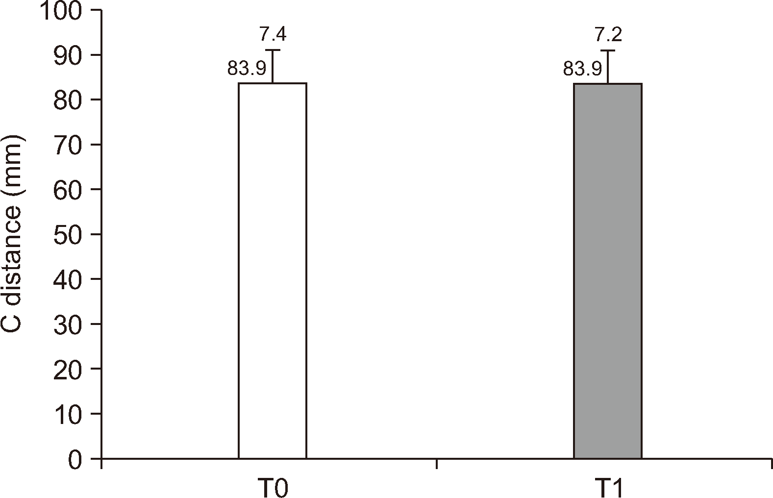
Regarding measurements performed in the coronal plane, mean β-angleR values at T0 and T1 were found to be 17.6° ± 10.4° and 14.1° ± 9.6°, respectively (P < 0.05), which indicated that the right condyle rotated outward postoperatively. Whereas, no significant change was observed between the mean β-angleL values measured at T0 (16.5° ± 9.7°) and T1 (18.5° ± 9.7°) (P > 0.05, Figure 7). Besides, regarding C distance no significant difference was observed in the mean values measured at T0 (83.9 ± 7.4 mm) and T1 (83.9 ± 7.2 mm) (P > 0.05, Figure 8), which indicated that the distance between the condyles did not change significantly postoperatively.
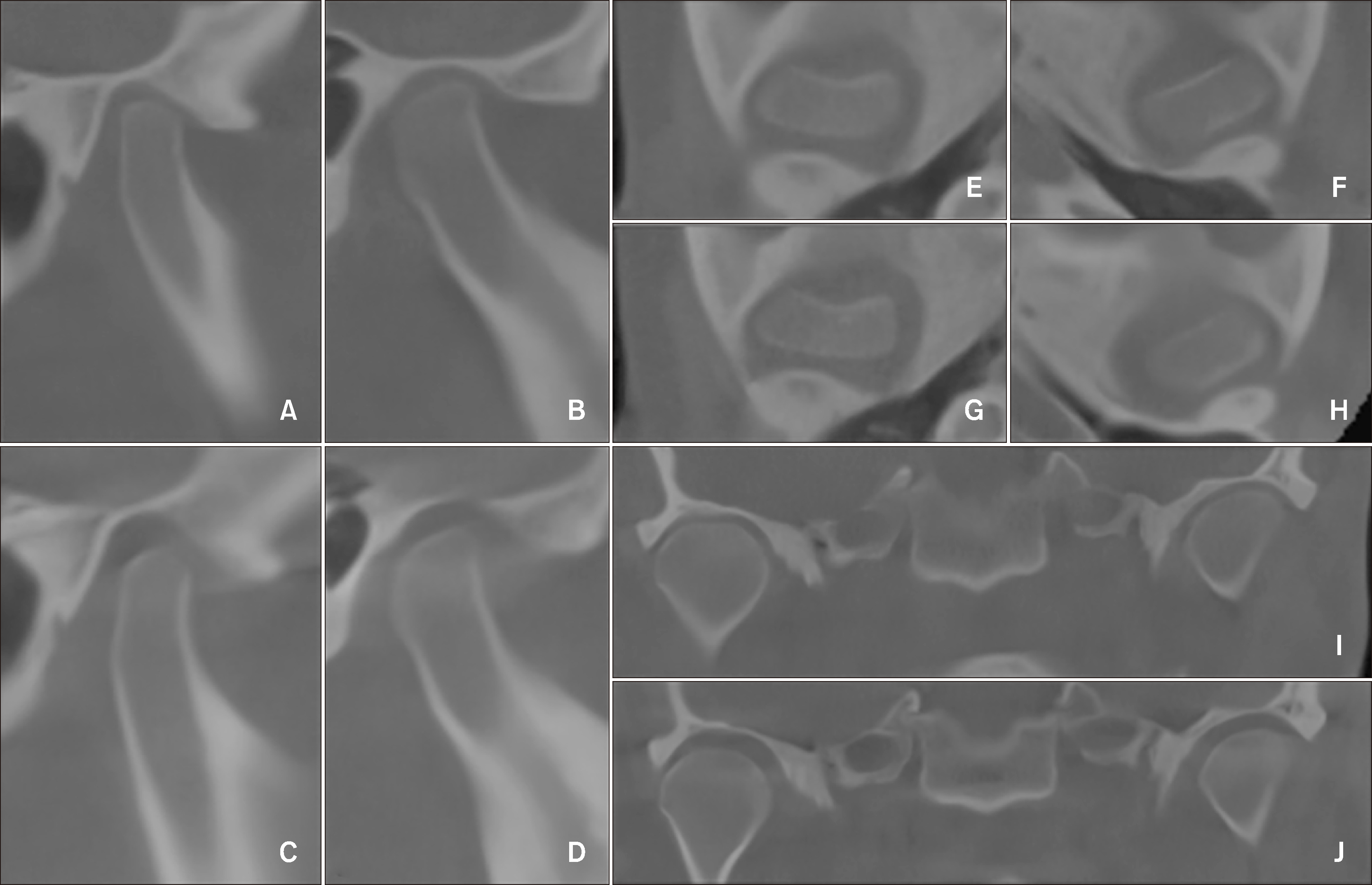
The preoperative and postoperative condylar positions were demonstrated on the CBCT sections of a representative patient (Figure 9). In sagittal sections, a vertical downward movement of both condyles was noted. In coronal sections, an inward rotation, especially in the left condyle, was observed in this patient.
The change in the mandibular condyle position in patients with mandibular prognathism and maxillary retrognathia who underwent BSSRO combined with Le Fort I with the mandibular condyle positioned manually was investigated. Despite the similarities with previously published studies, there needs to be more standardization in the study samples and evaluation methods. Therefore, this study aims to provide detailed information on the study design.
Although many methods have been described for condylar repositioning, the conventional method, manual condylar repositioning during fixation is one of the most common techniques.12 Gerressen et al.13 compared the Luhr positioning device with the manual positioning technique in BSSRO with or without Le Fort I using lateral cephalometrics with a mean follow-up period of 45 months and reported that the repositioning device did not improve long-term skeletal stability. Bethge et al.12 concluded that evaluation with magnetic resonance imaging revealed that sonographic condylar repositioning devices used intraoperatively were fast, comfortable, and cost-effective; however, they could not be proven more effective than manual repositioning. Sander et al.14 also concluded that manual condylar repositioning resulted in minimal changes. Most surgeons rely on manual repositioning to obtain the best mandibular proximal segment relationship with the condylar fossa.6 Although it is still controversial, in cases performed by an experienced surgeon, fixation provided by manual positioning is considered superior in terms of time management and postoperative results.
Hollender and Ridell15 showed anterior and inferior movements of the mandibular condyle after BSSRO, as evaluated by transcranial radiograms. Hu et al.16 reported backward movement and anterior rotation of the mandibular condyle on TMJ radiographs following mandibular setback with BSSRO. In addition, on CT images, Kawamata et al.17 reported a 1–2 mm backward movement with SSRO. Kim et al.18 noted in a report of two unilateral SSRO cases that although positional changes were observed in the mandibular condyle postoperatively, no significant changes would affect the patients clinically. Ueki et al.6 suggested that the most favorable postoperative condylar position, including the horizontal condylar angle, may not match the preoperative position but would not be dramatically different, except in cases with TMJ disorder or asymmetry. The ideal postoperative position should be where the remodeling volume of the TMJ induced by postoperative biomechanical stress is the most minor and degenerative changes are not caused. Conflicting findings on asymmetry have been reported in the literature. Choi et al.10 reported no significant difference in condylar position between groups with or without asymmetry, and BSSRO could be performed without substantial change in these cases.
In contrast, Lee et al.19 showed that mandibular prognathism with asymmetry was associated with bilateral differences in 3D morphology and orientation of the condyle. Regarding the correlation between the change in condylar position and the setback degree, Lee and Park20 found no significant changes after SSRO. In our study, to avoid sampling bias, TMJ disorders and facial asymmetry were excluded, and none of the patients developed postoperative symptoms or radiological signs of TMJ disorders. However, our study did not calculate the correlation between condylar position and maxillary or mandibular movements.
Ma et al.21 superimposed 3D models and examined them for one week, postoperative 1–2 weeks, and 3–12 months. They pointed out the inconsistency in the literature regarding the duration of stabilization of the TMJ position after surgery, and based on fused 3D images, they reported an initial change in the mandibular condyle position, which stabilized at 3 months postoperatively. They also noted that most condyles still needed to return to their preoperative position within a year fully. Podčernina et al.8 investigated condylar positional, structural, and volumetric changes after bimaxillary or only maxillary orthognathic surgeries in skeletal Class III patients using CBCT. The mandibular condyle position they provided using the bivector seating method did not report any significant difference between preoperative and one-year follow-up positions in any study groups. However, they reported condylar rotation in the axial and coronal planes in the bimaxillary surgery group. Accordingly, Sobouti et al.22 did not report any significant change in condyle position immediately after the operation or 8 months postoperatively. In our study, a follow-up period of 6 months was preferred, considering radiation protection principles.
Ueki et al.23 showed a mean α-angle (horizontal condylar angle) of 12.0° ± 7.9° and 11.8° ± 5.1° on the right and left, respectively, in Class III patients with facial symmetry. These findings were much lower than those reported in other studies, including our study. This might be attributed to the different populations and differences in the condylar shapes in the study populations.
Lee and Park,20 who also placed the posterior segment empirically during BSSRO and unlike our study, evaluated the TMJ region using CT instead of CBCT, 1–4 months postoperatively instead of 6 months, and measured condylar angulation as the acute angle of a right-angled triangle other than the α-angle reported an inward rotation of the condyle with a mean value of 4°. However, they did not report the side that demonstrated rotation. Choi et al.10 reported an increase in the α-angle postoperatively, which returned to the preoperative position after 6 months. Kim et al.24 reported an increase in the mandibular condyle angle of 2.23° and 2.18° on the right and left sides, respectively, which resulted in an inward rotation. Nishimura et al.25 stated that in patients with mandibular prognathism, the lateral part of the condyle rotates anteromedially due to internal fixation of the proximal segment, and there is less change with nonrigid fixation than with rigid fixation. Kim et al.26 reported that the condyles inclined outward insignificantly. Podčernina et al.8 evaluated TMJ position, and in axial condylar angle, they found similar values (T0: 19.6 ± 7.5 and T2-after 1 year: 22.2 ± 7.6) as in our study. However, they did not indicate whether these measurements were from the right or left side or the overall mean. Jung et al.,27 who used condylar positioning plate and performed evaluations on CT images reported significant differences in the left condylar axis angle (T0: 19.89° ± 5.40°, after 3-months: 21.21° ± 5.52°, after 1-year: 20.72° ± 5.44°), whereas no significance was demonstrated on the right side (T0: 20.93° ± 5.55°, after 3-months: 21.79° ± 6.04°, after 1-year: 21.24° ± 5.58°).
In our study, while α-angleL increased and demonstrated an inward rotation 6 months postoperatively, the increase in α-angleR was insignificant. This difference between the right and left condyles may be because the operator was always in the same operating position; thus, the direction and amount of force applied during the fixation phase differed. Inward rotation of the condylar head could also be due to screw insertion during rigid fixation procedures.
In addition, Omar and Bamber28 reported an unplanned rotation of the TMJ toward the right side in manual model surgery compared with a passive Robot Arm. Mediolateral errors toward the right side in manual model surgery may be due to the operator’s right-handedness. In our study, all surgical procedures were performed by a right-handed surgeon. The standing position and right-handedness of the operator could be the reasons for the inward rotation.
Choi et al.,10 who included patients with and without asymmetry, reported non-significant m/M ratio changes, representing horizontal movement pre- and postoperatively. Regarding the n/N ratio, which shows vertical movement, a significant decrease was reported immediately after surgery, indicating that the mandibular condyle had a perpendicular orientation to the inferior. However, at 6 months postoperatively, the n/N value also returned to the initial value, resulting in a non-significant positional change. Regarding the ‘m’ distance calculation, Choi et al.10 defined the distance between D1 and D3 points as the ‘m’ distance. Because these points are fixed anatomical points in which positional changes were not anticipated postoperatively, this might lead to an erroneous calculation that there will be no alteration in the sagittal ‘m/M’ ratio. Consequently, the horizontal movement change cannot be determined. In our research, ‘m’ distance was defined as the distance between D1 and D4, where D4 may change position due to surgical operation.
Ueki et al.,29 who used bent plates to maintain the condyle in its original position, reported no significant change in the anteroposterior direction after BSSRO on cephalograms. Lee and Park20 reported that the condyle moved down and forward, albeit in small amounts, which was non-significant at 6 months postoperatively. Kim et al.26 concluded that the superior movement of both mandibular condyles and anterior movement of the right condyle were substantial. Our study showed no significant change in either the anteroposterior or superoinferior direction. The manual positioning maneuver during BSSRO (directing the proximal part backward from the lower level of the vertical corpus osteotomy line and extraorally upward from the mandibular angulus) might be sufficient to provide the initial anteroposterior and superoinferior relationship of the condyle. No additional devices or processes during the operation are necessary for cost, time, or operation success.
Choi et al.10 reported that in patients with facial symmetry, the β-angle significantly decreased 1 month after BSSRO, which returned to the initial mean value 6 months postoperatively. Choi and Lee30 reported a decrease of 0.65° on the right and 0.5° on the left sides. Kim et al.26 reported that the condylar angle decreased by a mean of 0.92° on both sides. Compatible with the literature, our findings showed that the right β-angle decreased significantly. Besides, no significant change was observed on the left side at T1 time, which means that the right condyle rotated outward. Both the effects of the fixed standing position of the operator, which might also affect the axial plane, and the direction and amount of force during the fixation process could have contributed to this outward rotation.
Regarding intercondylar distance, Kawamata et al.17 reported a 2 mm increase following SSRO on CT images. Choi et al.10 reported that the distance between the condyles in the facial symmetry group (Me deviation < 4 mm) showed a significant increase at 1 month postoperatively and tended to return to the initial distance at 6 months postoperatively. Choi and Lee30 reported no significant differences, although the distance between the condyles tended to increase. In our study, no significant changes were observed, which is consistent with the results of previous studies.
One limitation was that there were two-time points; therefore, information regarding the later stages must be provided, considering radiation protection principles. Since TMJ remodeling is expected in these patients, changes in the condylar position could be better evaluated using landmarks designated by the condylar shape. Parameters such as condylar remodeling, asymmetry, TMJ disorders, and maxillary or mandibular movement, which might affect the condylar position, were not calculated. Although the data of the patients operated on by a single surgeon eliminates variables dependent on the operator, involving data obtained from patients operated on by other surgeons and verifying reproducibility between these surgeons is necessary.
Manual positioning of the mandibular condyles during BSSRO is an adequate and reproducible method. The left mandibular condyle demonstrated inward rotation in the axial plane, and the right condyle demonstrated outward rotation in the coronal plane. Owing to the lack of standardization of study samples and evaluation methods in the literature, further studies with similar evaluation parameters, including clinical findings with a long-term follow-up period, are needed to draw a valid conclusion.
Notes
AUTHOR CONTRIBUTIONS
Conceptualization: OK, EC. Data curation: OK, EC. Formal analysis: OK, YZA, EC. Investigation: OK, NE. Methodology: OK, NE, EC. Project administration: OK, EC. Resources: EC. Software: NE, YZA. Supervision: EC. Validation: NE, EC. Writing–original draft: OK. Writing–review & editing: NE, YZA, EC.
References
1. Kim YI, Cho BH, Jung YH, Son WS, Park SB. 2011; Cone-beam computerized tomography evaluation of condylar changes and stability following two-jaw surgery: le fort I osteotomy and mandibular setback surgery with rigid fixation. Oral Surg Oral Med Oral Pathol Oral Radiol Endod. 111:681–7. http://doi.org/10.1016/j.tripleo.2010.08.001. DOI: 10.1016/j.tripleo.2010.08.001. PMID: 21055977.

2. Hong M, Kim MJ, Shin HJ, Cho HJ, Baek SH. 2020; Three-dimensional surgical accuracy between virtually planned and actual surgical movements of the maxilla in two-jaw orthognathic surgery. Korean J Orthod. 50:293–303. https://doi.org/10.4041/kjod.2020.50.5.293. DOI: 10.4041/kjod.2020.50.5.293. PMID: 32938822. PMCID: PMC7500567.

3. Mda B, F ME, McZ D. 2022; Methods of mandibular condyle position and rotation center used for orthognathic surgery planning: a systematic review. J Stomatol Oral Maxillofac Surg. 123:345–52. https://doi.org/10.1016/j.jormas.2021.06.004. DOI: 10.1016/j.jormas.2021.06.004. PMID: 34237437.

4. Meriç G. 2010; A literature review of the sentric relation and registration methods up to date. J Dent Fac Atatürk Uni. 2010:54–9. https://dergipark.org.tr/en/pub/ataunidfd/issue/2481/31754.
5. Bettega G, Cinquin P, Lebeau J, Raphaël B. 2002; Computer-assisted orthognathic surgery: clinical evaluation of a mandibular condyle repositioning system. J Oral Maxillofac Surg. 60:27–34. discussion 34–5. https://doi.org/10.1053/joms.2002.29069. DOI: 10.1053/joms.2002.29069. PMID: 11757002.

6. Ueki K, Moroi A, Sotobori M, Ishihara Y, Marukawa K, Takatsuka S, et al. 2012; A hypothesis on the desired postoperative position of the condyle in orthognathic surgery: a review. Oral Surg Oral Med Oral Pathol Oral Radiol. 114:567–76. https://doi.org/10.1016/j.oooo.2011.12.026. DOI: 10.1016/j.oooo.2011.12.026. PMID: 22819333.

7. Savoldelli C, Chamorey E, Bettega G. 2018; Computer-assisted teaching of bilateral sagittal split osteotomy: learning curve for condylar positioning. PLoS One. 13:e0196136. https://doi.org/10.1371/journal.pone.0196136. DOI: 10.1371/journal.pone.0196136. PMID: 29694423. PMCID: PMC5918964. PMID: af8cc01d35b640559d4f5731ce013f9d.

8. Podčernina J, Urtāne I, Pirttiniemi P, Šalms Ģ, Radziņš O, Aleksejūnienė J. 2020; Evaluation of condylar positional, structural, and volumetric status in class III orthognathic surgery patients. Medicina (Kaunas). 56:672. https://doi.org/10.3390/medicina56120672. DOI: 10.3390/medicina56120672. PMID: 33291272. PMCID: PMC7762172. PMID: 5b5c5b09ed6d43728867478b33423c78.

9. Costa F, Robiony M, Toro C, Sembronio S, Polini F, Politi M. 2008; Condylar positioning devices for orthognathic surgery: a literature review. Oral Surg Oral Med Oral Pathol Oral Radiol Endod. 106:179–90. https://doi.org/10.1016/j.tripleo.2007.11.027. DOI: 10.1016/j.tripleo.2007.11.027. PMID: 18417381.

10. Choi BJ, Kim BS, Lim JM, Jung J, Lee JW, Ohe JY. 2018; Positional change in mandibular condyle in facial asymmetric patients after orthognathic surgery: cone-beam computed tomography study. Maxillofac Plast Reconstr Surg. 40:13. https://doi.org/10.1186/s40902-018-0152-6. DOI: 10.1186/s40902-018-0152-6. PMID: 29984220. PMCID: PMC6015790. PMID: 89bb133f32784a8f9487abb77af450ea.

11. Koo TK, Li MY. 2016; A guideline of selecting and reporting intraclass correlation coefficients for reliability research. J Chiropr Med. 15:155–63. https://doi.org/10.1016/j.jcm.2016.02.012. DOI: 10.1016/j.jcm.2016.02.012. PMID: 27330520. PMCID: PMC4913118.

12. Bethge LS, Ballon A, Mack M, Landes C. 2015; Intraoperative condyle positioning by sonographic monitoring in orthognathic surgery verified by MRI. J Craniomaxillofac Surg. 43:71–80. https://doi.org/10.1016/j.jcms.2014.10.012. DOI: 10.1016/j.jcms.2014.10.012. PMID: 25457463.

13. Gerressen M, Stockbrink G, Smeets R, Riediger D, Ghassemi A. 2007; Skeletal stability following bilateral sagittal split osteotomy (BSSO) with and without condylar positioning device. J Oral Maxillofac Surg. 65:1297–302. https://doi.org/10.1016/j.joms.2006.10.026. DOI: 10.1016/j.joms.2006.10.026. PMID: 17577492.
14. Sander AK, Martini M, Konermann AC, Meyer U, Wenghoefer M. 2015; Freehand condyle-positioning during orthognathic surgery: postoperative cone-beam computed tomography shows only minor morphometric alterations of the temporomandibular joint position. J Craniofac Surg. 26:1471–6. https://doi.org/10.1097/SCS.0000000000001781. DOI: 10.1097/SCS.0000000000001781. PMID: 26163838.
15. Hollender L, Ridell A. 1974; Radiography of the temporomandibular joint after oblique sliding osteotomy of the mandibular rami. Scand J Dent Res. 82:466–9. https://doi.org/10.1111/j.1600-0722.1974.tb00403.x. DOI: 10.1111/j.1600-0722.1974.tb00403.x. PMID: 4529372.

16. Hu J, Wang D, Zou S. 2000; Effects of mandibular setback on the temporomandibular joint: a comparison of oblique and sagittal split ramus osteotomy. J Oral Maxillofac Surg. 58:375–80. discussion 380–1. https://doi.org/10.1016/s0278-2391(00)90915-7. DOI: 10.1016/S0278-2391(00)90915-7. PMID: 10759116.

17. Kawamata A, Fujishita M, Nagahara K, Kanematu N, Niwa K, Langlais RP. 1998; Three-dimensional computed tomography evaluation of postsurgical condylar displacement after mandibular osteotomy. Oral Surg Oral Med Oral Pathol Oral Radiol Endod. 85:371–6. https://doi.org/10.1016/s1079-2104(98)90059-2. DOI: 10.1016/S1079-2104(98)90059-2. PMID: 9574943.

18. Kim MI, Kim JH, Jung S, Park HJ, Oh HK, Ryu SY, et al. 2015; Condylar positioning changes following unilateral sagittal split ramus osteotomy in patients with mandibular prognathism. Maxillofac Plast Reconstr Surg. 37:36. https://doi.org/10.1186/s40902-015-0036-y. DOI: 10.1186/s40902-015-0036-y. PMID: 26501042. PMCID: PMC4608983.

19. Lee JS, Xi T, Kwon TG. 2017; Three-dimensional analysis of mandibular condyle position in patients with deviated mandibular prognathism. Int J Oral Maxillofac Surg. 46:1052–8. https://doi.org/10.1016/j.ijom.2017.02.1272. DOI: 10.1016/j.ijom.2017.02.1272. PMID: 28302336.

20. Lee W, Park JU. 2002; Three-dimensional evaluation of positional change of the condyle after mandibular setback by means of bilateral sagittal split ramus osteotomy. Oral Surg Oral Med Oral Pathol Oral Radiol Endod. 94:305–9. https://doi.org/10.1067/moe.2002.126452. DOI: 10.1067/moe.2002.126452. PMID: 12324783.

21. Ma RH, Li G, Yin S, Sun Y, Li ZL, Ma XC. 2020; Quantitative assessment of condyle positional changes before and after orthognathic surgery based on fused 3D images from cone beam computed tomography. Clin Oral Investig. 24:2663–72. https://doi.org/10.1007/s00784-019-03128-z. DOI: 10.1007/s00784-019-03128-z. PMID: 31728734.

22. Sobouti F, Hadian H, Pakravan AH, Rahimi Z, Rakhshan V, Dadgar S. 2022; Short-term and long-term alterations of condylar position after bilateral sagittal split ramus osteotomy for mandibular setback: a preliminary before-after clinical trial. Dent Res J (Isfahan). 19:19. https://doi.org/10.4103/1735-3327.338782. DOI: 10.4103/1735-3327.338782. PMID: 35308442. PMCID: PMC8927962. PMID: 7235f4da86984ad086d74c1d59dcda92.

23. Ueki K, Nakagawa K, Takatsuka S, Shimada M, Marukawa K, Takazakura D, et al. 2000; Temporomandibular joint morphology and disc position in skeletal class III patients. J Craniomaxillofac Surg. 28:362–8. https://doi.org/10.1054/jcms.2000.0181. DOI: 10.1054/jcms.2000.0181. PMID: 11465144.

24. Kim YI, Jung YH, Cho BH, Kim JR, Kim SS, Son WS, et al. 2010; The assessment of the short- and long-term changes in the condylar position following sagittal split ramus osteotomy (SSRO) with rigid fixation. J Oral Rehabil. 37:262–70. https://doi.org/10.1111/j.1365-2842.2009.02056.x. DOI: 10.1111/j.1365-2842.2009.02056.x. PMID: 20113391.

25. Nishimura A, Sakurada S, Iwase M, Nagumo M. 1997; Positional changes in the mandibular condyle and amount of mouth opening after sagittal split ramus osteotomy with rigid or nonrigid osteosynthesis. J Oral Maxillofac Surg. 55:672–6. discussion 677–8. https://doi.org/10.1016/s0278-2391(97)90572-3. DOI: 10.1016/S0278-2391(97)90572-3. PMID: 9216497.
26. Kim JW, Lee DH, Lee SY, Kim JH, Lee SH. 2009; 3-D CT evaluation of condyle head position, mandibular width, and mandibular angle after mandibular setback surgery. J Korean Assoc Oral Maxillofac Surg. 35:229–39. https://www.jkaoms.org/journal/view.html?uid=374&vmd=Full.
27. Jung GS, Kim TK, Lee JW, Yang JD, Chung HY, Cho BC, et al. 2017; The effect of a condylar repositioning plate on condylar position and relapse in two-jaw surgery. Arch Plast Surg. 44:19–25. https://doi.org/10.5999/aps.2017.44.1.19. DOI: 10.5999/aps.2017.44.1.19. PMID: 28194343. PMCID: PMC5300918. PMID: 580d9d457aae41e59b8f4de365a4a3e8.

28. Omar EAZ, Bamber MA. 2010; Orthognathic model surgery by using of a passive robot arm. Saudi Dent J. 22:47–55. https://doi.org/10.1016/j.sdentj.2010.02.006. DOI: 10.1016/j.sdentj.2010.02.006. PMID: 24227912. PMCID: PMC3824638.

29. Ueki K, Hashiba Y, Marukawa K, Alam S, Nakagawa K, Yamamoto E. 2008; Skeletal stability after mandibular setback surgery: bicortical fixation using a 2.0-mm locking plate system versus monocortical fixation using a nonlocking plate system. J Oral Maxillofac Surg. 66:900–4. https://doi.org/10.1016/j.joms.2007.08.033. DOI: 10.1016/j.joms.2007.08.033. PMID: 18423278.
30. Choi KY, Lee SH. 1996; Evaluation of condylar position using computed tomograph following bilateral sagittal split ramus osteotomy. Maxillofac Plast Reconstr Surg. 18:570–93. https://kiss.kstudy.com/Detail/Ar?key=1956811.
Figure 1
Landmarks specified on the axial cone-beam computed tomography images to determine the condylar head position.
A1R, the lateral pole of the right condylar head; A2R, the medial pole of the right condylar head; A1L, the lateral pole of the left condylar head; A2L, the medial pole of the left condylar head; BR, the most posterior point of the right carotid canal; BL, the most posterior point of the left carotid canal; Line1R, the line passes through A1R and A2R points; Line1L, the line passes through A1L and A2L points; Line2, the line which is passing through BL and BR points; α-angleR, the angle formed between Line1R and Line2; α-angleL, the angle formed between Line1L and Line2.
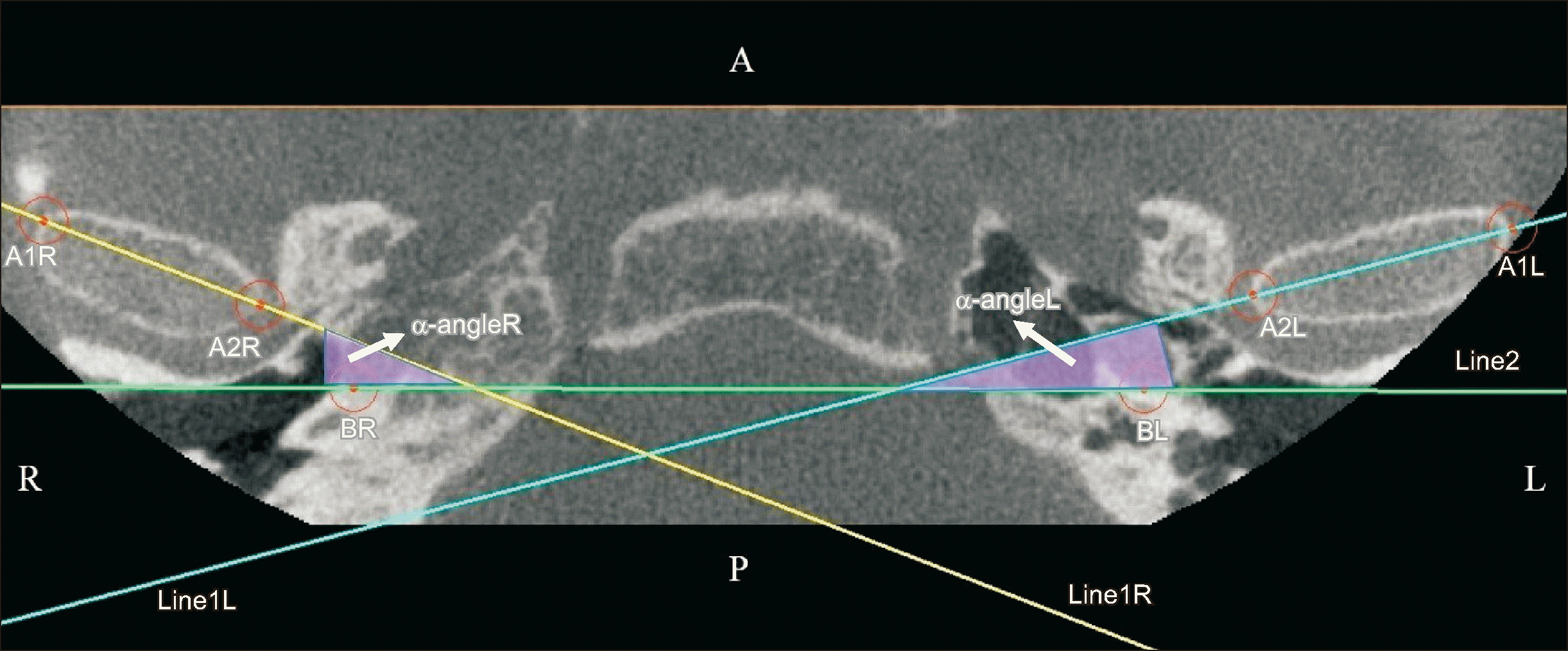
Figure 2
Landmarks specified on the sagittal cone-beam computed tomography images to determine the condylar head position.
C1, the highest point of the glenoid fossa; C2, the highest point of the condylar head; D1, the lowest point of the articular eminence; D2, the lowest point of the temporal squamotympanic fissure; D3, the point of the line drawn perpendicular to Line3 from point C1; D4, the point of the line drawn perpendicular to Line3 from C2; M, the distance between D1 and D2; m, the distance between D1 and D4; N, the distance between C1 and D3; n, the distance between C2 and D4.
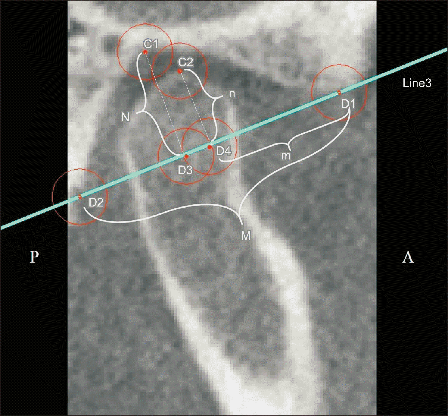
Figure 3
Landmarks specified on the coronal cone-beam computed tomography images to determine the condylar head position.
ER, the highest point of the right glenoid fossa; EL, the highest point of the left glenoid fossa; F1R, the lateral pole of the right condylar head; F1L, the lateral pole of the left condyle head; F2R, the medial pole of the right condylar head; F2L, the medial pole of the left condylar head; Line4, passes through ER and EL; Line5R, passes through F1R and F2R points, and Line5L passes through F1L and F2L points; β-angleR, the angle between Line4 and Line5R; β-angleL, the angle between Line4 and Line5L, and C is the distance between F2R and F2L points.
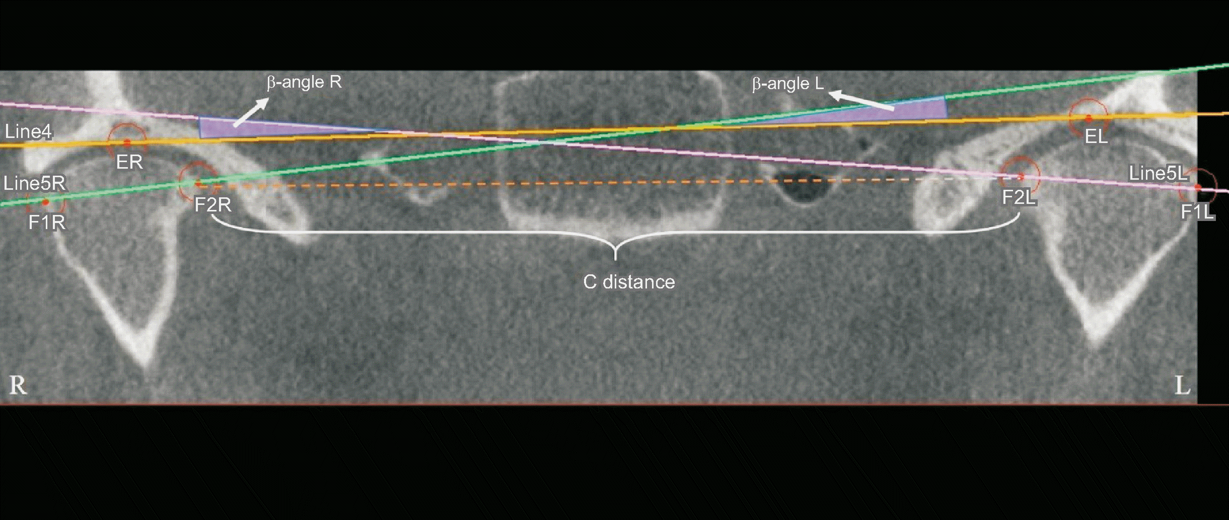
Figure 4
Comparison of the pre-and postoperative α-angle. It is the angle between Line1 (passing through the lateral and medial pole points of the condylar head) and Line2 (passing through the rearmost points of the right and left carotid canal) in the axial plane.
*Indicates statistical significance between the related pairs (P < 0.05).
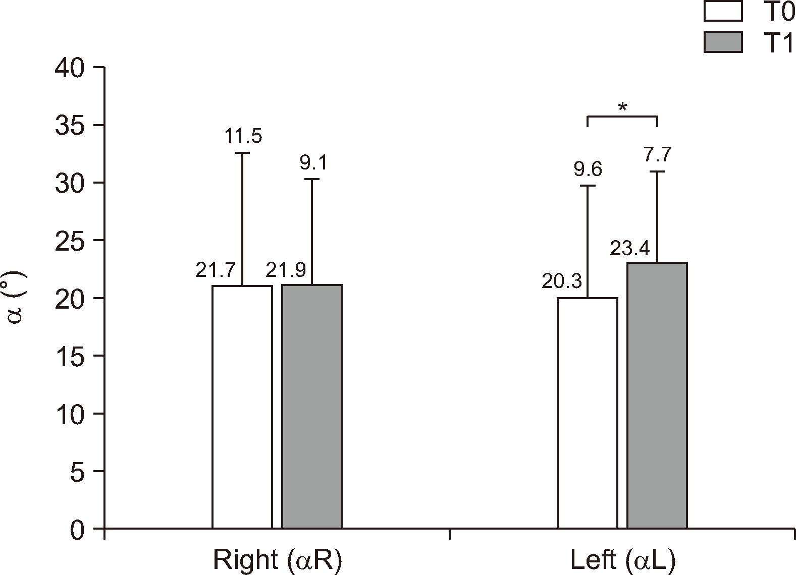
Figure 5
Comparison of the pre-and postoperative n/N ratio.
n/N, vertical evaluation rate of the mandibular condyle in the direction of the glenoid fossa.
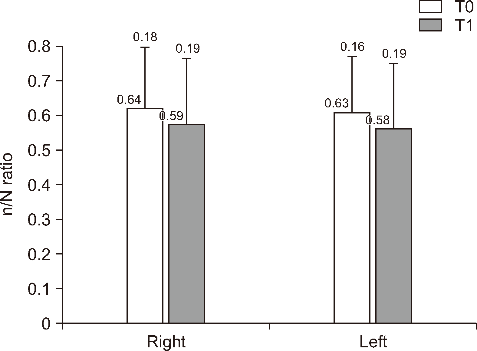
Figure 6
Comparison of the pre-and postoperative m/M ratio.
m/M, horizontal evaluation rate of the mandibular condyle in the direction of the glenoid fossa.
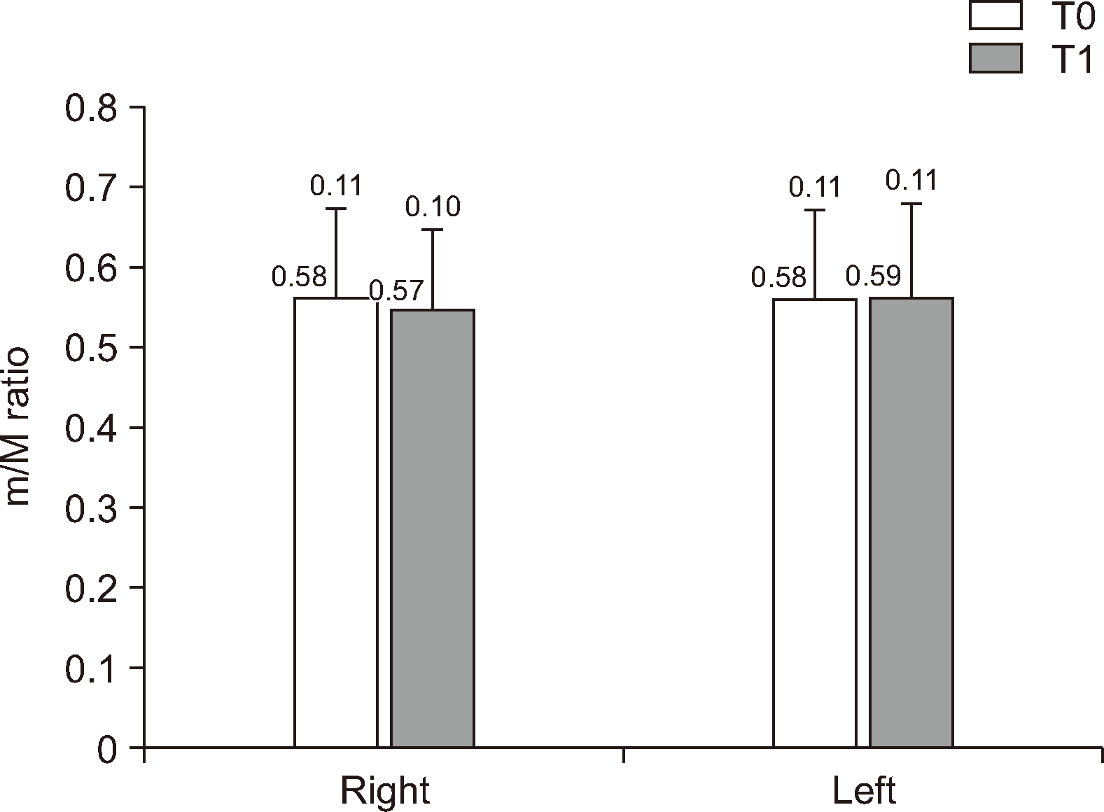
Figure 7
β-angle. It is the angle between the line passing through the lateral and medial pole points of the condylar head and the line passing through the highest points of the right and left glenoid fossa in the coronal plane.
*Indicates statistical significance between the related pairs (P < 0.05).
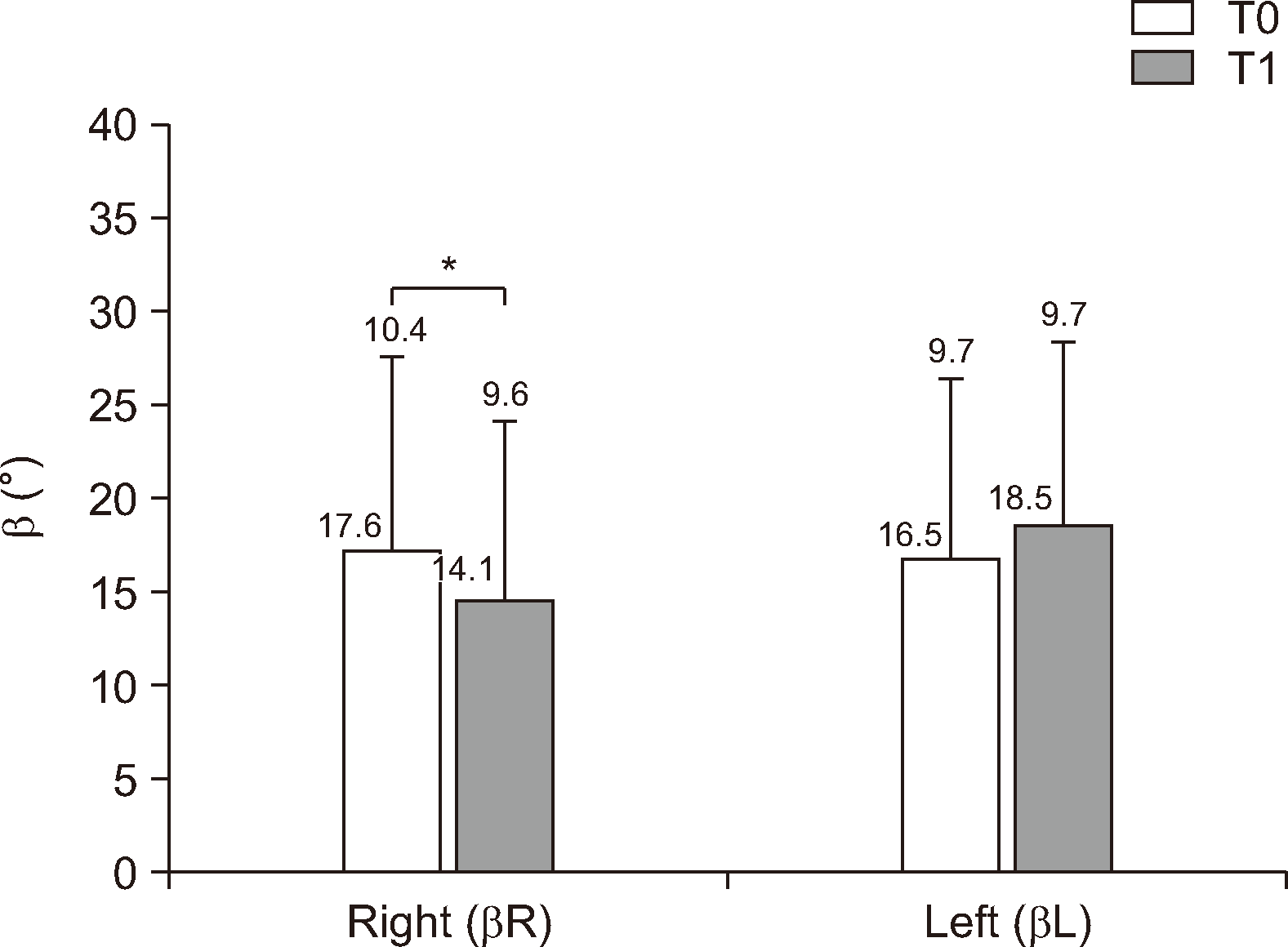
Figure 9
Cone-beam computed tomography sections of a representative patient demonstrating A, B, T0 sagittal right and left; C, D, T1 sagittal right and left; E, F, T0 axial right and left; G, H, T1 axial right and left; I, T0 coronal; J, T1 coronal. In this particular patient, the condyles’ vertical downward movement and inward rotation were noted.


 PDF
PDF Citation
 Print
Print


 XML Download