Journal List > Korean J Orthod > v.54(4) > 1516088009

Eliades and Eliades: Intraoral ageing of aligners and attachments: Adverse effects on clinical efficiency and release of biologically-active compounds

Abstract

The clinical application of aligners is accompanied by the ageing of the polymer appliances and the attachments used, which may result in inefficiency in reaching the predicted range of tooth movement, and release of compounds and microplastics in the oral cavity as a result of the friction, wear and attrition of the aligner and composite attachment. The purpose of this review is to present the mechanism and effects of in vivo ageing; describe the hydrolytic degradation of aligners and enzymatic degradation of composite attachments; examine the ageing pattern of aligners in vivo, under actual clinical scenarios; and identify a link to the discrepancy between predicted and actual clinical outcome. Lastly, strategies to deal with three potentially critical issues associated with the use of aligners, namely the necessity of weekly renewal, the dissimilar mechanical properties of aligner and attachment resulting in wear and plastic deformation of the aligner, and the development of integuments and biofilms with microbial colonization of the appliance, are discussed.

INTRODUCTION

Aligners have become an integral part of orthodontic treatment with more systems, new materials manufacturing/fabrication methods becoming available despite the lack of solid evidence on their efficiency compared to conventional treatment. Recently a number of studies have demonstrated that the basic characteristic of the majority of the class of these materials pertains to the notable lack of predictability in delivering the amount of movement predicted by the relevant software,1 introducing a necessity, almost in every case outside of minor movements, for the application of additional number of aligners compared to the tooth spatial orientation predicted at the initial stage, resulting in a longer treatment duration. It is the hypothesis of this review that this discrepancy is largely attributed to the failure to predict the performance of the polymeric materials involved in treatment, including aligner and composite resins, because of the false integral assumptions of the relevant software, which considers attachments and aligners as ideal materials. This fails to account for the alterations occurring in vivo, which introduce a deviation from the profile of the materials in the as-received form or before exposure to the oral cavity, with effects manifested in the roughness, mechanical properties, dimensional stability, and release of compounds.
The purpose of this article is therefore to
  • 1) examine the ageing pattern of aligners in vivo, under actual clinical scenarios;

  • 2) describe the alterations of the mechanical properties of the aligners induced by in vivo ageing, and identify a link to the discrepancy between predicted and actual clinical outcome;

  • 3) summarize the evidence on the composition and kinetics of the eluted compounds in vitro and in vivo; and propose ways to bypass the inefficiency of the system.

IN VIVO AGEING OF ALIGNERS AND COMPOSITES: SURFACE ALTERATIONS ACCOMPANYING THE EXPOSURE OF POLYMERS IN THE ORAL CAVITY

This section will describe the cascade of events accompanying the placement of an aligner and a composite attachment in the oral cavity by considering the fundamentals of integument formation and in vivo ageing of a biomaterial.
The molecular and elemental composition of a biomaterial surface determine the early response of the materials to the biological milieu they are placed. The material’s polar or non-polar nature, and hydrogen-bonding capacity play a significant role on the hydrophilic or hydrophobic character of the surface and the energetic state of the surfaces. In addition, surface electrical properties, such as the zeta and streaming potentials and surface charges, are also involved in interfacial interactions with biological fluids.2 Particularly for polymers of low modulus such as aligners, surface thermal mobility in the colloidal state or soft-chain segments of hard polymers, where the majority of the material or specific regions may demonstrate active Brownian motion, is important. These molecular regions are permeable to small molecules capable of modifying the conformation of biological fluids by steric or fluctuation forces. Crystallinity also determines surface mobility as rigid crystalline phases demonstrate higher adsorption energy compared to amorphous phases, with some exceptions found for materials exposed to high flow rates.3
Surface texture incorporates both ultrastructural and macroscopic features, and on an ultrastructural scale, vacancies, other structural imperfections, gas entrapment and adsorbed layers alter the surface composition. Macroscopically, surface roughness and porosity interfere with the biological fluid flow rate by producing secondary fluid motions of enhanced or reduced shear locally, which at high flow rates may affect the shape, distribution and aggregation of the attaching particles. At low flow rates or under static conditions, the grooves of rough surfaces may act as stagnation points, thereby promoting biofilm maturation.4 Because most surfaces are heterogeneous at a microscopic level, the regional surface distribution of the foregoing properties may produce domains with different characteristics, inducing a variable biological response.

Initial response-water bonding

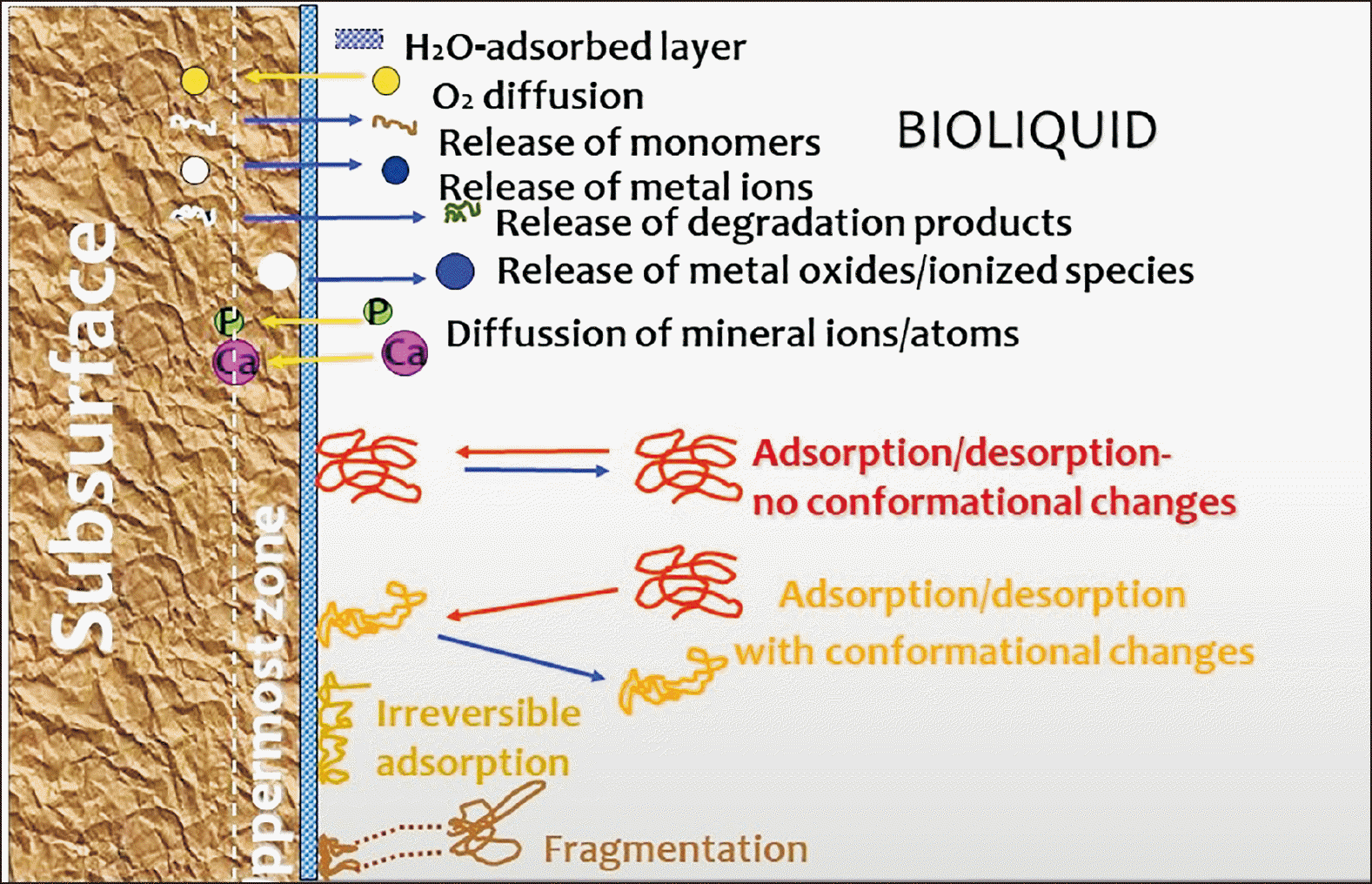
The biological responses to biomaterial surfaces involve a great variety of chemical species. A time sequence of the phenomena occurring on biomaterial surfaces placed in a biological environment has been described, based on experimental data and logical hypotheses (Figure 1).5,6
Initially, on a nanosecond scale, a water monolayer binds to a biomaterial surface by either oxygen or hydrogen bonding. Some water molecules may dissociate to hydroxyl groups, which may form surface hydroxyls. Then, a second water layer binds to the first monolayer. The orientation and density of water molecules in the first adsorbed monolayer may regulate the overall hydration state of the surface. On hydrophilic surfaces, for example, to which water molecules bind strongly, repulsive hydration (long-range) and steric (short-range) forces are generated when two such surfaces come in contact because of the energy required to dehydrate the surfaces; these forces are controlled by the presence of cations or pH. On hydrophobic surfaces on the other hand, the orientation of water molecules towards the surfaces is entropically unfavorable. Thus, in the event that two such surfaces approach each other, water is ejected into the bulk solution, reducing the total free energy of the system and establishing attractive long-range hydrophobic forces between the two surfaces.7

Protein adsorption

Protein adsorption is initiated within a few milliseconds after contact with saliva. The protein layers adsorbed are complex and may vary depending on the nature of the surface. In an aqueous environment, the interaction between hydrophilic surfaces with strongly attached water molecules and hydrated protein cores is weak, due to the development of hydrophilic repulsive forces. Thus, on hydrated surfaces, hydrophobic and electrostatic interactions are expected to govern protein adsorption. Heavily charged macromolecules are probably the first species to be adsorbed; however, the first protein adsorbed does not determine the potential for attachment and the overall response at later stages. In most situations competitive adsorption occurs, with rapidly decreasing protein affinity accompanying increasing surface occupancy. The result is a sequential adsortion/desorption process and exchange of proteins on surfaces, a phenomenon known as the Vroman effect.8 The adsorption/desorption sequence of proteins is not distinct since all proteins are adsorbed simultaneously at different rates, and displacement occurs according to their binding affinity; the latter is associated with the spreading capacity and the energy required to denature the proteins in the solution.9 Proteins with high carbohydrate content and relatively uniform atomic around the molecule show increased solubility, conformational stability, and minimal adsorption on hydrophobic and negatively charged surfaces, attributed to the carbohydrate shielding effect. However, when carbohydrate groups are localized, the protective effect diminishes. Lipoproteins comprise another important group having preferential adsorption on hydrophobic surfaces, especially on those possessing thermal mobility. It seems that lipid phase transition at body temperature and denaturing reactions control the adsorption process. Divalent ions, such as Ca2+, may exert a bridging effect, acting as aggregating factors in protein adsorption.

Protein-induced mineralization

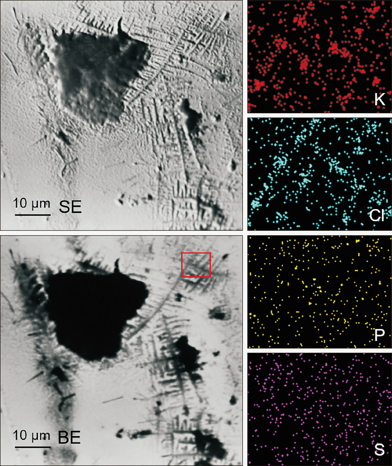
Adsorbed proteins may induce crystalline formation onto chemical groups of their structure that fulfill the stereochemical and surface charge requirements for crystal nucleation. If these conditions apply within a range sufficient for crystal nucleus stabilization and growth at a defined spatial orientation, then crystal growth occurs. Extracellular non-collagenous proteins are believed to be the principal components regulating crystal growth. Protein-induced mineralization in the oral environment proceeds by initial formation of KCl crystals,10 while calcium phosphate precipitation may take place at later stages. This mode of extracellular mineralization is involved in the formation of ectopic calcified deposits on tissues and materials, including teeth, orthodontic rackets, adhesives, headgear wires, NiTi wires, elastomers, and dental materials, and orthopaedic biomaterials.11-25

Pellicle formation

Pellicle formation involves selective protein adsorption, employing non-specific electrostatic and hydrophobic interactions and protein conformational changes, and it is affected by saliva flow rate and pH (Figures 2 and 3).26 It has been proposed that pellicle development proceeds through three phases.27 Initially, low molecular weight phosphoproteins are adsorbed at a high rate, producing a tightly bound layer. Then, competitive interactions take place mainly between phosphoproteins and low molecular weight glycoproteins, creating a second, less tightly bound, layer. Finally, high molecular weight glycoproteins are adsorbed, forming a third, loosely bound, layer.
Early microbial colonization of acquired pellicle involves non-specific forces that transfer microorganisms close to the substrate in order to establish specific bonding.28 During this phase, the detachment shear forces (originating from salivary flow, buccal and lingual tongue movements as well as hard food) and the surface energetic state of the substrate, as modified by pellicle, are the main factors controlling microorganism retention rate.29 Interactions of different microbial species and strains in the bioliquid (co-aggregation) or with surface-attached microorganisms (co-adhesion), release of biosurfactants, production of extracellular matrix polymers, and various competitive reactions are considered important factors for formation and rapid growth of plaque.28 After 18–48 hours in vivo, an equilibrium is reached, with plaque organized in cell microcolonies being entrapped in a dense polysaccharide-based matrix, interspersed by water channels.30,31
The important role of substrate surface roughness and surface free energy on in vivo plaque formation has been demonstrated in a series of studies on surfaces with well-defined characteristics. These studies showed that at supragingival regions the contribution of roughness is greater than that of the surface energy state, whereas at subgingival regions surface roughness is the determinant factor.32 The mechanism of roughness implication on plaque formation arises from the generation of flow stagnation points on surface irregularities (i.e., pores, grooves, cracks and scratches), which promote cell attachment, although the response of individual bacteria biofilms on rough substrates may vary.33 Moreover, it has been documented that on low-energy (hydrophobic) surfaces, the surface area of plaque accumulation is less than that on high-energy (hydrophilic) surfaces.1
A limited number of in vivo studies are available where such interactions between dental materials and developing plaque have been documented.34 However, there are in vivo studies illustrating the absence of any major structural and bacteriological differences in the early plaque formed on various dental materials.35 This finding may suggest that plaque formation is predominantly influenced by the oral environment rather than the type of the substrate. Nevertheless, such a mechanism fails to explain the low plaque retention clinically experienced with several materials (i.e., porcelain and other ceramics). Many in vitro plaque formation experiments, involving saliva incubation on dental material surfaces, have been performed as well, leading to contradictory conclusions. The results of these studies should be interpreted with caution, since shear force gradients are missing and the pellicles formed contain more intact protein species than those developed under in vivo conditions.36 It seems that some material surface properties are transferred through pellicle to plaque, but the detailed mechanism is not yet fully understood. For a thorough review of the processes and mechanisms of ageing the reader is referred to the chapter by Eliades et al.,6 chapter of the Dental materials in vivo: ageing and related phenomena.

WATER ATTACK AND HYDROLYTRIC DEGRADATION OF ALIGNERS AND ATTACHMENTS: EFFECTS ON THE MECHANICAL PROPERTIES, CLINICAL OUTCOME AND ELUTION OF COMPOUNDS

The foregoing section defined the nature of effectors which apart from water, can introduce significant changes in the structure and properties of the polymers, particularly aligners and composite resins. These include, plaque which can be mineralized, affecting the roughness and surface properties of the materials, and enzymes and microbial species, which can degrade polymers, thereby affecting the plasticization introduced by water. The cumulative effects of these factors on altering the hardness of the aligner and composite resin, as well as their dimensional stability and surface integrity are key factors for the explanation of the performance of the materials in clinical scenarios. In vitro research and clinical studies focusing exclusively on clinical outcome without examining the aligner or attachment ageing pattern, cannot provide a description of these effects, thus failing to formulate an underlying mechanism for the treatment outcome recorded.

Degradation of aligners

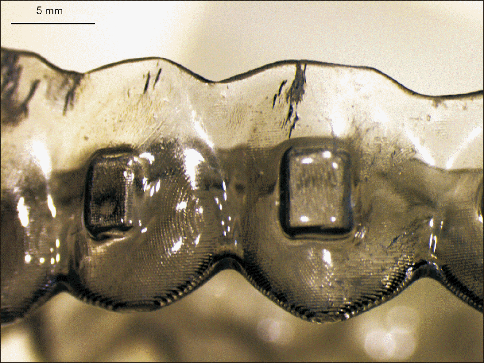
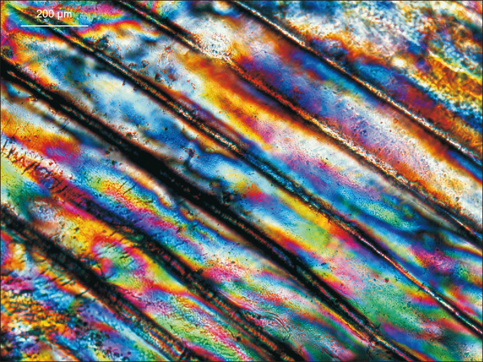
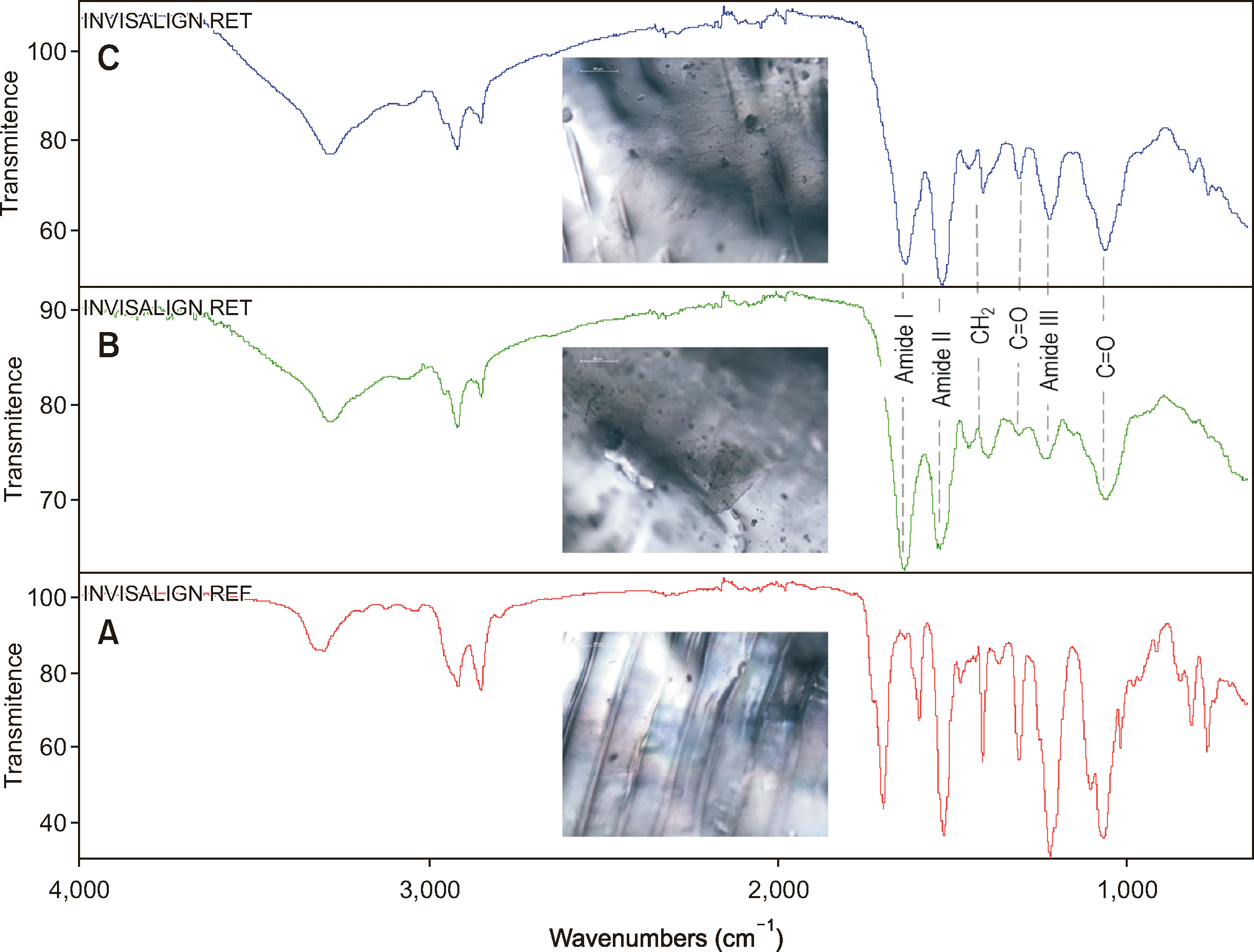
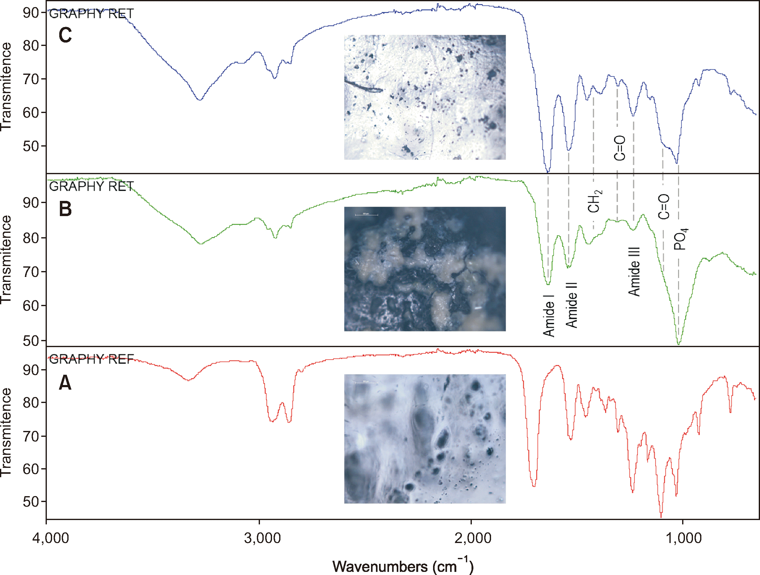
In polyurethanes, water molecules break up part of the weak hydrogen bonds between the urethane groups and replace them by water-urethane hydrogen bonds within 1 day of exposure to water-saturated air. Water molecules also reduce all other physical interactions between network chains because they solvate hydrophilic segments.37,38 With the first stage of uptake, water drastically decreases the aligner viscoelastic response. These results document pronounced plasticization, which decreases the hardness and modulus of the material, thereby effectively reducing the activation and range of movement predicted by the software. Moreover, a decrease in hardness further increases the apparent dissimilarity between the hardness of aligners which does not exceed 120 Martens Handness (HM) in the as-received condition, and may be as much as 40% reduced after in vivo application, and the composite resin attachments whose hardness may range between 400 and 650 HM, depending on the filler content and composition of the composite resin. Such large differences result in the attrition and wear of the aligner which decreases the projected range of tooth displacement and releases compounds and micro/nanoplastics in the oral environment as it has been documented by the decrease in roughness of used InvisalignTM (Tempe, Arizona, USA) appliances. Figures 47 demonstrate the extent of environmental effects as well as service-induced changes of aligners after use which indicate an ageing pattern characterized by the precipitation of proteinaceous species which are calcified at a later stage (Figures 6 and 7) along with cracking (Figure 4) or residual stresses of the aligner (Figure 5).
Polyethylene terephthalate glycol-based aligners very rarely undergo hydrolysis, and only after extended duration, that leads to chain scission in the amorphous phase and so a decrease of molar mass.39

Degradation of composite attachments

Saliva contains both inorganic components, such as calcium and magnesium, and organic components, such as protein. Among the protein components, some esterases are related to the biodegradation of resin-based materials.40 Human saliva-derived esterase (HSDE) is a salivary enzyme that exhibits vigorous degradation activity on bisphenol A-glycidyl methacrylate (Bis-GMA), one of the most common monomers in resin composites and adhesives. Cholesterol esterase (CE)-like activity and pseudocholinesterase (PCE)-like activity can be detected in saliva.41 Studies have shown that commercial CE and PCE can hydrolyze the ester bond in the resin matrix.40
Also, glycoproteins that widely exist in various tissues and have low specificity for acetylcholine can hydrolyze a part of the ester bonds in resin monomers.42 These two kinds of esterases can degrade Bis-GMA, triethylene glycol dimethacrylate (TEGDMA), 2-hydroxyethyl methacrylate (2-HEMA) and other monomers containing esters in resin composites.42 Although HSDE in the oral cavity is generally manifested as CE-like and PCE-like activity, CE and PCE only represent a small fraction of saliva components. There are also other complex proteins taking part in the degradation process of resin.
Streptococcus mutans has been also found to be the main bacteria species causing biodegradation.43,44 When co-cultured with saliva, Bis-GMA-based resins showed considerably enhanced roughness because of the S. mutans action.43 In addition, bacterial esterase can also increase the shrinkage of methacrylate resin and reduce the mechanical properties of the material, such as hardness, bending strength, radial tensile strength and elastic modulus.45 With an increase in the roughness of a resin attachment surface, adhesion of bacteria increases, and consequently the esterase that the bacteria produce enhanced biodegradation.
Another biological entity capable of inducing dental resin degradation is neutrophils which, in addition to phagocytizing, killing and dissolving bacteria can also exhibit esterase-like activities. Neutrophils mainly exist in the gingival crevicular fluid in the oral environment, and can directly contact the edge of the attachment, especially ones placed close to the gingiva. The esterase-like activity of neutrophils has been shown to induce degradation of a Bis-GMA-based composite.46
The sequence of phemomena occurring includes a disruption of the integrity of the bonding interface, which produces microcracks. At the same time, the surface roughness of the resin increases, and more cariogenic bacteria colonize the material, thus accelerating the degradation of resin. Esterase activity sets in and therefore, the attachment layer is slowly degraded, which results in further bacterial attachment and invasion. In addition, the acidic microenvironment which derives from the metabolism of cariogenic bacteria enhances the alterations of the composite and contributes to their reproduction.
Apart from that the leaching substances, there is still an issue of micro-, and nanoplastics (MNPs) which has recently received high importance with studies assigning systemic cardiovascular effects to the circulating polymer particles, especially the one with size between 1 to 5 microns.47-49
The first evidence related to the aligners50 albeit in vitro and with no attachments bonded to the teeth, which would increase the reactivity of the surfaces, enhancing friction and potentially resulting in more particulates produced, concluded that particles were generated with a mean size much higher than that categorized as microplastics, with less of those belonging to the 1–5-micron class. More research under realistic conditions and with the use of attachments should explore the variation of particle size distribution of microplastics.

PROBLEM-SOLVING APPROACHES IN ADDRESSING THE SHORTCOMINGS OF THE SYSTEM

This section deals with the management of the challenges described in the previous sections and includes strategies to eliminate unwanted effects of the aligners and adhesives during use. Three main problems are posed as the main limiting factors of clinical efficiency and potential unwanted effects of reactivity of polymers through the release of biologically active compounds.

Problem 1

Short application of aligners for 1 week causes a constantly renewed pool of compounds to be released intarorally

Aligners are changed weekly to reduce force relaxation and wear, however, this increases the number of aligners per treatment with effects on the potential of compound release owing to the repeated renewal of the appliance.

Strategy

Shape memory polymers without force relaxation for a wider range of movements, which will reduce the number of aligners needed resulting in reduced elution capacity (400% range of movement) could greatly reduce the necessity to replace aligners weekly. Along with this advancement, aligner enhancement in areas where loads are developing because of the movement planned could facilitate reduced force relaxation and resistance on the mechanical ageing of the aligner material in the region of activation.
Alternatively, new fillers, such as graphene particles can be introduced along with aligner resin to increase the resistance to wear, reduced attrition, and increase hardness and modulus.

Problem 2

Dissimilar hardness of aligners and attachments results in plastic deformation, wear and attrition

Aligners have a range of hardness 4 to 7 times lower than the corresponding hardness of composite resin attachment which generates wear and attrition of the aligner, eliminates predictability of tooth movement since the activation of the aligner over the bonded attachment is dissipated as indentation, plastic deformation of the aligner, and leads to generation of MNPs which reach the gastrointestinal track.

Strategy

Attachments made from biomedical polymers which will be bonded to enamel with an adhesive as opposed to bulk composite material blocks. These polymers will present hardness within the range of aligner material to avoid indentation, wear and attrition of the aligner by the much harder composite resin currently being utilized for this purpose. As a result, the generation of MNPs from the attrition of the aligner surfaces will be reduced while, at the same time, the release of bisphenol A, various monomers (mainly Bis-GMA TEGDMA, urethane dimethacrylate), degradation products of these monomers, and hydrolized molecules deriving from polymerization initiators documented to be present as released compounds in vitro and in vivo, will be greatly reduced or vanished.
Aligner-integrated hinges to provide rotation in the form of sequential pivoting generating a moment from the anchor of a stiffer aligner element could be also utilized to increase efficiency of movement.

Problem 3

Aligners attract microbia

Aligners accumulate integuments and attract microbial, which are expected to be increased with a more mature biofilm when the projected advent of shape memory polymers, which would present a range of tooth movement of over 400% per set.

Strategy

Hydrophobic aligner surfaces to reduce accumulation of plaque or microbial attachment on aligners can be employed as a strategy to reduce biofilm formation on aligners when these are brought to the level of reliably delivering a bigger range of tooth movement per aligner set as discussed previously (one aligner per month with quadrupled tooth movement planned). In addition, antimicrobial agents in the form of additives in the resin such as extracts of plants or biomimetic approaches as self-cleaning materials can be adopted. Antimicrobial films are not expected to survive owing to the tight fit of the aligners and the resultant friction between the incisal edges of crown with the much softer aligner. This friction and the documented reduction of the roughness of the aligner after use, leads to material loss in the inside surface of the aligner, which comes in contact with the teeth, which in essence removes the antimicrobial film. A bulk antimicrobial agent on the other hand, mixed with the resin before polymerization would provide a sustainable action as this would be integrated in the bulk material provided that a controlled release occurs, which is not attainable with the currently available materials and techniques.

CONCLUSIONS

The intraoral ageing of aligners and attachment, which follows the pattern identified in other biomedical materials characterized of water adsorption, followed by protein deposition, which is later calcified, includes aspects, which are capable of modifying the reactivity and mechanical properties of polymers. These include increase of roughness of attachments and decrease of roughness of aligners owing to the friction, attrition and wear; a reduction of the hardness and elastic modulus, which induces softening thereby affecting the efficiency of delivering the predicted tooth movement; and hydrolytic degradation attributed to the action of microbial, neutrophils and the conditions of the microenvironment which results in the release of biologically active compounds from composite resins with proven adverse reaction on the human body including monomers, bisphenol-A, and degradation products of the resin constituents.
This review provides a hypothesis for the documented inefficiency of aligners to provide the tooth movement range predicted by the software, and suggests three major strategies to bypass the obstacles associated with the relaxation of the polymers, the release of substances from composites and the micorobial attachment onto aligners.

ACKNOWLEDGEMENTS

Parts of the text of the first section and the Figure 1 of this article derive from the chapter Eliades G, Eliades T, Vavuranakis M. General aspects of biomaterial surface alterations following exposure to biologic fluids. In: Eliades G, Eliades T, Brantley WA, Watts DC, eds. Dental materials in vivo: aging and related phenomena. Chicago: Quintessence Publishing Co.; 2003. p. 3-22 and are reproduced with permission.

Notes

AUTHOR CONTRIBUTIONS

Conceptualization: All authors. Project administration: TE. Writing–original draft: All authors. Writing–review & editing: All authors.

CONFLICTS OF INTEREST

No potential conflict of interest relevant to this article was reported.

FUNDING

None to declare.

References

1. Papageorgiou SN, Koletsi D, Iliadi A, Peltomaki T, Eliades T. 2020; Treatment outcome with orthodontic aligners and fixed appliances: a systematic review with meta-analyses. Eur J Orthod. 42:331–43. https://doi.org/10.1093/ejo/cjz094. DOI: 10.1093/ejo/cjz094. PMID: 31758191.
crossref
2. Quirynen M, Marechal M, Busscher HJ, Weerkamp AH, Arends J, Darius PL, et al. 1989; The influence of surface free-energy on planimetric plaque growth in man. J Dent Res. 68:796–9. https://doi.org/10.1177/00220345890680050801. DOI: 10.1177/00220345890680050801. PMID: 2715472.
crossref
3. Merrill EW. 1987; Distinctions and correspondences among surfaces contacting blood. Ann N Y Acad Sci. 516:196–203. https://doi.org/10.1111/j.1749-6632.1987.tb33041.x. DOI: 10.1111/j.1749-6632.1987.tb33041.x. PMID: 3439726.
crossref
4. Karino T, Goldsmith HL, Motomiya M, Mabuchi S, Sohara Y. 1987; Flow patterns in vessels of simple and complex geometries. Ann N Y Acad Sci. 516:422–41. https://doi.org/10.1111/j.1749-6632.1987.tb33063.x. DOI: 10.1111/j.1749-6632.1987.tb33063.x. PMID: 3439740.
crossref
5. Kasemo B, Lausmaa J. Davies JE, editor. 1991. The biomaterial-tissue interface and its analogues in surface science and technology. Bone-bio material interface. University of Toronto Press;Toronto: p. 19–32. https://doi.org/10.3138/9781442671508-005. DOI: 10.3138/9781442671508-005. PMID: 1960574.
crossref
6. Eliades G, Eliades T, Vavuranakis M. Eliades G, Eliades T, Brantley WA, Watts DC, editors. 2003. General aspects of biomaterial surface alterations following exposure to biologic fluids. Dental materials in vivo: aging and related phenomena. Quintessence Publishing Co.;Chicago: p. 3–22. https://www.quintessence-publishing.com/gbr/en/product/dental-materials-in-vivo-aging-and-related-phenomena. DOI: 10.1016/s0889-5406(03)00538-9.
7. Israelachvili J. 2011. Intermolecular and surface forces. 3rd ed. Academic Press;San Diego: p. 275–86. https://doi.org/10.1016/C2009-0-21560-1. DOI: 10.1016/C2009-0-21560-1.
crossref
8. Brash JL. Missirlis YF, Lemm W, editors. 1991. Studies of protein adsorption relevant to blood compatible materials. Modern aspects of protein adsorption on biomaterials. Kluwer Academic Press;Dordrecht: p. 39–47. https://doi.org/10.1007/978-94-011-3752-2_5. DOI: 10.1007/978-94-011-3752-2_5.
crossref
9. Andrade JD, Hlady V. 1987; Plasma protein adsorption: the big twelve. Ann N Y Acad Sci. 516:158–72. https://doi.org/10.1111/j.1749-6632.1987.tb33038.x. DOI: 10.1111/j.1749-6632.1987.tb33038.x. PMID: 3439723.
crossref
10. Wood SR, Kirkham J, Marsh PD, Shore RC, Nattress B, Robinson C. 2000; Architecture of intact natural human plaque biofilms studied by confocal laser scanning microscopy. J Dent Res. 79:21–7. https://doi.org/10.1177/00220345000790010201. DOI: 10.1177/00220345000790010201. PMID: 10690656.
crossref
11. Zinelis S, Polychronis G, Papadopoulos F, Kokkinos C, Economou A, Panayi N, et al. 2022; Mechanical and electrochemical characterization of 3D printed orthodontic metallic appliances after in vivo ageing. Dent Mater. 38:1721–7. https://doi.org/10.1016/j.dental.2022.09.002. DOI: 10.1016/j.dental.2022.09.002. PMID: 36123188.
crossref
12. Papadopoulou AK, Cantele A, Polychronis G, Zinelis S, Eliades T. 2019; Changes in roughness and mechanical properties of Invisalign® appliances after one- and two-weeks use. Materials (Basel). 12:2406. https://doi.org/10.3390/ma12152406. DOI: 10.3390/ma12152406. PMID: 31357697. PMCID: PMC6696190. PMID: db94c46c52ee400fa864a2d4fd2053a5.
crossref
13. Schätzle M, Zinelis S, Markic G, Eliades G, Eliades T. 2017; Structural, morphological, compositional, and mechanical changes of palatal implants after use: a retrieval analysis. Eur J Orthod. 39:579–85. https://doi.org/10.1093/ejo/cjx001. DOI: 10.1093/ejo/cjx001. PMID: 28199512.
14. Gugger J, Pandis N, Zinelis S, Patcas R, Eliades G, Eliades T. 2016; Retrieval analysis of lingual fixed retainer adhesives. Am J Orthod Dentofacial Orthop. 150:575–84. https://doi.org/10.1016/j.ajodo.2016.06.012. DOI: 10.1016/j.ajodo.2016.06.012. PMID: 27692414.
crossref
15. Hersche S, Sifakakis I, Zinelis S, Eliades T. 2017; Elemental, microstructural, and mechanical characterization of high gold orthodontic brackets after intraoral aging. Biomed Tech (Berl). 62:97–102. https://doi.org/10.1515/bmt-2015-0205. DOI: 10.1515/bmt-2015-0205. PMID: 27049606.
crossref
16. Soteriou D, Ntasi A, Papagiannoulis L, Eliades T, Zinelis S. 2014; Decomposition of Ag-based soldering alloys used in space maintainers after intra-oral exposure. A retrieval analysis study. Acta Odontol Scand. 72:130–8. https://doi.org/10.3109/00016357.2013.812745. DOI: 10.3109/00016357.2013.812745. PMID: 23822908.
crossref
17. Eliades T, Zinelis S, Papadopoulos MA, Eliades G. 2009; Characterization of retrieved orthodontic miniscrew implants. Am J Orthod Dentofacial Orthop. 135:10.e1–7. discussion 10–1. https://doi.org/10.1016/j.ajodo.2008.06.019. DOI: 10.1016/j.ajodo.2008.06.019. PMID: 19121491.
18. Zinelis S, Eliades T, Dimitrakopoulos I, Silikas N, Eliades G. 2009; Surface characterization and force relaxation of retrieved silk sutures. J Biomed Mater Res B Appl Biomater. 89:551–7. https://doi.org/10.1002/jbm.b.31248. DOI: 10.1002/jbm.b.31248. PMID: 18985772.
crossref
19. Schuster S, Eliades G, Zinelis S, Eliades T, Bradley TG. 2004; Structural conformation and leaching from in vitro aged and retrieved Invisalign appliances. Am J Orthod Dentofacial Orthop. 126:725–8. https://doi.org/10.1016/j.ajodo.2004.04.021. DOI: 10.1016/j.ajodo.2004.04.021. PMID: 15592222.
20. Eliades T, Papadopulos JS, Eliades G, Silikas N, Watts DC. 2003; Multi-technique characterization of retrieved bone cement from revised total hip arthroplasties. J Mater Sci Mater Med. 14:967–72. https://doi.org/10.1023/a:1026350616079. DOI: 10.1023/A:1026350616079. PMID: 15348509.
21. Eliades T, Athanasiou AE. 2002; In vivo aging of orthodontic alloys: implications for corrosion potential, nickel release, and biocompatibility. Angle Orthod. 72:222–37. https://pubmed.ncbi.nlm.nih.gov/12071606/.
22. Eliades T, Eliades G, Watts DC. 2001; Intraoral aging of the inner headgear component: a potential biocompatibility concern? Am J Orthod Dentofacial Orthop. 119:300–6. https://doi.org/10.1067/mod.2001.111402. DOI: 10.1067/mod.2001.111402. PMID: 11244424.
crossref
23. Eliades T, Eliades G, Athanasiou AE, Bradley TG. 2000; Surface characterization of retrieved NiTi orthodontic archwires. Eur J Orthod. 22:317–26. https://doi.org/10.1093/ejo/22.3.317. DOI: 10.1093/ejo/22.3.317. PMID: 10920564.
crossref
24. Eliades T, Eliades G, Watts DC. 1999; Structural conformation of in vitro and in vivo aged orthodontic elastomeric modules. Eur J Orthod. 21:649–58. https://doi.org/10.1093/ejo/21.6.649. DOI: 10.1093/ejo/21.6.649. PMID: 10665194.
crossref
25. Magnissalis EA, Eliades G, Eliades T. 1999; Multitechnique characterization of articular surfaces of retrieved ultrahigh molecular weight polyethylene acetabular sockets. J Biomed Mater Res. 48:365–73. https://doi.org/10.1002/(sici)1097-4636(1999)48:3<365::aid-jbm22>3.0.co;2-t. DOI: 10.1002/(SICI)1097-4636(1999)48:3<365::AID-JBM22>3.0.CO;2-T.
crossref
26. Vassilakos N. 1992. Some biophysical aspects of salivary film formation. Studies of salivary adsorption at solid/liquid and air/liquid interfaces [PhD dissertation]. Lund University;Lund: DOI: 10.1016/0003-9969(92)90137-W. PMID: 1332664.
27. Embery G, Hogg SD, Heaney TG, Stanbuty JB, Green RDJ. ten Cate JM, Leach SA, Arends J, editors. 1984. Some considerations on dental pellicle formation and early bacterial colonization: the role of high and low molecular weight proteins of the major and minor salivary glands. Bacterial adhesion and preventive dentistry. IRL Press;Oxford: p. 73–84.
28. Busscher HJ, van der Mei HC. 1997; Physico-chemical interactions in initial microbial adhesion and relevance for biofilm formation. Adv Dent Res. 11:24–32. https://doi.org/10.1177/08959374970110011301. DOI: 10.1177/08959374970110011301. PMID: 9524439.
crossref
29. Christersson CE, Dunford RG, Glantz PO, Baier RE. 1989; Effect of critical surface tension on retention of oral microorganisms. Scand J Dent Res. 97:247–56. https://doi.org/10.1111/j.1600-0722.1989.tb01609.x. DOI: 10.1111/j.1600-0722.1989.tb01609.x. PMID: 2740836.
crossref
30. Bowden GH, Li YH. 1997; Nutritional influences on biofilm development. Adv Dent Res. 11:81–99. https://doi.org/10.1177/08959374970110012101. DOI: 10.1177/08959374970110012101. PMID: 9524446.
31. Vasin SL, Rosanova IB, Sevastianov VI. 1998; The role of proteins in the nucleation and formation of calcium-containing deposits on biomaterial surfaces. J Biomed Mater Res. 39:491–7. https://doi.org/10.1002/(sici)1097-4636(19980305)39:3<491::aid-jbm21>3.0.co;2-c. DOI: 10.1002/(SICI)1097-4636(19980305)39:3<491::AID-JBM21>3.0.CO;2-C. PMID: 9468061.
crossref
32. Quirynen M, Marechal M, Busscher HJ, Weerkamp AH, Darius PL, van Steenberghe D. 1990; The influence of surface free energy and surface roughness on early plaque formation. An in vivo study in man. J Clin Periodontol. 17:138–44. https://doi.org/10.1111/j.1600-051x.1990.tb01077.x. DOI: 10.1111/j.1600-051X.1990.tb01077.x. PMID: 2319000.
crossref
33. Yamauchi M, Yamamoto K, Wakabayashi M, Kawano J. 1990; In vitro adherence of microorganisms to denture base resin with different surface texture. Dent Mater J. 9:19–24. https://doi.org/10.4012/dmj.9.19. DOI: 10.4012/dmj.9.19. PMID: 2098207.
34. Siegrist BE, Brecx MC, Gusberti FA, Joss A, Lang NP. 1991; In vivo early human dental plaque formation on different supporting substances. A scanning electron microscopic and bacteriological study. Clin Oral Implants Res. 2:38–46. https://doi.org/10.1034/j.1600-0501.1991.020105.x. DOI: 10.1034/j.1600-0501.1991.020105.x. PMID: 1807421.
crossref
35. Hannig M. 1999; Transmission electron microscopy of early plaque formation on dental materials in vivo. Eur J Oral Sci. 107:55–64. https://doi.org/10.1046/j.0909-8836.1999.eos107109.x. DOI: 10.1046/j.0909-8836.1999.eos107109.x. PMID: 10102751.
crossref
36. Yao Y, Grogan J, Zehnder M, Lendenmann U, Nam B, Wu Z, et al. 2001; Compositional analysis of human acquired enamel pellicle by mass spectrometry. Arch Oral Biol. 46:293–303. https://doi.org/10.1016/s0003-9969(00)00134-5. DOI: 10.1016/S0003-9969(00)00134-5. PMID: 11269863.
crossref
37. Possart W, Zimmer B. 2024; Water in polyurethane networks: physical and chemical ageing effects and mechanical parameters. Contin Mech Thermodyn. 36:261–87. https://doi.org/10.1007/s00161-022-01082-y. DOI: 10.1007/s00161-022-01082-y.
crossref
38. Kozakiewicz J, Rokicki G, Przybylski J, Sylwestrzak K, Parzuchowski PG, Tomczyk KM. 2010; Studies of the hydrolytic stability of poly(urethane-urea) elastomers synthesized from oligocarbonate diols. Polym Degrad Stab. 95:2413–20. https://doi.org/10.1016/j.polymdegradstab.2010.08.017. DOI: 10.1016/j.polymdegradstab.2010.08.017.
39. Arhant M, Le Gall M, Le Gac PY, Davies P. 2019; Impact of hydrolytic degradation on mechanical properties of PET- towards an understanding of microplastics formation. Polym Degrad Stab. 161:175–82. https://doi.org/10.1016/j.polymdegradstab.2019.01.021. DOI: 10.1016/j.polymdegradstab.2019.01.021.
crossref
40. Shokati B, Tam LE, Santerre JP, Finer Y. 2010; Effect of salivary esterase on the integrity and fracture toughness of the dentin-resin interface. J Biomed Mater Res B Appl Biomater. 94:230–7. https://doi.org/10.1002/jbm.b.31645. DOI: 10.1002/jbm.b.31645. PMID: 20524199.
crossref
41. Finer Y, Jaffer F, Santerre JP. 2004; Mutual influence of cholesterol esterase and pseudocholinesterase on the biodegradation of dental composites. Biomaterials. 25:1787–93. https://doi.org/10.1016/j.biomaterials.2003.08.029. DOI: 10.1016/j.biomaterials.2003.08.029. PMID: 14738842.
crossref
42. Marashdeh MQ, Gitalis R, Levesque C, Finer Y. 2018; Enterococcus faecalis hydrolyzes dental resin composites and adhesives. J Endod. 44:609–13. https://doi.org/10.1016/j.joen.2017.12.014. DOI: 10.1016/j.joen.2017.12.014. PMID: 29397213. PMCID: PMC5869143.
crossref
43. Bourbia M, Ma D, Cvitkovitch DG, Santerre JP, Finer Y. 2013; Cariogenic bacteria degrade dental resin composites and adhesives. J Dent Res. 92:989–94. https://doi.org/10.1177/0022034513504436. DOI: 10.1177/0022034513504436. PMID: 24026951. PMCID: PMC3797536.
crossref
44. Nedeljkovic I, De Munck J, Ungureanu AA, Slomka V, Bartic C, Vananroye A, et al. 2017; Biofilm-induced changes to the composite surface. J Dent. 63:36–43. https://doi.org/10.1016/j.jdent.2017.05.015. DOI: 10.1016/j.jdent.2017.05.015. PMID: 28554609.
crossref
45. Rios-Madrigal AM, Orea-Vega DC, Vega-González M, Espinosa-Cristóbal LF, Arenas-Arrocena MC, Castro-Ruiz JE, et al. 2021; Effect of Streptococcus mutans on surface-topography, microhardness, and mechanical properties of contemporary resin composites. J Appl Biomater Funct Mater. 19:22808000211065260. https://doi.org/10.1177/22808000211065260. DOI: 10.1177/22808000211065260. PMID: 34915756. PMID: 495821ad3ad346579b3fead6e9c57606.
46. Gitalis R, Zhou L, Marashdeh MQ, Sun C, Glogauer M, Finer Y. 2019; Human neutrophils degrade methacrylate resin composites and tooth dentin. Acta Biomater. 88:325–31. https://doi.org/10.1016/j.actbio.2019.02.033. DOI: 10.1016/j.actbio.2019.02.033. PMID: 30807874. PMCID: PMC6491217.
47. Ahmadieh S, Kim HW, Weintraub NL. 2020; Potential role of perivascular adipose tissue in modulating atherosclerosis. Clin Sci (Lond). 134:3–13. https://doi.org/10.1042/CS20190577. DOI: 10.1042/CS20190577. PMID: 31898749. PMCID: PMC6944729.
crossref
48. Zhu X, Wang C, Duan X, Liang B, Genbo Xu E, Huang Z. 2023; Micro- and nanoplastics: a new cardiovascular risk factor? Environ Int. 171:107662. https://doi.org/10.1016/j.envint.2022.107662. DOI: 10.1016/j.envint.2022.107662. PMID: 36473237.
crossref
49. Marfella R, Prattichizzo F, Sardu C, Fulgenzi G, Graciotti L, Spadoni T, et al. 2024; Microplastics and nanoplastics in atheromas and cardiovascular events. N Engl J Med. 390:900–10. https://doi.org/10.1056/NEJMoa2309822. DOI: 10.1056/NEJMoa2309822. PMID: 38446676.
crossref
50. Quinzi V, Orilisi G, Vitiello F, Notarstefano V, Marzo G, Orsini G. 2023; A spectroscopic study on orthodontic aligners: first evidence of secondary microplastic detachment after seven days of artificial saliva exposure. Sci Total Environ. 866:161356. https://doi.org/10.1016/j.scitotenv.2022.161356. DOI: 10.1016/j.scitotenv.2022.161356. PMID: 36603638.

Figure 1
Schematic representation of the possible interactions occurring at the biomaterial surfaces exposed to a biological environment. Adapted from the article of Eliades et al. (2003)6 with original copyright holder’s permission.
kjod-54-4-199-f1.tif
Figure 2
Dark- (A) and bright-field (B) reflected light microscopic images of 1 hr-pellicle developed intraorally on ultra-smooth Ge crystal surface. The integument formed is composed of amorphous proteinaceous material with dendritic arrangement of microbial aggregates. Arrows show early evidence of crystalline development onto the adsorbed species.
kjod-54-4-199-f2.tif
Figure 3
Secondary (SE) and atomic number contrast backscattered electron images (BE) of 1 hr-pellicle developed intraorally on ultra-smooth Ge crystal surface. Crystalline structures are identified onto the adsorbed biofilm (black areas of low mean atomic number) mainly composed of K and Cl. The X-ray elemental mapping of the area in the insert of the BE image demonstrated that K and Cl have similar distribution even on small dendritic arrangements with low atomic number, indicating KCl formation, whereas P and S are uniformly distributed. No Ca was traced.
kjod-54-4-199-f3.tif
Figure 4
Reflected light stereomicroscopic image of a retrieved polyethylene terephthalate glycol aligner with evidence of cracking.
kjod-54-4-199-f4.tif
Figure 5
Polarized light microscopic image of a polyester-urethan aligner with a birefringence pattern indicating residual stresses.
kjod-54-4-199-f5.tif
Figure 6
Attenuated total reflection Fourier-transform infrared spectroscopy (FTIR) spectra of a polyester-urethane aligner (InvisalignTM, Tempe, Arizona, USA), before (A) and after 1-week intraoral exposure (B, C), with the corresponding reflected light images. The original aligner structure is covered to various extents by proteinaceous material, which creates an amorphous film adsorbed on the aligner surface, as illustrated in the corresponding images.
kjod-54-4-199-f6.tif
Figure 7
Attenuated total reflection Fourier-transform infrared spectroscopy (FTIR) spectra of a 3D-printed acrylate/urethane dimethacrylate aligner (GraphyTM, Seoul, Korea), before (A) and after 1-week intraoral exposure (B, C), with the corresponding reflected light images. The original aligner structure (A) is covered to various extents by an amorphous proteinaceous material, which clearly demonstrates evidence of mineralization, as illustrated in the spectra and the corresponding images.
kjod-54-4-199-f7.tif
TOOLS
Similar articles