Abstract
Background
Radiofrequency ablation is an effective treatment modality in the symptomatic treatment of knee osteoarthritis. Our aim was to compare the efficacy of radiofrequency ablation of the superomedial and inferomedial genicular nerves (2 branches) with the superolateral, superomedial, and inferomedial genicular nerves (3 branches) and to show whether the 2-branch procedure is inferior to the 3-branch procedure.
Methods
This study is a prospective, randomized, single-blind clinical study. Eligible participants were randomized into 2 groups group A, which applied the procedure to the superomedial and inferomedial genicular nerves, and group B, which applied it to the superomedial, superolateral and inferomedial genicular nerves. Pain was evaluated with the numerical rating scale, quality of life with the Short Form-36 (SF-36), and disability with the Western Ontario and McMaster Universities (WOMAC) Osteoarthritis Index before, and at 1 and 3 months after the procedure.
Results
A total of 41 patients were included. There were no differences between the groups except for the SF-36 physical health sub-score at baseline. A significant improvement was seen in the numeric rating scale (NRS) score, SF-36 sub-scores, WOMAC Index total, as well as pain and physical function scores in both groups, though no significant difference was detected between the groups during follow-up.
Conclusions
Although we were unable to establish the noninferiority of conventional radiofrequency ablation (CRFA) applied to 2 branches to CRFA applied to 3 branches, in this trial, significant and similar improvement was observed in NRS, WOMAC total, pain, and physical function and SF-36 scores in both groups.
Osteoarthritis is a degenerative joint disease characterized by articular cartilage erosion, subchondral bone and joint margin changes, capsular thickening, and synovial inflammation [1]. The goals of treatment are to reduce pain, improve patient function and quality of life, and halt or slow the progression of cartilage damage. To this end, patient education, diet, exercise, physical therapy modalities, and topical/systemic medications are recommended as conservative treatment. Interventional procedures such as intra-articular injections, nerve blocks, and radiofrequency ablation (RFA) are used in patients who do not respond to conservative treatments. Conventional radiofrequency ablation (CRFA) is one of the interventional procedures used in the symptomatic treatment of eligible patients. The purpose of the CRFA procedure is to reduce pain associated with osteoarthritis by partially denervating the anterior knee capsule [2]. Although the neural stimulation of the anterior knee region has not been clearly elucidated, the nerves to which CRFA is standardly applied in this region are the superolateral (SLGN), superomedial (SMGN), and inferomedial (IMGN) genicular nerves. These nerves are named after the area of the anterior knee for which they are responsible for sensation. The first study demonstrating that CRFA treatment of the SLGN, SMGN, and IMGN by targeting anatomical landmarks under fluoroscopy as an alternative method in the treatment of knee osteoarthritis and significant pain reduction was published by Choi et al. [2] in 2011. In the following years, many studies have been published reporting that CRFA procedure performed under fluoroscopy or ultrasonography on these 3 genicular nerves is effective, due to providing improvement in pain and function [3–6].
In knee osteoarthritis, the medial tibiofemoral and patellofemoral joints are more commonly affected, and isolated lateral tibiofemoral joint involvement is rare. The painful area of the knee can give an indication of the affected area. In medial tibiofemoral joint involvement, pain is more common in the anteromedial or medial part of the knee, while in patellofemoral joint involvement, pain is more likely to be in the anterior part of the knee [7]. In a study conducted in patients with medial tibiofemoral compartment involvement, a significant improvement in pain and function was demonstrated after application of pulsed radiofrequency treatment to the SMGN and IMGN, which receive sensation from the medial part of the anterior capsule of the knee joint. This study suggested that these 2 nerves are predominantly responsible for pain in medial compartment osteoarthritis of the knee [8]. On the other hand, to the best of the authors’ knowledge, there is no study in the literature on the effect of not applying CRFA to the SLGN, one of the 3 standard targeted branches in medial compartment knee osteoarthritis, on treatment outcomes. At the same time, although minor side effects are generally mentioned in genicular CRFA treatment, considering the occurrence of serious side effects such as septic arthritis, hemarthrosis, third-degree skin burns (albeit at the case report level), increased radiation exposure, and prolonged procedure time, it would be easier and safer to perform CRFA on fewer nerves [9–13].
Considering all these data, the primary aim of this study was to show whether CRFA applied to the SMGN and IMGN (2 branches), which is less invasive and safer, can be used instead of CRFA applied to the SLGN, SMGN, and IMGN (3 branches), and whether it is non-inferior to CRFA applied to all 3 branches. Our secondary objective was to evaluate both groups in terms of quality of life, functional outcomes, and the presence of possible procedure-related side effects.
This prospective, randomized, single-blind clinical trial was conducted between January 2022 and August 2022 in patients who were admitted to the Division of Pain Medicine, Department of Physical Medicine and Rehabilitation in the Marmara University Faculty of Medicine with complaints of knee pain and were diagnosed with knee osteoarthritis after clinical, laboratory, and radiological evaluation. Inclusion criteria were age 50–80 years, knee pain due to osteoarthritis for at least 3 months, non-response to weight control recommendations, exercise program, medical treatment, and other conservative treatment methods (physical therapy or intra-articular injections), pain scored 6 or more points on the numerical rating scale (NRS), Kellgren–Lawrence stage 3 or 4 osteoarthritis, and predominantly medial tibiofemoral compartment involvement on plain radiography. Patients with a history of knee surgery, uncontrolled diabetes mellitus, a pacemaker or defibrillator, a history of allergic reaction to the drugs to be administered, lumbar radicular pain, active local or systemic infection, bleeding diathesis, history of intra-articular injection in the knee within 3 months, secondary causes of knee osteoarthritis, genu valgum deformity, chronic widespread pain syndrome (fibromyalgia syndrome, chronic fatigue syndrome, etc.), and uncontrolled psychiatric diseases were excluded. All patients gave written informed consent to participate in the study, and all procedures conformed to the 1975 Declaration of Helsinki. The clinical trial was approved by the Research Ethics Review Committee of the Marmara University Faculty of Medicine (approval number: 09.2021.654) and was registered at http://www.clinicaltrials.gov (NCT05447624) before the start of patient enrollment.
Patients were randomized into 2 groups using a computerized randomization program. In the first group (group A), CRFA was performed on 2 genicular nerves (the SMGN and IMGN), while in the second group (group B), CRFA was performed on 3 genicular nerves (the SMGN, IMGN, and SLGN). The randomization numbers were kept in sealed envelopes and the envelopes were opened on the day of the procedure by the clinician (C1) who performed the CRFA. Randomization was performed by a clinician (C2) independent of the study period. The clinician (C3) who evaluated the outcome measures during patient follow-up was blinded to the groups in which the patients were enrolled.
The CRFA procedure was performed under fluoroscopic guidance (Ziehm Vision R; Ziehm) by a pain medicine specialist with at least 10 years of experience in this field. Patients were placed in the supine position on the fluoroscopy table and monitored with intravenous access. The knee was flexed 25˚–30˚ with a pillow placed under the knee joint. The surgical site was cleaned 3 times with antiseptic solution (povidone-iodine solution) and covered with a sterile drape. The knee joint was then visualized in an anteroposterior view with a fluoroscope. A cranial angle of 5˚–10˚ was given to the fluoroscope for the SLGN and SMGN, and a caudal angle of 5˚–10˚ for the IMGN. Then, the entry points of the CRFA cannula into the skin were determined for each nerve, and 1 mL of 2% lidocaine was applied to the skin and subcutaneous tissue at these points. The femoral condyle-shaft junction for the SMGN and SLGN and the tibial condyle-shaft junction for the IMGN were then targeted with a 20-gauge RF cannula (UnifiedTM EchoRFTM; Boston Scientific Neuromodulation Corporation) with a total length of 6 cm and a 5-mm active tip using a coaxial technique. Lateral images were taken and the cannula tip was advanced to approximately mid-femoral width for the SMGN and the SLGN and mid-tibial width for the IMGN. Prior to initiating RFA, stimulation was performed at a frequency of 50 Hz and less than 0.6 V to verify the proximity of the cannula to the sensory nerve. Sensory stimulation was successfully terminated after the patient confirmed the occurrence of sensory complaints such as numbness, tingling, or pain in a manner and location similar to knee pain. To verify that the cannula was away from the motor nerve fibers, a 2 V stimulation at a frequency of 2 Hz was performed and it was confirmed that the patient had no fasciculation of the lower limb muscles. Before starting the CRFA procedure, 1 mL of 2% lidocaine was applied to the lesion area to reduce the pain the patient would feel during the procedure. The CRFA procedure was then performed at 80°C for 90 seconds. At the end of the time, one-third of a mixture of 1 mL of triamcinolone (40 mg) and 2 mL of 0.5% bupivacaine (1 mL mixture per lesion) was applied to the lesion area to reduce the risk of post-procedure neuralgia/neuritis. These procedures were performed on 2 (the SMGN and IMGN) or 3 (the SMGN, IMGN, and SLGN) genicular nerve branches, depending on the patient group. After the procedure, the patients were taken to the observation room and observed for 2 hours for possible complications.
Demographic data such as age, sex, body mass index, marital status, educational status, and occupation; clinical data such as duration of pain, history of treatment for knee osteoarthritis, number and type of analgesics used, side of the knee with pain (right, left, or both), and comorbidities; and stages of knee osteoarthritis according to the Kellgren–Lawrence staging system were recorded. At baseline, before the CRFA procedure, pain was assessed with the NRS, quality of life with the Short Form-36 (SF-36), and functionality with the Western Ontario and McMaster Universities (WOMAC) Osteoarthritis Index. No changes were made to the patients' existing analgesic treatment, and no additional analgesic treatment was initiated during the follow-up period. Patients with severe pain in both knees (NRS ≥ 6) underwent CRFA of both knees if they met the criteria. At baseline, these patients completed the WOMAC Osteoarthritis Index and SF-36 forms separately for each knee. The worse knee, in terms of pain and function, was identified, and the WOMAC Osteoarthritis Index and SF-36 were assessed on that knee during follow-up. Pain (NRS) was assessed separately for each knee [14,15].
All assessment methods were repeated 1 and 3 months after the CRFA. Patients were asked about possible side effects during and after the procedure and at all follow-up visits. The proportion of knees with a decrease in NRS score of ≥ 2 points from the pretreatment value was considered a clinically significant change (CSC) [15–17]. A decrease of ≥ 12% in WOMAC total score was considered a minimally clinically significant change (MCSC) [14,18]. The primary outcome measures of the study were the change in NRS scores after treatment and the proportion of knees with a CSC. Secondary outcome measures were post-treatment changes in functional status and quality of life, development of adverse events, and the proportion of knees with a 50% improvement in NRS scores.
The power analysis required to determine the number of patients to be included in the study was performed using the G Power 3.1 program. In a study evaluating changes in NRS score, a MCSC was defined as 1 point and a CSC (much better) was defined as 2 points [16]. Our study was planned with a non-inferior design and our aim was to investigate whether 2 genicular nerve ablations (the SMGN, IMGN) are non-inferior to 3 genicular nerve ablations (the SMGN, IMGN, SLGN) in terms of pain relief. In this context, the non-inferiority margin for the NRS score was set at 1 and the standard deviation at 1.3, taking into account the MCSC, to show that there is no difference between these two methods [17]. Since the minimal clinically important difference (MCID) is considered to be an important parameter for noninferiority studies to determine the margin that can be declared noninferior, the noninferiority margin in this study was determined based on the MCID value determined in the literature and the clinical judgment of expert opinions [19]. A sample size of 44 knees was determined to be sufficient to detect non-inferiority between the two treatment groups with α = 0.05 and 80% power. Assuming a dropout rate of 20%, the sample size was calculated to be 27 knees per group for a total of 54 knees.
SPSS version 23 (IBM Corp.) was used for statistical analysis. The Shapiro–Wilk normality test was used to evaluate the normal distribution of the data. In descriptive statistical analysis, for continuous variables, data that fit the normal distribution were expressed as mean ± standard deviation (SD), and data that did not fit were expressed as median (interquartile range [IR]). Categorical variables were expressed as numbers (percentages). For comparison between groups, the chi-squared test was used for categorical data, and for independent variables, the independent samples t-test was used if the data were normally distributed, and the Mann–Whitney U-test if not. For within-group comparisons, normally distributed data were evaluated using the one-way repeated measures analysis of variance test, and non-normally distributed data were evaluated using the Friedman test. When statistical significance was determined with the Bonferroni correction for multiple comparisons, the Wilcoxon signed-rank test and the t-test for dependent samples were used for pairwise comparisons. For the between-group difference, non-inferiority was considered met if the lower bound of the one-sided 95% confidence interval (CI) in NRS change scores compared with pretreatment was greater than 1 at 1 and 3 months.
A P value of < 0.017 was considered statistically significant with Bonferroni correction, and a P value of < 0.05 was considered statistically significant for all other analyses.
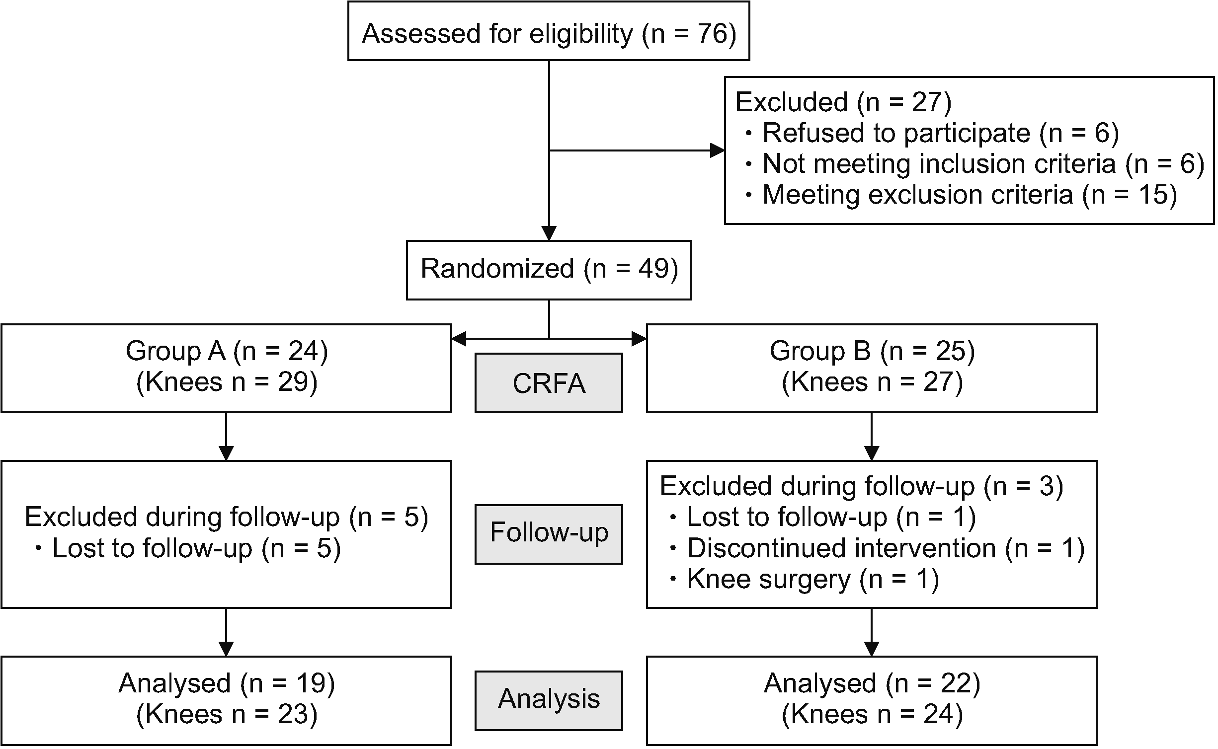
Of the 76 patients evaluated for knee pain, 49 patients who met the inclusion criteria were included in the study. Patients were randomized into group A (n = 24) and group B (n = 25). In group A, 19 patients (17 females, 2 males) completed the follow-up period at 1 and 3 months, while in group B, 23 patients completed the follow-up period at 1 month and 22 patients completed the follow-up period at 3 months (21 females, 1 male) (Fig. 1). The mean age was 63.42 ± 9.90 in group A and 63.54 ± 6.29 in group B. There was no significant difference in age, sex, body mass index, duration of symptoms, or clinical and demographic data between the groups. There was no statistically significant difference between the groups except for the physical health score of SF-36 at baseline (Table 1).
Patients were taking acetaminophen or nonsteroidal anti-inflammatory drugs for knee pain before CRFA procedure, but none were taking opioid analgesics or disease-modifying osteoarthritis drugs. There was no difference in the median weekly medication use before CRFA procedure between the two groups.
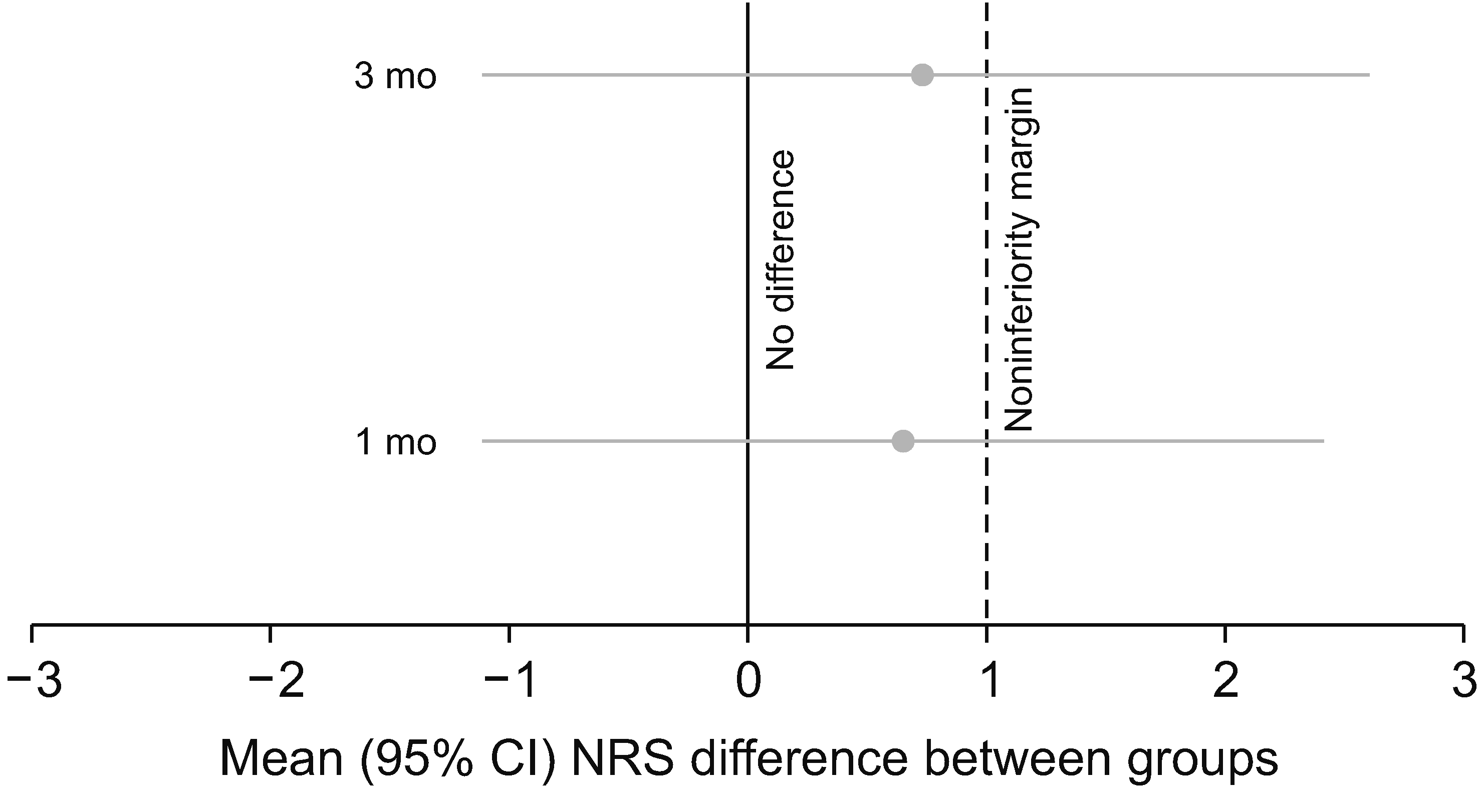
In the study, pain was assessed separately for 47 knees using the NRS. A significant decrease in NRS scores was observed in both groups at month 1 and month 3 compared to baseline. There was no significant difference in baseline, 1st and 3rd month NRS scores between the groups (Table 2). However, the non-inferiority of group A to B was not established because the mean difference in NRS change scores (95% CI) between the groups was 0.65 (–1.10 to 2.41) and 0.73 (–1.10 to 2.56) at 1 and 3 months, respectively, and the 95% CI exceeded the non-inferiority margin of 1 (Table 3 and Fig. 2).
The number of knees with CSC (≥ 2 points) on the NRS during follow-up was 18 (78.3%) at 1 month and 15 (65.2%) at 3 months in group A, and 19 (79.2%) at 1 month and 17 (70.8%) at 3 months in group B. There was no statistical difference between the groups. And the number of knees with 50% improvement in NRS scores was 9 (39.1%) at 1 month and 6 (26.1%) at 3 months in group A and 14 (58.3%) at 1 and 3 months in group B, with a statistical difference between groups at 3 months (Table 4).
At baseline, the SF-36 emotional health score was similar in both groups, whereas the physical health score was significantly lower in group A than in group B (P = 0.005). In the within-group assessments, a significant decrease in the physical and emotional health scores was observed in both groups at the 1st and 3rd month follow-up compared to baseline (group A and B, P < 0.001, P = 0.002; P = 0.005, P < 0.001, respectively). There was no difference between groups in either of the SF-36 subgroup scores at months 1 and 3 (Table 5).
In the within-group analysis of the WOMAC scale, statistically significant improvements in total score, pain score, and physical function score were found in both groups at months 1 and 3 compared to pre-treatment data. While there was no significant difference in the joint stiffness score in group A, a significant improvement was observed in group B only at month 1 compared to baseline (P = 0.013). There was no significant difference between the groups in any of the WOMAC subscores at 1 and 3 months (Table 6). The number of patients who achieved MCSC in the WOMAC total score was 14 (73.7%) at month 1 and 12 (63.2%) at month 3 in group A, while in group B it was 16 (72.7%) at month 1 and 17 (77.3%) at month 3. It was found that there was no significant difference between the groups (P values at month 1 and 3 were 0.945 and 0.322, respectively).
No significant complications were observed in any of the patients. In group B, 1 patient developed sudden hypotension during the procedure, which was resolved with clinical monitoring without further intervention, but the patient was not included in the final analysis because the procedure was not completed. Approximately 2 weeks after the procedure, 1 patient in group B developed mild (NRS 3/10) "lightning-like" pain in brief episodes that began deep to the skin entry points of the CRFA electrodes and radiated to the anterior aspect of the knee. No additional treatment was planned for the patient, and the pain completely resolved at the 1-month follow-up.
The purpose of this study was to evaluate the effect of excluding the SLGN, one of the 3 standard targeted geniculate nerve branches in the CRFA procedure, on treatment outcomes in patients with knee osteoarthritis. Although the authors were unable to demonstrate the non-inferiority of excluding the SLGN from the CRFA procedure in this study, significant improvements in NRS, WOMAC total, pain and physical function, and SF-36 scores were observed after treatment in both patient groups (group A; SMGN, IMGN, group B; SMGN, IMGN, SLGN). The proportion of knees with CSC (NRS ≥ 2 reduction) at 3 months was 65.2% in group A and 70.8% in group B, with no statistically significant difference between the groups. Similarly, no difference was observed between groups in terms of post-treatment change in NRS scores, number of patients achieving a MCSC (≥ 12% reduction) in WOMAC total score, SF-36 score, and presence of adverse events.
This study is consistent with the results of many studies in the literature that reported a decrease in pain and an increase in functionality and quality of life in patients treated with CRFA targeting 3 standard genicular nerves (the SMGN, IMGN, and SLGN) [5,15,20]. In addition, the results of this study were found to be consistent with the results of studies in the literature that treated the nerves innervating the medial knee [8,21].
When the treatment results were compared between the groups, the number of knees with CSC at 3 months was found to be 65.2% in group A and 70.8% in group B, but no significant difference was found between the groups. In the study by Choi et al. [2], this rate was reported to be 59% at 3 months. The fact that this rate was lower compared to our study may be explained by the fact that an NRS ≥ 50% reduction was considered the primary treatment outcome criterion in that study. In our study, when the proportion of patients with 50% improvement in NRS score was examined, it was observed that the proportion of patients in group B was similar to the literature, while the proportion in group A was lower, and although this suggests that CRFA treatment applied to the 3 genicular nerves seems to be more effective, it has been clearly demonstrated in studies that a 2-point change in NRS score is a CSC, and this rate is similar between the two groups. Although SF-36 scores increased after treatment in both groups, no significant difference was found between the groups. In the WOMAC scores, a significant improvement in all subscores except joint stiffness was observed in both groups after treatment, but no significant difference was found between the groups. Improvement in the joint stiffness score was observed only in group B at the first month of follow-up, while no significant difference was found at the third month. Reviewing 2 studies in the literature that evaluated joint stiffness after radiofrequency procedure, the joint stiffness score decreased during all follow-ups in the study by El-Hakeim et al. [3], while in the study by Santana-Pineda et al. [22] the joint stiffness score decreased significantly only at month 1. The high initial joint stiffness score in the study by El-Hakeim et al. [3] may have facilitated the statistical detection of clinical improvement (baseline joint stiffness scores were 2.08 and 3.64 in the groups in the current study, 3.05 in the study by Santana-Pineda et al. [22] and 7.87 in the study by El-Hakeim et al. [3]). However, the fact that conventional RFA treatment, which aims to reduce pain by ablating sensory nerves, does not target different biomolecular pathways that may be the cause of joint stiffness may underlie this situation.
There are few studies in the literature evaluating the results of radiofrequency treatment of only the genicular nerve branches innervating the medial knee in a group of patients with osteoarthritis diagnosed with anteromedial knee pain. One study with a non-randomized design targeted the medial retinacular nerve and the infrapatellar branch of the saphenous nerve [23]. A significant decrease in visual analog scale (VAS) score was observed in the CRFA group during the 3-month follow-up period compared to the control group that underwent the genicular block. In another study, Kesikburun et al. [8] applied ultrasound-guided pulsed RF to the SMGN and IMGN and observed a significant decrease in VAS and WOMAC scores at the 3-month follow-up. These studies, similar to our study, suggest that targeting only the nerves innervating the medial capsule in patients with anteromedial knee pain may result in a decrease in pain and an increase in function.
Regardless of the quadrant in which the knee pain occurs, it is highly likely that targeting more nerves will result in a greater improvement in clinical outcomes. However, due to the complex and variable anatomy of the anterior knee capsule innervation, many studies have emphasized that a procedure to completely cover this area should include all 10 genicular nerve branches [24–26]. As the number of nerves treated increases, it is a natural consequence that the procedure time is prolonged and the possibility of side effects increases. In addition, it would not be wrong to assume that the radiation dose to patients, physicians, and other health care personnel will also increase for procedures performed under fluoroscopy. Considering all these limitations, a more reasonable approach would be to select the nerves to be treated according to the patient's symptoms and radiographic findings and perform an individualized procedure. In the present study, in patients with anteromedial knee pain and medial compartment involvement on plain radiographs, similar treatment results were obtained by ablating the SMGN and IMGN, which are only responsible for the medial innervation of the knee, as opposed to the standard approach, supporting our hypothesis.
The treatment results obtained in the current study were discussed above. However, the advantages and disadvantages of the groups over each other should also be mentioned. Although there are no data on procedure time and radiation doses in the current study, less radiation exposure as a natural consequence of CRFA application to fewer nerves, shorter procedure time, and reduced complication rate are important advantages for group A. In contrast, the higher proportion of patients with ≥ 50% improvement in NRS scores is an important advantage in favor of group B. However, similar results in terms of other treatment outcomes suggest that it may be a good idea to select genicular nerves associated with pain localization.
To the authors’ knowledge, this is the first study in the literature to investigate the effects of excluding the SLGN genicular nerve from the CRFA procedure in patients with anteromedial knee pain. However, the study has several limitations. First, the non-inferiority margin was not determined on the basis of statistical grounds, but clinically based on the results of studies in the literature and the opinions of experts with more than 15 years of experience in this field. Secondly, prognostic blocks were not used to predict treatment success prior to ablation. However, the role of prognostic blocks in predicting RFA treatment success is controversial, and there is still no clear consensus in the literature on which critical threshold should be used (NRS ≥ 50% or 80%). Thirdly, radiation doses and procedure times were not recorded, but it is easy to estimate that the group with fewer nerves ablated had less radiation dose and a shorter procedure time. Although patients were enrolled according to the sample size calculated by the noninferiority margin, the study may still be underpowered, suggesting that this may be associated with inconclusive results. Further studies including larger numbers of patients are needed to demonstrate noninferiority. Other limitations of the study are that the follow-up period was limited to 3 months and most of the patients were female. Finally, the lack of a control group without CRFA makes it difficult to assess treatment efficacy.
In conclusion, this study suggests that in patients with osteoarthritis of the knee with anteromedial knee pain and medial tibofemoral compartment involvement, exclusion of the SLGN from the CRFA procedure is neither non-inferior nor inferior to the standard approach in terms of treatment outcomes. Genicular CRFA is a safe treatment option for patients with anteromedial knee pain and osteoarthritis with medial tibiofemoral joint involvement, providing pain relief and improving quality of life and functionality, and when selecting genicular nerves for CRFA, it may be a more appropriate option to opt for individualized approaches that specifically target the genicular nerves thought to be responsible for the patient's knee pain. Further multicenter, long-term, double-blind studies are needed to determine the exact impact of this approach on treatment outcomes.
Notes
DATA AVAILABILITY
Data files are available from Harvard Dataverse: https://doi.org/10.7910/DVN/GQ6NJF.
REFERENCES
1. Glyn-Jones S, Palmer AJ, Agricola R, Price AJ, Vincent TL, Weinans H, et al. 2015; Osteoarthritis. Lancet. 386:376–87. DOI: 10.1016/S0140-6736(14)60802-3. PMID: 25748615.
2. Choi WJ, Hwang SJ, Song JG, Leem JG, Kang YU, Park PH, et al. 2011; Radiofrequency treatment relieves chronic knee osteoarthritis pain: a double-blind randomized controlled trial. Pain. 152:481–7. DOI: 10.1016/j.pain.2010.09.029. PMID: 21055873.

3. El-Hakeim EH, Elawamy A, Kamel EZ, Goma SH, Gamal RM, Ghandour AM, et al. 2018; Fluoroscopic guided radiofrequency of genicular nerves for pain alleviation in chronic knee osteoarthritis: a single-blind randomized controlled trial. Pain Physician. 21:169–77. DOI: 10.36076/ppj.2018.2.169.
4. Qudsi-Sinclair S, Borrás-Rubio E, Abellan-Guillén JF, Padilla Del Rey ML, Ruiz-Merino G. 2017; A comparison of genicular nerve treatment using either radiofrequency or analgesic block with corticosteroid for pain after a total knee arthroplasty: a double-blind, randomized clinical study. Pain Pract. 17:578–88. DOI: 10.1111/papr.12481. PMID: 27641918.

5. Jadon A, Jain P, Motaka M, Swarupa CP, Amir M. 2018; Comparative evaluation of monopolar and bipolar radiofrequency ablation of genicular nerves in chronic knee pain due to osteoarthritis. Indian J Anaesth. 62:876–80. DOI: 10.4103/ija.IJA_528_18. PMID: 30532324. PMCID: PMC6236782.

6. Yildiz G, Perdecioglu GRG, Yuruk D, Can E, Akkaya OT. 2023; Comparison of the efficacy of genicular nerve phenol neurolysis and radiofrequency ablation for pain management in patients with knee osteoarthritis. Korean J Pain. 36:450–7. DOI: 10.3344/kjp.23200. PMID: 37732409. PMCID: PMC10551393.

7. Creamer P, Lethbridge-Cejku M, Hochberg MC. 1998; Where does it hurt? Pain localization in osteoarthritis of the knee. Osteoarthritis Cartilage. 6:318–23. DOI: 10.1053/joca.1998.0130. PMID: 10197166.

8. Kesikburun S, Yaşar E, Uran A, Adigüzel E, Yilmaz B. 2016; Ultrasound-guided genicular nerve pulsed radiofrequency treatment for painful knee osteoarthritis: a preliminary report. Pain Physician. 19:E751–9. DOI: 10.36076/ppj/2019.19.E751.
9. Ajrawat P, Radomski L, Bhatia A, Peng P, Nath N, Gandhi R. 2020; Radiofrequency procedures for the treatment of symptomatic knee osteoarthritis: a systematic review. Pain Med. 21:333–48. DOI: 10.1093/pm/pnz241. PMID: 31578561.

10. Conger A, McCormick ZL, Henrie AM. 2019; Pes anserine tendon injury resulting from cooled radiofrequency ablation of the inferior medial genicular nerve. PM R. 11:1244–7. DOI: 10.1002/pmrj.12155. PMID: 30859692.

11. Khan D, Nagpal G, Conger A, McCormick ZL, Walega DR. 2020; Clinically significant hematoma as a complication of cooled radiofrequency ablation of the genicular nerves; a case series. Pain Med. 21:1513–5. DOI: 10.1093/pm/pnz319. PMID: 31880783.
12. McCormick ZL, Walega DR. 2018; Third-degree skin burn from conventional radiofrequency ablation of the inferiomedial genicular nerve. Pain Med. 19:1095–7. DOI: 10.1093/pm/pnx204. PMID: 29025030.

13. Khanna A, Knox N, Sekhri N. 2019; Septic arthritis following radiofrequency ablation of the genicular nerves. Pain Med. 20:1454–6. DOI: 10.1093/pm/pny308. PMID: 30698785.

14. Deyle GD, Allen CS, Allison SC, Gill NW, Hando BR, Petersen EJ, et al. 2020; Physical therapy versus glucocorticoid injection for osteoarthritis of the knee. N Engl J Med. 382:1420–9. DOI: 10.1056/NEJMoa1905877. PMID: 32268027.
15. McCormick ZL, Korn M, Reddy R, Marcolina A, Dayanim D, Mattie R, et al. 2017; Cooled radiofrequency ablation of the genicular nerves for chronic pain due to knee osteoarthritis: six-month outcomes. Pain Med. 18:1631–41. DOI: 10.1093/pm/pnx069. PMID: 28431129.

16. Salaffi F, Stancati A, Silvestri CA, Ciapetti A, Grassi W. 2004; Minimal clinically important changes in chronic musculoskeletal pain intensity measured on a numerical rating scale. Eur J Pain. 8:283–91. DOI: 10.1016/j.ejpain.2003.09.004. PMID: 15207508.

17. Farrar JT, Young JP Jr, LaMoreaux L, Werth JL, Poole MR. 2001; Clinical importance of changes in chronic pain intensity measured on an 11-point numerical pain rating scale. Pain. 94:149–58. DOI: 10.1016/S0304-3959(01)00349-9. PMID: 11690728.

18. Angst F, Aeschlimann A, Stucki G. 2001; Smallest detectable and minimal clinically important differences of rehabilitation intervention with their implications for required sample sizes using WOMAC and SF-36 quality of life measurement instruments in patients with osteoarthritis of the lower extremities. Arthritis Rheum. 45:384–91. DOI: 10.1002/1529-0131(200108)45:4<384::AID-ART352>3.0.CO;2-0. PMID: 11501727.

19. Lin CJ, Saver JL. 2020; The minimal clinically important difference for achievement of substantial reperfusion with endovascular thrombectomy devices in acute ischemic stroke treatment. Front Neurol. 11:524220. DOI: 10.3389/fneur.2020.524220. PMID: 33123069. PMCID: PMC7569750.

20. Davis T, Loudermilk E, DePalma M, Hunter C, Lindley D, Patel N, et al. 2018; Prospective, multicenter, randomized, crossover clinical trial comparing the safety and effectiveness of cooled radiofrequency ablation with corticosteroid injection in the management of knee pain from osteoarthritis. Reg Anesth Pain Med. 43:84–91. DOI: 10.1097/AAP.0000000000000690. PMID: 29095245. PMCID: PMC5768219.
21. Uematsu H, Osako S, Hakata S, Kabata D, Shintani A, Kawazoe D, et al. 2021; A double-blind, placebo-controlled study of ultrasound-guided pulsed radiofrequency treatment of the saphenous nerve for refractory osteoarthritis-associated knee pain. Pain Physician. 24:E761–9. DOI: 10.36076/ppj.2021.24.E761.

22. Santana-Pineda MM, Vanlinthout LE, Santana-Ramírez S, Vanneste T, Van Zundert J, Novalbos-Ruiz JP. 2021; A randomized controlled trial to compare analgesia and functional improvement after continuous neuroablative and pulsed neuromodulative radiofrequency treatment of the genicular nerves in patients with knee osteoarthritis up to one year after the intervention. Pain Med. 22:637–52. DOI: 10.1093/pm/pnaa309. PMID: 33179073.

23. Ikeuchi M, Ushida T, Izumi M, Tani T. 2011; Percutaneous radiofrequency treatment for refractory anteromedial pain of osteoarthritic knees. Pain Med. 12:546–51. DOI: 10.1111/j.1526-4637.2011.01086.x. PMID: 21463469.
24. Tran J, Peng PWH, Lam K, Baig E, Agur AMR, Gofeld M. 2018; Anatomical study of the innervation of anterior knee joint capsule: implication for image-guided intervention. Reg Anesth Pain Med. 43:407–14. DOI: 10.1097/AAP.0000000000000778. PMID: 29557887.
25. Conger A, Cushman DM, Walker K, Petersen R, Walega DR, Kendall R, et al. 2019; A novel technical protocol for improved capture of the genicular nerves by radiofrequency ablation. Pain Med. 20:2208–12. DOI: 10.1093/pm/pnz124. PMID: 31131850.

26. McCormick ZL, Cohen SP, Walega DR, Kohan L. 2021; Technical considerations for genicular nerve radiofrequency ablation: optimizing outcomes. Reg Anesth Pain Med. 46:518–23. DOI: 10.1136/rapm-2020-102117. PMID: 33483425.
Fig. 2
Between-group difference in NRS score change (group B - group A) at 1 and 3 months compared with pretreatment. NRS: numeric rating scale, CI: confidence interval.

Table 1
Comparison of demographic data and baseline assessment measures
Table 2
Intragroup and intergroup evaluation of NRS scores
Table 3
Comparison of changes in NRS scores between groups
| NRS change | Group A | Group B | P value | Mean difference (95% CI) |
|---|---|---|---|---|
| Baseline & 1 mo | 3.93 (2.69) | 4.04 (3.25) | 0.745 | 0.65 (–1.10 to 2.41) |
| Baseline & 3 mo | 3.39 (3.31) | 4.13 (2.91) | 0.808 | 0.73 (–1.10 to 2.56) |
Table 4
Number of knees with clinically significant change (≥ 2 point improvement) and 50% improvement in NRS score
Table 5
Intragroup and intergroup evaluation of SF-36 sub-scores
Physical health score was calculated by summing 4 sub-scores (bodily pain, physical functioning, role limitations due to physical health problems, general health perceptions). Emotional health score was calculated by summing 4 sub-scores (vitality, social functioning, role limitations due to emotional problems, mental health).
Table 6
Intragroup and intergroup evaluation of WOMAC sub-scores


 PDF
PDF Citation
 Print
Print


 XML Download