TO THE EDITOR
We want to congratulate Dr. Yildiz et al. [
1] for their recent study on comparing the efficacy of genicular nerve phenol neurolysis and radiofrequency ablation for pain management. In addition, we would like to provide some insights.
First, the conventional anatomical targets we initially learnt have changed significantly. Numerous studies now describe that the targets most papers depict as the superolateral and superomedial genicular nerves are associated with the lateral and medial branches of the nerve to the vastus intermedius [
2–
4]. In contrast, the former were much more distal and closer to the lateral and medial epicondyles of the femur. We recommend updating anatomical targets for future studies,
Figs. 1–
6.
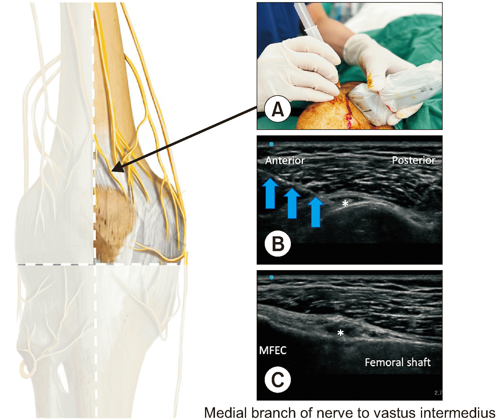 | Fig. 1Medial branch of nerve to vastus intermedius. Fig. 1A indicates the probe position. Fig. 1B is transverse view of ultrasound image. Fig. 1C is coronal view. Note the importance of bi-plane confirmation of this final needle position. The needle tip is at around 30% of the anterior cortex of femoral shaft. MFEC: medial femoral epicondyle, Asterisk: final needle position with phenol, Blue arrows: needle. 
|
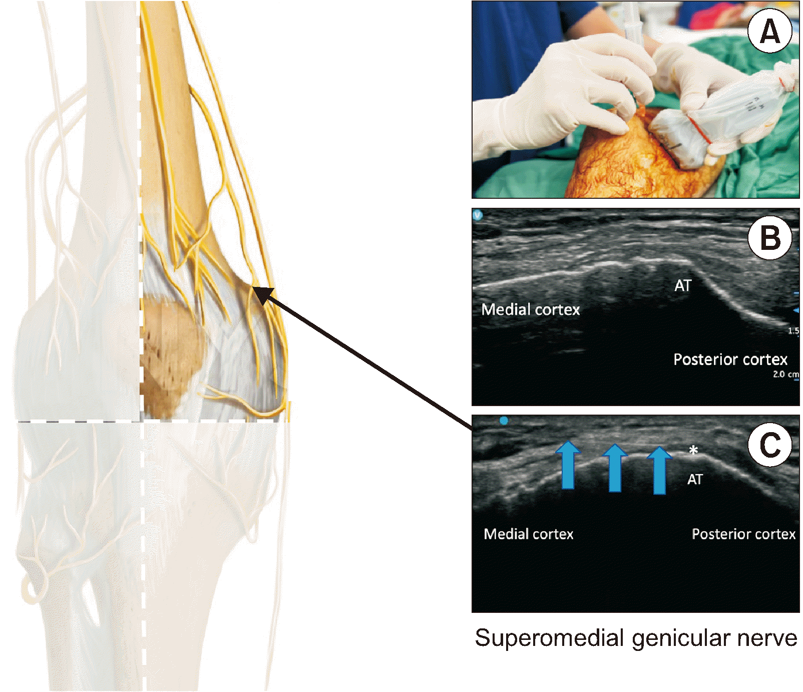 | Fig. 2Superomedial genicular nerve. Fig. 2A indicates the probe position. Fig. 2B is transverse view of the adductor tubercle (AT) as the landmark. Fig. 2C showing that the actual plane of needle insertion is slightly proximal to the AT until the AT becomes less sharp. This modification can reduce the risk of adductor tendon irritation by phenol. Asterisk: final needle position with phenol, Blue arrows: needle. 
|
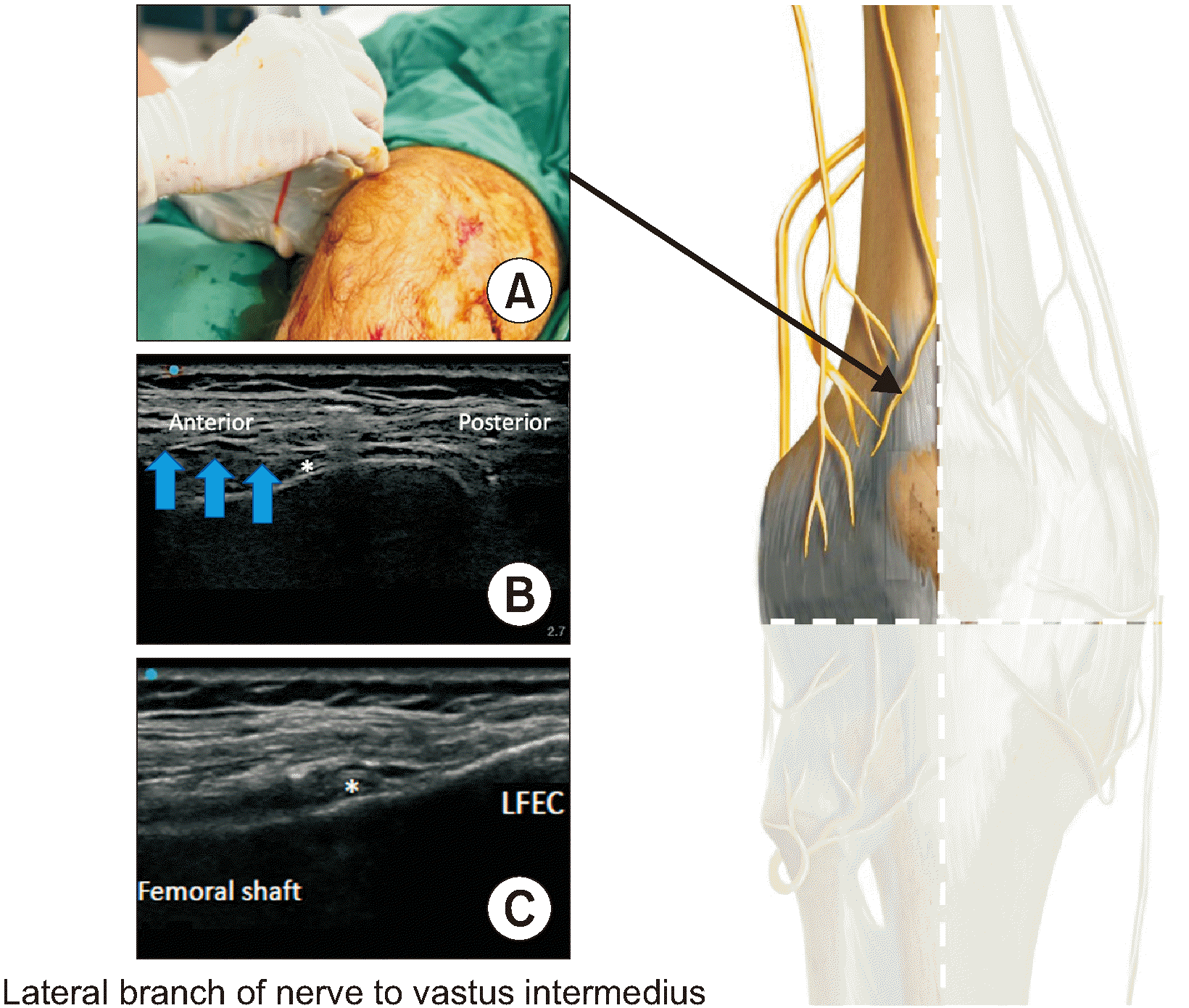 | Fig. 3Lateral branch of nerve to vastus intermedius. Fig. 3A indicates the probe position. Fig. 3B is transverse view of ultrasound image. Fig. 3C is coronal view. It is a mirror image of that of the medial branch of nerve to vastus intermedius. LFEC: lateral femoral epicondyle, Asterisk: final needle position with phenol, Blue arrows: needle. 
|
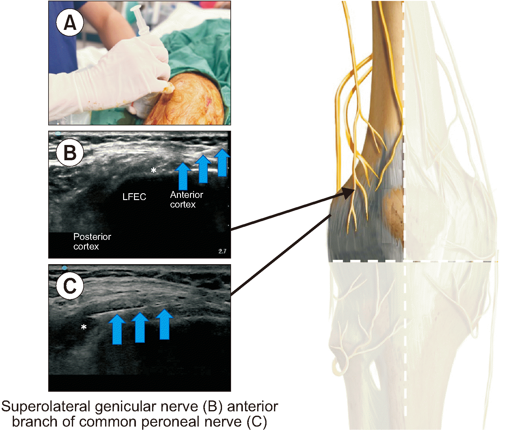 | Fig. 4Superolateral genicular nerve and anterior branch of common peroneal nerve. Fig. 4A indicates the probe position. Fig. 4B is transverse view of ultrasound image for the superolateral genicular nerve. Fig. 4C showing that the needle trajectory is redirected to the posterior cortex for the anterior branch of common peroneal nerve, this distal branch is not depicted in the diagram because is posterior. The landmark is the lateral femoral epicondyle (LFEC). Blue arrows: needle, Asterisk: final needle position with phenol. 
|
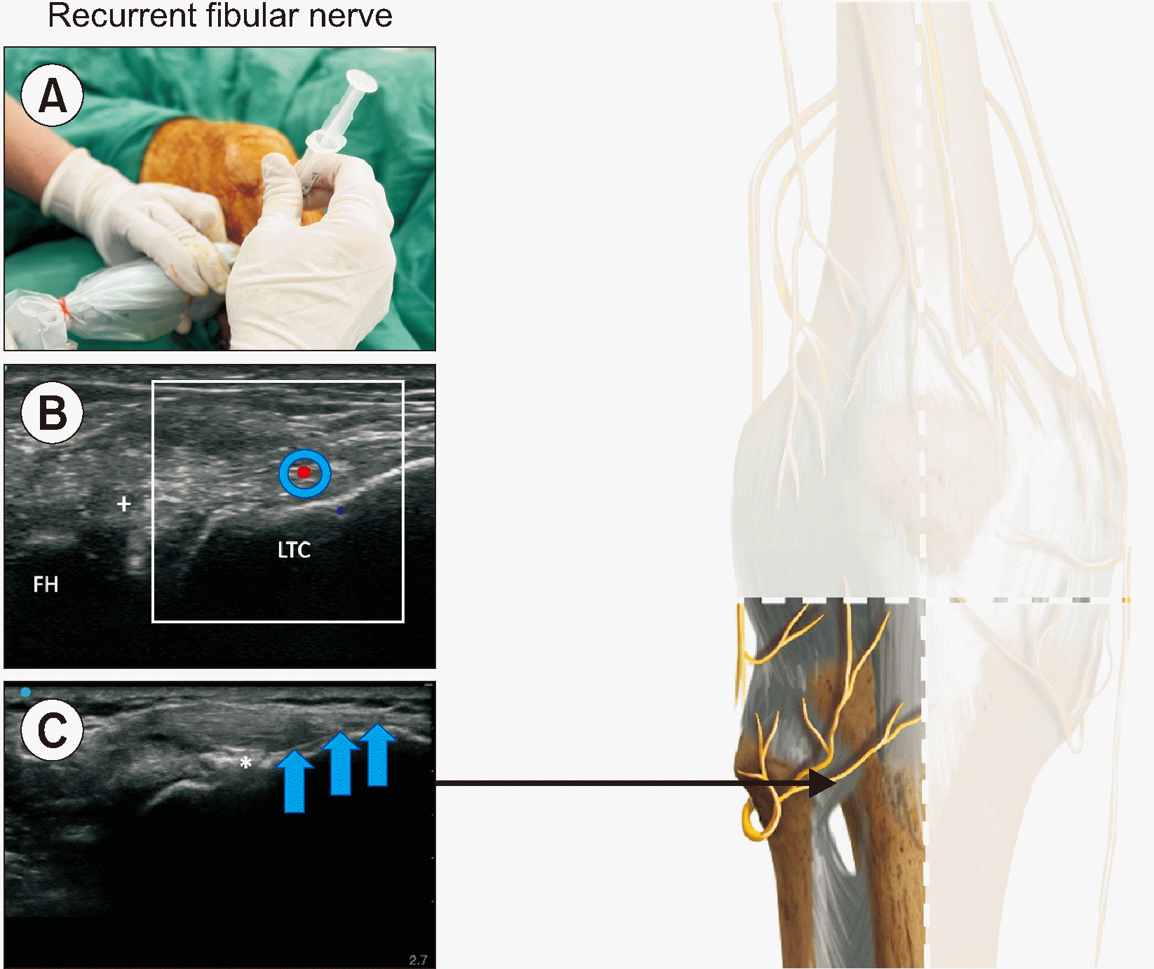 | Fig. 5Recurrent fibular nerve. Fig. 5A indicates the probe position. Fig. 5B is doppler image of transverse view depicting the anterior tibial recurrent artery. Fig. 5C showing that the needle trajectory is along the bony surface of the lateral tibial condyle (LTC) to minimize the chance of phenol extravasation to the superficial structure, particularly skin. The final needle position is around the midway of the LTC. FH: fibular head, Asterisk: final needle position with phenol, Blue arrows: needle, (+): anterior tibiofibular ligament, Red dot: anterior tibial recurrent artery. 
|
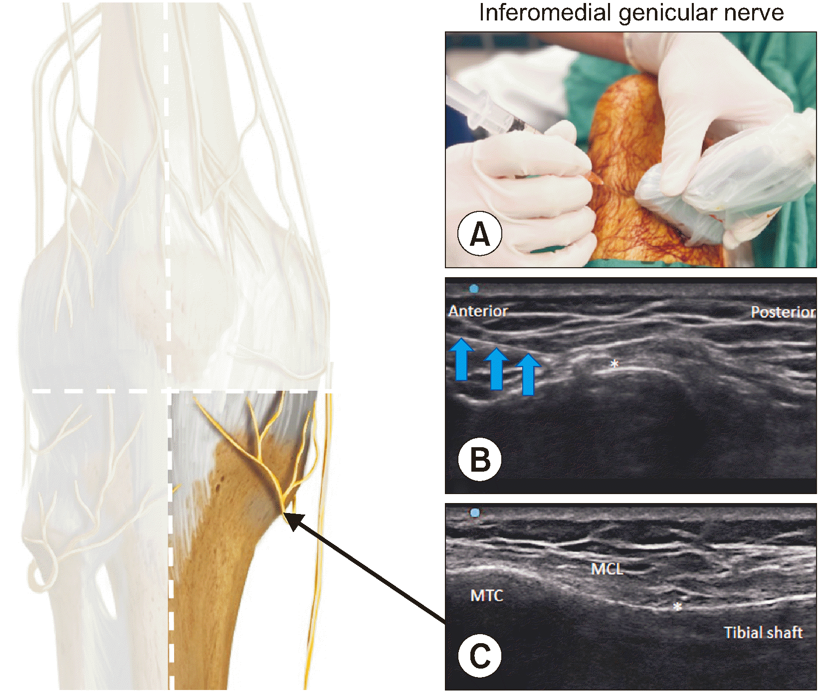 | Fig. 6Inferomedial genicular nerve. Fig. 6A indicates the probe position. Fig. 6B is transverse view of ultrasound image with needle tip around 50% of the anterior cortex of tibial shaft. Fig. 6C is coronal view, showing needle tip underneath the medial collateral ligament (MCL). Inferomedial genicular nerve. Note the dual-plane confirmation of the final needle position. MTC: medial tibial condyle, Asterisk: final needle position, Blue arrows: needle. 
|
Secondly, determining the number of targets has emerged as a new frontier, and the traditional practice of denervating only 3 targets is likely outdated. This may explain cases in which patients do not respond to previous diagnostic blocks (as described in this paper), since it might be necessary to cover more areas of pain [
5]. Forero et al. [
6] has proposed a 6-target denervation approach for the anterior capsule, while Ng et al. [
4] has proposed a posterior target for the posterior knee capsule involvement. The question that remains is: which targets are the most appropriate to establish a standardized procedure? We recommend expanding the approach based on the patient's pain, considering 6 nerves instead of 3, by dividing the anterior joint space into superomedial, superolateral, inferomedial, and inferolateral quadrants. The following nerves should be taken into account: the medial branch of the nerve to the vastus intermedius, the superomedial genicular nerve, the lateral branch of the nerve to the vastus intermedius, the superolateral genicular nerve, the anterior branch of the common peroneal nerve, the inferomedial genicular nerve, and the recurrent fibular nerve (RFN). Although there is a theoretical concern that extensive denervation could potentially lead to neurogenic arthropathy (Charcot joint), we disagree because, compared to chronic generalized neuropathies, these are controlled lesions. Hence, our recommendation is to thoroughly examine the patient to map the pain and to look for specific locations to denervate within the tricompartmental knee joint without causing excessive lesions (
Figs. 1–
6). Based on the anatomical considerations, we recommend targeting the RFN over the lateral condyle of the tibia, specifically medial to the superior tibiofibular joint. This location offers a distinct and optimal target area for intervention, minimizing the risk of inadvertent injury to nearby structures. At this level, the common peroneal nerve is positioned on the lateral aspect of the fibula head, providing a natural anatomical barrier against the extravasation of chemical agents towards the RFN target. Additionally, the anterior superior tibiofibular ligament (upper middle bundle), fibularis longus muscle, and extensor digitorum muscle further safeguard against inadvertent exposure of the common peroneal nerve. Moreover, a study conducted by Forero et al. [
6], corroborates the safety and efficacy of lesioning at the specified RFN target. This study provides valuable evidence supporting the rationale behind our recommendation.
Additionally, we do not recommend routinely applying neurolytic agents to the infrapatellar branch of the saphenous nerve because its terminal branch is very superficial, increasing the risk of skin necrosis through either radiofrequency ablation or chemical neurolysis. While it is possible to target the nerve more proximally along its course to minimize the risk of skin necrosis, there remains a potential for skin allodynia due to its dermatomal innervation. Another challenge arises from the highly variable course of this nerve [
7]. Studies have shown that this branch does not frequently emit an articular branch, but it consistently maintains associations with skin innervation [
8]. Therefore, it is highly recommended to engage in a thorough discussion with patients before attempting to denervate this nerve, especially in cases of persistent concordant residual pain following the denervation of other targets.
Another critical issue is the size of the lesion. Regarding phenol and alcohol, this depends on the volume used for every target and the fascial plane in which it is deposited [
9]. In this study, a volume of 2 mL was employed, but previous research has achieved favorable outcomes with less volume, for instance, 1–1.5 mL [
10]. Considering the difference in surrounding structures, the optimal volume for different targets should also vary. Many studies indicate anatomical variations in the course of genicular nerves [
11], thus suggesting that more extensive lesions could be the best option. Nevertheless, the key mechanical difference between conventional and cooled radiofrequency is the cooling effect and the larger diameter of the cannula, which aids in enlarging the lesion size. The challenge when comparing radiofrequency lesions (electrical field) to chemical denervation (liquid agent) is that the size and shape are not as precise as those created by an electrical field. The larger the lesion the greater the risk of complications, and this applies not only to chemical lesions but also to radiofrequency procedures [
12]. The factors that affect heat lesion size have been previously described in detail but are not the same with phenol [
13]. Therefore, our recommendation is to always use the minimal volume for the diagnostic block and then apply the same volume for the neurolytic procedure.
Nevertheless, we must also acknowledge the advantages of chemical neurolysis. First, compared to cooled radiofrequency, it is cheaper and more accessible, and the distribution of the agent allows the capture of most of the nerve fibers. Besides, the continuous evolution of technology in terms of high-definition images and the constant rediscovery of precise anatomical targets are contributing to the performance of more accurate lesions.
Another issue concerning neurolytic blocks is the safety evaluation. When using neurolytic agents, secondary outcomes such as complications are probably as important as the primary outcome related to pain control. Complications are divided into two phases: early and late. Typically, early complications are well known and described in most case reports, such us local pain and swelling, however late complications must also be described to understand the incidence of abortive regeneration, inappropriate direction, and hyperinnervation events. In this paper, paresthesia was reported but the duration and the pattern of the paresthesia were not. Is it possible that the relatively high volume of injectate to certain targets contributed to this paresthesia?
Finally, an unsolved mystery revolves around the timing. Should we repeat the procedure? And if so, how many repetitions should be performed? Because we must concern ourselves with the biotensegrity and fascia integrity, graded rehabilitation exercises need to be carried out to strengthen the stabilizing knee muscles involved in ambulation. This can potentially reduce the loading stress to the knee while increasing the protection of the knee joint through its stabilizing muscles [
14,
15]. It may then prolong the treatment effect of denervation. However, probably only studies with long follow-up periods can propose a clinical approach to answer these questions. The future lies in personalizing the treatment.
Go to :


 PDF
PDF Citation
 Print
Print


 XML Download