Journal List > J Stroke > v.26(2) > 1516087924

Heo, Yun, Kim, Jung, Yoo, Kim, and Nam: Cancer-Associated Stroke: Thrombosis Mechanism, Diagnosis, Outcome, and Therapeutic Strategies

Abstract

Cancer can induce hypercoagulability, which may lead to stroke. This occurs when tumor cells activate platelets as part of their growth and metastasis. Tumor cells activate platelets by generating thrombin and expressing tissue factor, resulting in tumor cell-induced platelet aggregation. Histopathological studies of thrombi obtained during endovascular thrombectomy in patients with acute stroke and active cancer have shown a high proportion of platelets and thrombin. This underscores the crucial roles of platelets and thrombin in cancer-associated thrombosis. Cancer-associated stroke typically occurs in patients with active cancer and is characterized by distinctive features. These features include multiple infarctions across multiple vascular territories, markedly elevated blood D-dimer levels, and metastasis. The presence of cardiac vegetations on echocardiography is a robust indicator of cancer-associated stroke. Suspicion of cancer-associated stroke during endovascular thrombectomy arises when white thrombi are detected, particularly in patients with active cancer. Cancer-associated stroke is almost certain when histopathological examination of thrombi shows a very high platelet and a very low erythrocyte composition. Patients with cancer-associated stroke have high risks of mortality and recurrent stroke. However, limited data are available on the optimal treatment regimen for stroke prevention in these patients. Thrombosis mechanism in cancer is well understood, and distinct therapeutic targets involving thrombin and platelets have been identified. Therefore, direct thrombin inhibitors and/or antiplatelet agents may effectively prevent stroke recurrence. Additionally, this strategy has potential benefits in cancer treatment as accumulating evidence suggests that aspirin use reduces cancer progression, metastasis, and cancer-related mortality. However, clinical trials are necessary to assess the efficacy of this strategy involving the use of direct thrombin inhibitors and/or antiplatelet therapies.

Introduction

Systemic cancer and stroke are the most common causes of mortality and disability worldwide. In the United States, approximately 40% of the population faces a lifetime risk of developing cancer, with over 50% of these cases occurring in individuals aged 65 years and older [1]. In Korea, the annual incidence rate of cancer is approximately 3.6% [2]. Similarly, the estimated worldwide lifetime risk of stroke from the age of 25 years is 25% [3]. Notably, approximately 1 in 10 patients with ischemic stroke also have comorbid cancer [4]. As cancer treatment continues to improve, the incidence of stroke in patients with cancer is expected to increase. Given the prevalence of these two conditions, their coexistence is common. However, recognizing that cancer can also directly cause stroke is important. According to data from the Korean National Health Insurance Service National Sample Cohort database, patients with cancer have a higher risk of ischemic stroke, with a subdistribution hazard ratio (HR) of 1.17 [5].
The potential mechanisms through which cancer can contribute to stroke include tumor-induced hypercoagulability, direct invasion or compression of arteries, infection, and the secondary effects of radiation therapy or chemotherapy [6-8]. Among these, hypercoagulability is the most common mechanism of stroke in patients with cancer.
Patients with stroke attributable to hypercoagulability, referred to as cancer-associated stroke, have an elevated risk of recurrence and mortality. However, uncertainties remain regarding strategies for preventing stroke recurrence, including the selection of the best or optimal antithrombotic therapies [9,10]. Understanding the mechanism of thrombosis in cancer-associated stroke could potentially guide in identifying the optimal antithrombotic treatment to prevent stroke recurrence. Numerous studies have demonstrated the mechanisms and roles of hypercoagulability in tumor growth and metastasis. Recent histopathological studies of fresh thrombi obtained from the cerebral artery in patients with acute stroke have provided valuable insights into the thrombosis mechanisms in cancer-associated stroke.
This review primarily focuses on the mechanisms of thrombosis, histopathological features of cerebral thrombi, clinical characteristics, outcomes, the current status of antithrombotic use, and perspectives on treatment strategies for stroke prevention in cancer-associated stroke.

Thrombosis mechanisms in cancer-associated stroke

Platelets play a crucial role in tumor growth and metastasis. The increased tendency of thrombosis in cancer is a consequence of these platelet-mediated processes. Tumor cell-induced platelet activation and aggregation are well-established phenomena that have been extensively reviewed [11-17]. Herein, we briefly explore the mechanism of cancer-associated thrombosis.
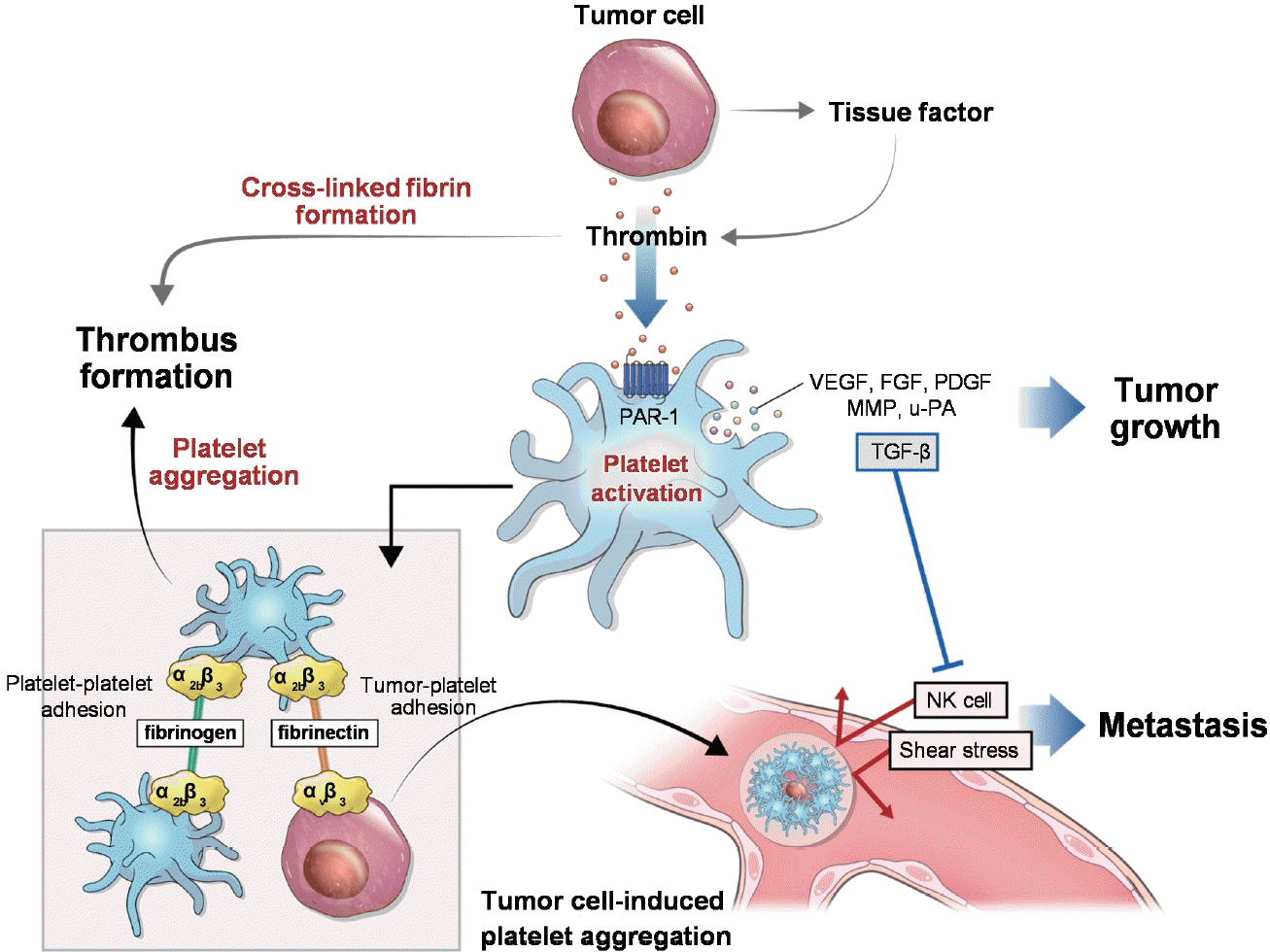
Tumor cells activate platelet alpha granules, secreting growth factors, chemokines, and proteinases including vascular endothelial growth factor, transforming growth factor-β, fibroblast growth factor, platelet-derived growth factor, and matrix metalloproteinase. These molecules contribute to tumor growth and metastasis. Tumor angiogenesis not only provides nutrients and oxygen for tumor growth but also facilitates the entry of tumor cells into the bloodstream, enabling metastasis. Vascular endothelial growth factor is the most critical factor in tumor angiogenesis, while fibroblast growth factor and platelet-derived growth factor also contribute to this process [18,19]. Extracellular matrix remodeling is required for angiogenesis and tumor invasion, with matrix metalloproteinase-9 playing an important role in this process (Figure 1) [20].
Interaction between platelets and tumor cells is essential for tumor cell survival in the blood stream. Activated platelets form aggregates with tumor cells, a phenomenon known as tumor cell-induced platelet aggregation (TCIPA). Platelet glycoprotein IIb/IIIa (GPIIb/IIIa) binds each other via a fibrinogen bridge and tumor cell integrin αvβ3 via a fibronectin bridge [21]. These platelets surrounding tumor cells in TCIPA protect tumor cells from natural killer (NK) cell-mediated cytotoxicity and shear-induced damage in circulation. Platelets also suppress NK cell-mediated cytotoxicity by releasing transforming growth factor-β, which counteracts NK cell activity. Consequently, the formation of TCIPA promotes the survival and spread of tumor cells in the bloodstream [11,22,23]. Platelet-mediated thrombosis is induced during this process (Figure 1).
Platelets also facilitate the adhesion and arrest of tumor cells on the endothelium and their extravasation [24]. TCIPA promotes rolling and weak adhesion to the vascular endothelium via P-selectin, which is expressed on platelets [25]. Subsequently, the platelet GPIb-IX-V receptor complex mediates the firm arrest of TCIPA on the endothelium. In the presence of high shear stress in arterial blood flow, platelet GPIbα interacts with von Willebrand factor, mediating their arrest on the endothelium [19]. This may explain the development of vegetations, known as nonbacterial thrombotic endocarditis (NBTE), on the cardiac valves, where blood flow is very rapid.
Thrombin plays a crucial role in platelet activation induced by tumor cells, as these cells directly generate thrombin [26]. This thrombin generation is further enhanced by tissue factor expressed on tumor cells. Tissue factor initiates the extrinsic coagulation pathway by binding to factor VIIa to form factor X, which leads to thrombin generation [14]. Thrombin, generated from prothrombin, is one of the most potent factors in coagulation and thrombosis. Thrombin is not only the most potent activator of platelets but also converts soluble fibrinogen into insoluble crosslinked fibrin during clot formation and retraction. Furthermore, thrombin activates coagulation factors V, VIII, XI, and XII, amplifying the coagulation response and perpetuating the generation of thrombin. Thus, thrombin-related platelet activation is maximized in cancer. Tumor cells activate platelets by generating thrombin for their growth and survival. The inhibition of thrombin using hirudin, a specific thrombin inhibitor, has been shown to inhibit tumor growth and metastasis in mice [27]. However, this process is often accompanied by local and sometimes extensive intravascular thrombus formation.

Thrombus histopathology in cancer-associated stroke

A thrombus represents the endpoint of thrombosis and reflects, to some extent, the underlying mechanisms of thrombosis and stroke etiology. Understanding the mechanisms of thrombosis plays a pivotal role in determining the appropriate treatment strategies. Endovascular thrombectomy (EVT) has proven effective in patients with acute stroke due to large vessel occlusion in both the anterior and posterior circulations [28,29]. The successful implementation of EVT in patients with acute ischemic stroke has allowed for the retrieval of fresh thrombi from the cerebral arteries, enabling histopathological analysis and research [30,31].
In a previous study, thrombus composition was compared among patients with active cancer, patients with inactive cancer, and patients without cancer [32]. Patients with active cancer exhibited higher platelet fractions and lower erythrocyte fractions than the other patient groups. Thrombus composition was further compared according to stroke etiology. Notably, patients with NBTE had high platelet and low erythrocyte fractions. Patients with active cancer and cryptogenic etiology also showed high platelet and low erythrocyte fractions, whereas those with conventional etiology showed high erythrocyte and low platelet fractions [32]. The platelet-rich features of thrombi in patients with stroke and active cancer were also demonstrated in a subsequent study [33].
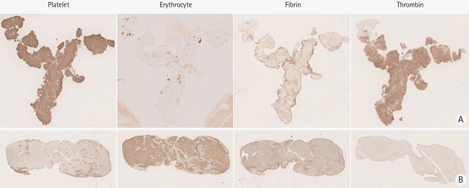
A recent study compared the expression of coagulation factors in thrombi between patients with cancer-associated stroke and those without cancer, who were matched using propensity scores. The cancer-associated stroke group exhibited a higher platelet fraction and a lower erythrocyte fraction than the control group. Furthermore, among the coagulation factors, thrombin and tissue factor were significantly elevated in the cancer group. A positive correlation between thrombin and platelets was observed in the cancer group [34]. Histopathological studies of cerebral thrombi revealed distinct features of cancer-associated stroke, characterized by compositions rich in platelets and thrombin and low in erythrocytes (Figure 2). This finding confirms previous experimental evidence of a tumor cell-specific interaction between thrombin and platelets.
Thrombus examination is crucial for diagnosing cancer-associated stroke. Thrombi in cancer-associated stroke are visibly white, allowing for suspicion of cancer as the underlying cause upon thrombus retrieval in the angiographic suite [35,36]. On hematoxylin and eosin-stained thrombi, a fibrin/platelet proportion of ≥65% accurately predicted cancer-associated stroke, with an area under the curve of 0.84 [33]. Although platelets cannot often be accurately differentiated from fibrin on hematoxylin and eosin staining, substantial fractions of fibrin are present in the thrombi of conventional stroke etiologies. Therefore, immunohistochemical assessments may offer more precise information for the diagnosis of cancer-associated stroke. A machine-learning model using immunohistochemically stained platelet slides achieved a highly accurate diagnosis of cancer-associated stroke, with areas under the curve ranging from 0.946 to 0.986. Moreover, it demonstrated the ability to predict occult cancer, with probabilities ranging from 88.5% to 99.2% [37,38].

Diagnosis of cancer-associated stroke

Patients with stroke are often diagnosed with cancer. The diagnosis of cancer-associated stroke primarily applies to patients with stroke and active cancer. Active cancer is defined as a newly diagnosed cancer within the past 6 months, cancer requiring chemotherapy or surgical treatment within the past 6 months, or cases of recurrent, metastatic, or inoperable cancer [39]. However, the presence of active cancer does not necessarily implicate cancer as the actual cause of the stroke, and in some cases, cancer may be an incidental finding. Because patients with cancer-associated stroke are often treated with anticoagulation therapy and are at a higher risk of recurrent stroke, an accurate diagnosis of stroke attributed to cancer is crucial.
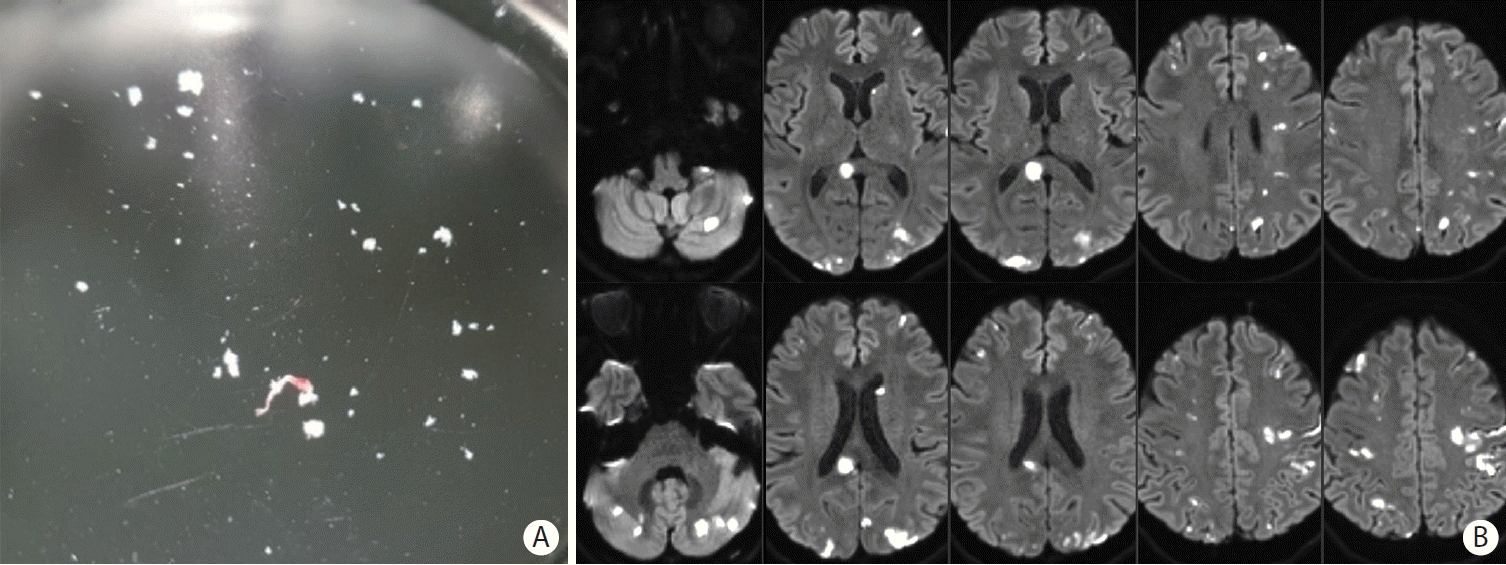
Patients with cancer-associated stroke exhibit distinct characteristics (Table 1). Thrombi retrieved during EVT tend to fragment easily (Figure 3A). Thromboembolic events often occur in a high-flow state under hypercoagulable conditions, resulting in multiple infarctions on diffusion-weighted imaging. These infarctions are commonly observed bilaterally in both anterior and posterior circulations (Figure 3B).
D-dimer levels in the blood are markedly elevated in patients with cancer-associated strokes. D-dimer is produced during the degradation of cross-linked fibrin as part of fibrinolysis and serves as an indirect marker of intravascular thrombosis and fibrinolysis. Its production involves three key enzymes: thrombin, coagulation factor XIII, and plasmin. Thrombin converts soluble fibrinogen into fibrin monomers that subsequently form fibrin polymers. Factor XIII reinforces these fragile fibrin networks by cross-linking the D-domains of adjacent fibrin monomers and the opposing α-chains. D-dimer and fibrin degradation products are produced when fibrin networks are degraded by plasmin, which is converted from plasminogen by tissue- and urokinase-type plasminogen activators [40,41]. Plasminogen activators are released locally from endothelial cells in response to injury, including thrombosis. D-dimer levels can increase in various pathological conditions, including venous thrombosis. However, D-dimer levels are notably elevated in cancer due to the generation of thrombin and urokinase-type plasminogen activator by tumor cells.
Cancer-associated thrombosis may be closely associated with metastasis because tumor cell-induced platelet activation and the resulting intravascular thrombosis typically occur during metastasis. In patients with stroke and cancer, after excluding those with a conventional stroke etiology, a significant majority (69.2%–83.3%) show evidence of metastasis [42-45]. Therefore, the presence of metastasis can serve as an indicator that raises suspicion of a cancer-associated stroke. Conversely, evaluation of metastasis should be considered in patients with suspected cancer-associated stroke.
A diagnosis of cancer-associated stroke is highly probable when cardiac vegetation (NBTE) is detected on echocardiography, particularly in the absence of a conventional stroke etiology. NBTE is characterized by sterile valvular vegetations, typically wart-like and small, on cardiac valves, chordae tendineae, or endocardium [46,47]. Cancer is the predominant cause of NBTE, although it may develop in connective tissue diseases [48]. In an autopsy study of 65 patients with NBTE, 51 (78.5%) had concurrent cancers [49]. NBTE is pathologically characterized by the presence of platelet-fibrin thrombi and may result from interactions between tumor cells and platelets in the circulation during the metastatic process. A previous study of patients with cancer-associated stroke revealed that all 20 patients with NBTE had metastasis, and NBTE was not detected on transesophageal echocardiography in patients without metastasis [45].
In patients undergoing EVT with obtained thrombi, cancer-associated stroke is suspected when the thrombi are predominantly white, particularly in the presence of active cancer (Figure 2A and 3A). Cancer-associated stroke can be confirmed by histopathological examination of thrombi, which typically show high platelet and low erythrocyte fractions [32,38].

Outcomes in cancer-associated stroke

Patients with cancer-associated stroke are at a high risk of mortality and recurrent stroke (Table 2). Mortality and the risk of thromboembolism/ischemic stroke have been retrospectively assessed in patients with stroke and active cancer. In studies involving 230 patients with cancer (69% with metastatic cancer) experiencing stroke of any etiology, the median survival was 84 days (interquartile range [IQR], 24–419 days). Recurrent thromboembolism occurred in 34% of patients, and ischemic stroke recurred in 15.7% [50]. In studies involving patients with active cancer and stroke of any etiology, the mortality rates were 13.0%– 18.3% at 1 month, 26.1%–60.1% at 6 months, and 71.6% at 1 year [45,51-53]. Ischemic stroke recurred in 11.0% at 1 month and 22.4% at 6 months [45,54].
In studies including patients with active cancer and cryptogenic stroke etiology, the mortality rates were 12.4% at 1 month, 56% at 3 months, and 54.2% at 6 months, with ischemic stroke recurrence rates of 9.9% at 1 month and 25% at 6 months [45,55]. Patients with NBTE had notably higher mortality and stroke recurrence rates, with 80% mortality and a 50% recurrence of stroke within a 6-month follow-up period [45].
Patients with stroke and cancer often receive reperfusion therapy. In the multicenter randomized clinical trial of endovascular treatment for acute ischemic stroke in the Netherlands (MR CLEAN) substudy, among patients with any stroke etiology receiving EVT, the mortality rate was 52.2% and stroke recurred in 4% during a 3-month follow-up period [56]. In the National Readmission Database of United States for large vessel occlusion, inhospital mortality after EVT was significantly higher in patients with metastatic cancer than those without cancer (26% vs. 14%, P<0.001) [57].
Outcomes were more severe in patients with cryptogenic stroke etiology. Following reperfusion therapy, the 6-month mortality rate was significantly higher in patients with cryptogenic etiology than in those with any other stroke etiology (85.7% vs. 24.3%, P<0.001). Additionally, the rate of functional dependence or death at 3 months was more common in patients with cryptogenic etiology than in those with a determined etiology (90.5% vs. 47.1%, P=0.003) [58].
Patients with active solid cancer and ischemic stroke were prospectively followed up. During a median follow-up of 48 days (mean, 278 days), 42 of the 50 enrolled patients (86%) developed major thromboembolic events, including 26% with ischemic stroke, or died. The cumulative incidence of major thromboembolic events or death was 52% (95% confidence interval [CI], 37%–66%) at 90 days, 62% (95% CI, 47%–75%) at 180 days, and 72% (95% CI, 58%–84%) at 360 days. The cumulative incidence of recurrent ischemic stroke was 18% (95% CI, 9%–31%) at 90 days, 20% (95% CI, 10%–34%) at 180 days, and 20% (95% CI, 10%–34%) at 360 days. In another group of 50 patients with active cancer but without stroke, 30% experienced major thromboembolism, including 2% with ischemic stroke, and 18% died. Among an additional 50 patients with stroke but without cancer, 32% experienced major thromboembolism with no deaths [59].
Overall, patients with stroke and active cancer have higher mortality and stroke recurrence rates than those with stroke but without cancer. Patients with active cancer and cryptogenic stroke are more likely to have cancer-associated stroke. These patients tend to experience worse outcomes. The prognosis is poor when NBTE is detected on echocardiography, often indicating TCIPA and accompanying metastasis.

Antiplatelet agents in cancer treatment

Approximately three decades ago, the potential benefits of aspirin in preventing colorectal cancer emerged from retrospective and prospective observational studies and randomized clinical trials [60-66]. In 2012, a systematic review and meta-analysis confirmed these benefits, revealing a 38% reduction in the risk of colorectal cancer and a 42% reduction in the 20-year risk of colorectal cancer-related death in aspirin users. Moreover, consistent reductions in the risks of gastrointestinal (gastric, biliary, and esophageal) and breast cancers were observed, along with a 31% reduction in distant metastasis [67]. Notably, the incidence of hepatocellular cancer decreased by 31% among aspirin users with hepatitis B or C, without an increased 10-year risk of gastrointestinal bleeding [68].
In a recent systematic review and meta-analysis, aspirin use reduced the overall risk of cancer by 21% and cancer-related mortality by 20% [69]. Other recent meta-analyses have consistently supported aspirin’s role in reducing various cancer risks and cancer-related deaths [70-72]. Importantly, the benefits of aspirin extended to patients with post-diagnosis colorectal cancer, as indicated by a meta-analysis involving 237,245 patients. The findings showed improvement in colorectal cancer-specific survival by 26% [73].
In contrast to aspirin, evidence regarding the effects of other antiplatelet agents on cancer is limited. Concerns arose regarding thienopyridine derivatives (prasugrel and clopidogrel) following the trial to assess improvement in therapeutic outcomes by optimizing platelet inhibition with prasugrel–thrombolysis in myocardial infarction (TRITON–TIMI)-38 [74]. However, a UK population-based cohort study involving patients with colorectal, breast, and prostate cancers found no evidence of increased cancer-specific mortality with clopidogrel [75]. Furthermore, a study conducted using Taiwan’s National Health Insurance Research Database showed a reduced colorectal cancer risk with aspirin, clopidogrel, and dual antiplatelet therapy. The adjusted HRs for the risk of colorectal cancer during 13 years of follow-up were 0.59 (95% CI, 0.56–0.61) for aspirin, 0.77 (95% CI, 0.68–0.87) for clopidogrel, and 0.37 (95% CI, 0.33–0.40) for dual antiplatelet cohorts [76]. A nested case–control study further revealed risk reductions of 17% with aspirin, 20% with clopidogrel, and similar effects with dual antiplatelet therapy [77].
Preclinical studies using the P2Y12 inhibitors ticagrelor and clopidogrel have shown reduced tumor growth and metastasis in murine models of ovarian and melanoma cancers, suggesting their potential benefits in cancer [78-80]. These clinical and preclinical findings suggest that P2Y12 inhibitors, including clopidogrel, may also be beneficial in cancer.
Over the past three decades, substantial and consistent evidence has supported the role of aspirin in reducing cancer risk, metastasis, and cancer-related mortality, even after cancer diagnosis. While the use of other antiplatelet agents, including P2Y12 inhibitors, for cancer treatment is promising, further evidence is required. Platelets are implicated in tumorigenesis, and tumor-induced platelet activation promotes tumor growth and metastasis. Therefore, aspirin and other antiplatelet agents have the potential to suppress tumorigenesis and metastasis. Additionally, the use of aspirin as an anti-inflammatory agent may contribute to cancer prevention by mitigating repeated inflammation-induced damage to normal tissues. Aspirin inhibits cyclooxygenase-1 in platelets and cyclooxygenase-2 in endothelial cells, which are associated with tumorigenesis and tumor spread through involvement in various signaling pathways [81]. Consequently, TCIPA has emerged as a target for cancer treatment [21]. The use of antiplatelet and antithrombotic agents has been suggested as an adjunctive treatment for cancer [82].

Antithrombotic use in cancer-associated stroke

Few studies have compared the effects of various antithrombotic agents on patients with acute stroke and active cancer (Table 3). In a retrospective observational study involving 263 patients with stroke and active cancer, 117 experienced recurrent thromboembolism, including 36 with recurrent ischemic stroke. The rates of recurrent thromboembolism were similar between patients receiving antiplatelet therapy and those receiving anticoagulant therapy [50].
D-dimer levels were compared between enoxaparin and warfarin in 79 patients with active cancer and acute ischemic stroke that were not explained by conventional stroke mechanisms. During a mean follow-up period of 4.9 months, stroke recurred in 1 of 29 (3.4%) patients treated with enoxaparin and in 8 of 50 (16.0%) patients treated with warfarin. Follow-up D-dimer levels were measured at a median of 8 days after the initial assessments in both groups. D-dimer levels significantly decreased in the enoxaparin group (3.88 μg/mL) compared with the warfarin group (17.42 μg/mL) (P=0.026), despite similar initial D-dimer levels. These results suggest the potential superiority of enoxaparin over warfarin based on the short-term decline in D-dimer levels [44].
In a study of 48 patients with ischemic stroke and active cancer, excluding those with conventional stroke, no significant differences were observed in cardio-cerebrovascular recurrence (57% vs. 49%, P=0.096) and the occurrence of new territory lesions (57% vs. 57.9%, P=1.000) between seven patients treated with non-vitamin K oral anticoagulant (NOAC) and 41 patients treated with low-molecular-weight heparin (LMWH) [43].
Among the 59 patients with ischemic stroke and active cancer, 19 were treated with subcutaneous heparin. Notably, the 10 patients who received long-term subcutaneous heparin therapy did not experience stroke recurrence. However, three of the nine patients who discontinued subcutaneous heparin had recurrent stroke. The diagnosis of cancer-associated stroke was uncertain in this study, as 11 out of the 59 patients had a final diagnosis of conventional stroke mechanisms other than cancer [83]. In another study involving 59 patients with cryptogenic stroke and active cancer and venous thromboembolism, ischemic stroke recurred less frequently in patients treated with subcutaneous unfractionated heparin (4%, 1/24 patients) than in those treated with oral direct factor Xa inhibitors (DiXaIs, 31%, 9/29 patients) during a 30-day follow-up period (P=0.008). The incidence of major bleeding was similar between the two groups [84].
In an open-label, pilot, randomized clinical trial, enoxaparin was compared with aspirin in patients with stroke within 4 weeks and active cancer. Patients with other conventional etiologies were excluded because the exclusion criteria included clear indications for anticoagulation or antiplatelet therapy and symptomatic carotid stenosis. The primary outcome was feasibility. Of 49 eligible patients, 20 were enrolled in this study. Among the 10 patients randomized to receive enoxaparin, 4 (40%) switched to aspirin because of discomfort with injections. No differences were found in major bleeding, thromboembolic events, or survival between the two groups. Although the sample size was too small to compare efficacy, this study suggests that comparing aspirin with NOACs can be considered instead of parenteral heparins [85].
This group is planning a new trial to compare apixaban and aspirin in patients with embolic stroke of an undetermined source (ESUS) and active cancer. A recent open-label randomized pilot trial, comparing edoxaban and enoxaparin in 40 patients with cancer-associated stroke, found no significant changes of D-dimer levels or microembolic signals on transcranial Doppler [86].
A subgroup analysis of the new approach rivaroxaban inhibition of factor Xa in a global trial versus aspirin to prevent embolism in embolic stroke of undetermined source (NAVIGATE ESUS) trial included 543 patients with a history of cancer. The rate of recurrent ischemic stroke did not differ between the rivaroxaban (7.7%) and aspirin (5.4%) groups, and the rate of major bleeding was not significantly higher in the rivaroxaban group than in the aspirin group (rivaroxaban 2.9% vs. aspirin 1.1%). All-cause mortality was also similar between the groups (rivaroxaban 3.7% vs. aspirin 3.3%) [87]. However, only 49 patients (9%) were diagnosed with cancer less than 1 year before the index stroke in this study. Therefore, most of them might have had inactive cancer, and cancer might not have been the cause of the stroke, but rather a bystander.
Previous studies provided limited guidance in determining the optimal antithrombotic regimen to prevent stroke recurrence in patients with cancer-associated stroke. These studies investigated the efficacy of various drugs in non-controlled studies with small sample sizes. The empirical use of LMWH is common; however, it lacks clinical evidence and is not based on the thrombosis mechanisms of cancer-associated stroke. The 2021 American Heart Association/American Stroke Association guidelines state that there is a paucity of data on the best treatment regimen for patients who have had a stroke attributed to hypercoagulability from cancer, and that the potential benefit of LMWH in preventing stroke remains unknown [9].

Antithrombotic use in patients with stroke, cancer, and atrial fibrillation

Cancer often coexists with atrial fibrillation (AF), with a prevalence of approximately 20% in patients with cancer [88,89]. Conversely, the incidence of cancer in patients with AF is 30%–40% higher than that in the general population [90]. The introduction of NOAC has led to increased use in patients with cancer and AF [91]. Several studies have compared NOAC with warfarin in this context [92-94].
A recent systematic review and meta-analysis analyzed patients with cancer and AF from three randomized controlled trials comparing NOAC and warfarin and five retrospective cohort studies. In the randomized controlled trials involving 2,657 participants, no significant differences were found between NOAC and warfarin in stroke/systemic embolism (odds ratio [OR], 0.69; 95% CI, 0.45–1.06; P=0.09), venous thromboembolism (OR, 0.91; 95% CI, 0.33–2.52; P=0.86), myocardial infarction (OR, 0.74; 95% CI, 0.44–1.23; P=0.24), or major bleeding (OR, 0.81; 95% CI, 0.61–1.06; P=0.12). However, observational cohorts with approximately 22,008 participants showed that NOAC users had significantly lower risks of ischemic stroke (OR, 0.51; 95% CI, 0.28–0.92; P=0.02), venous thromboembolism (OR, 0.50; 95% CI, 0.41–0.60; P<0.00001), major bleeding (OR, 0.28; 95% CI, 0.14–0.55; P=0.0002), and intracranial or gastrointestinal bleeding (OR, 0.59; 95% CI, 0.37–0.92; P=0.02) compared with warfarin users [95].
A nationwide retrospective cohort study from Taiwan, comparing 6,274 patients with cancer and AF on NOAC with 1,681 patients on warfarin, found that NOAC was associated with lower risks of major adverse cardiovascular events (defined as ischemic stroke/systemic embolism or acute myocardial infarction) (HR, 0.63; 95% CI, 0.50–0.80; P=0.0001), major adverse limb events (HR, 0.41; 95% CI, 0.24–0.70; P=0.001), venous thrombosis (HR, 0.37; 95% CI, 0.23–0.61; P<0.001), and major bleeding (HR, 0.73; 95% CI, 0.56–0.94; P=0.017). This effect was consistent regardless of stroke history, cancer stage, or NOAC type/dosage [96].
In a retrospective observational study involving 40,271 patients with active cancer and AF [95], apixaban showed the lowest risk of stroke/systemic embolism (HR, 0.59; 95% CI, 0.45–0.78) compared with dabigatran (HR, 0.88; 95% CI, 0.54–1.41) and rivaroxaban (HR, 0.82; 95% CI, 0.62–1.08). The risk of major bleeding was reduced in apixaban users (HR, 0.58; 95% CI, 0.50–0.68) and dabigatran users (HR, 0.76; 95% CI, 0.57–1.01), but not in rivaroxaban users (HR, 0.95; 95% CI, 0.85–1.06) [97].
Based on this evidence, the 2021 American Heart Association/American Stroke Association guidelines recommend considering NOAC over warfarin for anticoagulation in patients with ischemic stroke or transient ischemic attack in the setting of AF and cancer (class of recommendation 2a and level of evidence B-NR) [9]. However, these studies primarily included patients with cancer and AF, and their findings may not be directly applicable to patients with stroke, cancer, and AF. Therefore, determining whether stroke is associated with cancer-associated thrombosis in patients with stroke, active cancer, and AF is crucial. NOAC is the preferred choice for patients for whom cancer is considered an incidental condition. However, in cases in which the risk of stroke associated with cancer is significant, the selection of antithrombotic agents should be directed toward preventing cancer-associated thrombosis.

Perspective on antithrombotic use in cancer-associated stroke

Accumulating preclinical and in vitro evidence has highlighted the crucial role of platelet activation and thrombin generation in tumor cell growth and metastasis. Clinical studies have consistently demonstrated the effectiveness of aspirin and other antiplatelet drugs in reducing the risks of cancer, metastasis, and cancer-related mortality. Recent histopathological studies of cerebral artery thrombi in patients with stroke and active cancer have further validated the involvement of platelets and thrombin in cancer-associated stroke. This suggests that the inhibition of platelets and thrombin may be an effective strategy for preventing cancer-associated stroke and attenuating cancer progression and metastasis.
Drugs that target the coagulation pathway can suppress thrombin generation. LMWH has empirically been used to prevent recurrence in cancer-associated strokes, but it often faces low compliance due to discomfort associated with injections [85]. Recently, NOAC has also been used in cancer-associated stroke. However, upstream thrombin inhibitors, such as heparin, LMWH, warfarin, and factor Xa inhibitors, may be insufficient to prevent tumor-induced thrombin generation. This insufficiency occurs because tumor cells generate thrombin directly and indirectly through the activation of the coagulation pathway by tissue factor expression. Direct thrombin inhibitors may be more effective than other upstream anticoagulants because they can inhibit both direct and indirect mechanisms of thrombin generation in cancer.
Traditionally, platelet inhibitors have not been primarily considered for preventing recurrence in cancer-associated stroke. However, with increasing evidence of tumor cell involvement in platelet activation and findings of platelet-rich thrombi in cancer-associated stroke, antiplatelet agents could potentially serve as an alternative or additional treatment alongside thrombin inhibitors. Furthermore, antiplatelet agents may offer additional benefits by suppressing tumor growth and metastasis [21]. Considering that most of the evidence regarding platelet inhibitors in cancer is based on aspirin users, aspirin might be the preferred choice among various antiplatelet agents.
Selection of antithrombotic agents should be guided by the underlying thrombosis mechanism. Thrombosis in cancer, involving thrombin and platelets, is well understood. Patients with cancer-associated stroke have a higher short-term risk of recurrence and mortality, emphasizing the need for potent thrombosis inhibition.
Therefore, a short-term strategy targeting both thrombin and platelets may effectively prevent early stroke recurrence. However, an assessment of individualized bleeding risk is needed because some patients with cancer may have a higher risk of bleeding. Although previous evidence supports the use of direct thrombin inhibitors and/or antiplatelet agents for stroke prevention, clinical trials are required to validate this approach.

Notes

Funding statement

This work was supported by the National Research Foundation of Korea (NRF) grant funded by the Korean government (RNF-2021R1A2C2003658) and the Korea Health Technology R&D Project through the Korea Health Industry Development Institute (KHIDI), funded by the Ministry of Health & Welfare, Republic of Korea (RS-2023-00265497).

Conflicts of interest

The authors have no financial conflicts of interest.

Author contribution

Conceptualization: JHH. Study design: JHH. Data collection: JHH, JY, JWJ, KHK. Investigation: JHH, JY, JWJ, KHK. Writing— original draft: JHH, JY, JWJ, KHK. Writing—review & editing: JY, YDK, HSN. Funding acquisition: JHH, YDK. Approval of final manuscript: all authors.

ACKNOWLEDGMENTS

We would like to express our appreciation to the Medical Illustration & Design (MID) team, a part of the Medical Research Support Services at Yonsei University College of Medicine, for their exceptional assistance with the medical illustrations.

References

1. White MC, Holman DM, Boehm JE, Peipins LA, Grossman M, Henley SJ. Age and cancer risk: a potentially modifiable relationship. Am J Prev Med. 2014; 46(3 Suppl 1):S7–S15.
2. Kang MJ, Jung KW, Bang SH, Choi SH, Park EH, Yun EH, et al. Cancer statistics in Korea: incidence, mortality, survival, and prevalence in 2020. Cancer Res Treat. 2023; 55:385–399.
crossref
3. Feigin VL, Nguyen G, Cercy K, Johnson CO, Alam T, Parmar PG, et al. Global, regional, and country-specific lifetime risks of stroke, 1990 and 2016. N Engl J Med. 2018; 379:2429–2437.
crossref
4. Sanossian N, Djabiras C, Mack WJ, Ovbiagele B. Trends in cancer diagnoses among inpatients hospitalized with stroke. J Stroke Cerebrovasc Dis. 2013; 22:1146–1150.
crossref
5. Jang HS, Choi J, Shin J, Chung JW, Bang OY, Kim GM, et al. The long-term effect of cancer on incident stroke: a nationwide population-based cohort study in Korea. Front Neurol. 2019; 10:52.
crossref
6. Navi BB, Iadecola C. Ischemic stroke in cancer patients: a review of an underappreciated pathology. Ann Neurol. 2018; 83:873–883.
crossref
7. Bang OY, Chung JW, Lee MJ, Seo WK, Kim GM, Ahn MJ. Cancer-related stroke: an emerging subtype of ischemic stroke with unique pathomechanisms. J Stroke. 2020; 22:1–10.
crossref
8. Dardiotis E, Aloizou AM, Markoula S, Siokas V, Tsarouhas K, Tzanakakis G, et al. Cancer-associated stroke: pathophysiology, detection and management (review). Int J Oncol. 2019; 54:779–796.
crossref
9. Kleindorfer DO, Towfighi A, Chaturvedi S, Cockroft KM, Gutierrez J, Lombardi-Hill D, et al. 2021 Guideline for the prevention of stroke in patients with stroke and transient ischemic attack: a guideline from the American Heart Association/ American Stroke Association. Stroke. 2021; 52:e364–e467.
10. Woock M, Martinez-Majander N, Seiffge DJ, Selvik HA, Nordanstig A, Redfors P, et al. Cancer and stroke: commonly encountered by clinicians, but little evidence to guide clinical approach. Ther Adv Neurol Disord. 2022; 15:17562864221106362.
crossref
11. Gay LJ, Felding-Habermann B. Contribution of platelets to tumour metastasis. Nat Rev Cancer. 2011; 11:123–134.
crossref
12. Goubran HA, Burnouf T, Radosevic M, El-Ekiaby M. The platelet-cancer loop. Eur J Intern Med. 2013; 24:393–400.
crossref
13. Mege D, Aubert M, Lacroix R, Dignat-George F, Panicot-Dubois L, Dubois C. Involvement of platelets in cancers. Semin Thromb Hemost. 2019; 45:569–575.
crossref
14. Plantureux L, Mège D, Crescence L, Dignat-George F, Dubois C, Panicot-Dubois L. Impacts of cancer on platelet production, activation and education and mechanisms of cancer-associated thrombosis. Cancers (Basel). 2018; 10:441.
crossref
15. Plantureux L, Crescence L, Dignat-George F, Panicot-Dubois L, Dubois C. Effects of platelets on cancer progression. Thromb Res. 2018; 164(Suppl 1):S40–S47.
crossref
16. Mezouar S, Frère C, Darbousset R, Mege D, Crescence L, Dignat-George F, et al. Role of platelets in cancer and cancerassociated thrombosis: experimental and clinical evidences. Thromb Res. 2016; 139:65–76.
crossref
17. Connolly GC, Phipps RP, Francis CW. Platelets and cancer-associated thrombosis. Semin Oncol. 2014; 41:302–310.
crossref
18. Ma L, Perini R, McKnight W, Dicay M, Klein A, Hollenberg MD, et al. Proteinase-activated receptors 1 and 4 counterregulate endostatin and VEGF release from human platelets. Proc Natl Acad Sci U S A. 2005; 102:216–220.
crossref
19. Haemmerle M, Stone RL, Menter DG, Afshar-Kharghan V, Sood AK. The platelet lifeline to cancer: challenges and opportunities. Cancer Cell. 2018; 33:965–983.
crossref
20. Kwon MJ. Matrix metalloproteinases as therapeutic targets in breast cancer. Front Oncol. 2023; 12:1108695.
crossref
21. Strasenburg W, Józ´wicki J, Durs´lewicz J, Kuffel B, Kulczyk MP, Kowalewski A, et al. Tumor cell-induced platelet aggregation as an emerging therapeutic target for cancer therapy. Front Oncol. 2022; 12:909767.
crossref
22. Egan K, Cooke N, Kenny D. Living in shear: platelets protect cancer cells from shear induced damage. Clin Exp Metastasis. 2014; 31:697–704.
crossref
23. Leblanc R, Peyruchaud O. Metastasis: new functional implications of platelets and megakaryocytes. Blood. 2016; 128:24–31.
crossref
24. Weber MR, Zuka M, Lorger M, Tschan M, Torbett BE, Zijlstra A, et al. Activated tumor cell integrin αvβ3 cooperates with platelets to promote extravasation and metastasis from the blood stream. Thromb Res. 2016; 140(Suppl 1):S27–S36.
crossref
25. Qi C, Wei B, Zhou W, Yang Y, Li B, Guo S, et al. P-selectinmediated platelet adhesion promotes tumor growth. Oncotarget. 2015; 6:6584–6596.
crossref
26. Heinmöller E, Weinel RJ, Heidtmann HH, Salge U, Seitz R, Schmitz I, et al. Studies on tumor-cell-induced platelet aggregation in human lung cancer cell lines. J Cancer Res Clin Oncol. 1996; 122:735–744.
crossref
27. Hu L, Lee M, Campbell W, Perez-Soler R, Karpatkin S. Role of endogenous thrombin in tumor implantation, seeding, and spontaneous metastasis. Blood. 2004; 104:2746–2751.
crossref
28. Abdalkader M, Finitsis S, Li C, Hu W, Liu X, Ji X, et al. Endovascular versus medical management of acute basilar artery occlusion: a systematic review and meta-analysis of the randomized controlled trials. J Stroke. 2023; 25:81–91.
crossref
29. Morsi RZ, Elfil M, Ghaith HS, Aladawi M, Elmashad A, Kothari S, et al. Endovascular thrombectomy for large ischemic strokes: a living systematic review and meta-analysis of randomized trials. J Stroke. 2023; 25:214–222.
crossref
30. Heo JH, Nam HS, Kim YD, Choi JK, Kim BM, Kim DJ, et al. Pathophysiologic and therapeutic perspectives based on thrombus histology in stroke. J Stroke. 2020; 22:64–75.
crossref
31. Aliena-Valero A, Baixauli-Martín J, Torregrosa G, Tembl JI, Salom JB. Clot composition analysis as a diagnostic tool to gain insight into ischemic stroke etiology: a systematic review. J Stroke. 2021; 23:327–342.
crossref
32. Park H, Kim J, Ha J, Hwang IG, Song TJ, Yoo J, et al. Histological features of intracranial thrombi in stroke patients with cancer. Ann Neurol. 2019; 86:143–149.
33. Fu CH, Chen CH, Lin YH, Lee CW, Tsai LK, Tang SC, et al. Fibrin and platelet-rich composition in retrieved thrombi hallmarks stroke with active cancer. Stroke. 2020; 51:3723–3727.
crossref
34. Yoo J, Kwon I, Kim S, Kim HM, Kim YD, Nam HS, et al. Coagulation factor expression and composition of arterial thrombi in cancer-associated stroke. Stroke. 2023; 54:2981–2989.
crossref
35. Ikeda H, Ishibashi R, Kinosada M, Uezato M, Hata H, Kaneko R, et al. Factors related to white thrombi in acute ischemic stroke in cancer patients. Neuroradiol J. 2023; 36:453–459.
crossref
36. Sun YE, Na HK, Kwak S, Kim YD, Nam HS, Heo JH. Different thrombus histology in a cancer patient with deep vein thrombosis and recurrent strokes. J Stroke. 2022; 24:300–302.
crossref
37. Heo J, Seog Y, Lee H, Lee IH, Kim S, Baek JH, et al. Automated composition analysis of thrombus from endovascular treatment in acute ischemic stroke using computer vision. J Stroke. 2022; 24:433–435.
crossref
38. Heo J, Lee H, Seog Y, Kim S, Baek JH, Park H, et al. Cancer prediction with machine learning of thrombi from thrombectomy in stroke: multicenter development and validation. Stroke. 2023; 54:2105–2113.
crossref
39. Lee AY, Levine MN, Baker RI, Bowden C, Kakkar AK, Prins M, et al. Low-molecular-weight heparin versus a coumarin for the prevention of recurrent venous thromboembolism in patients with cancer. N Engl J Med. 2003; 349:146–153.
crossref
40. Johnson ED, Schell JC, Rodgers GM. The D-dimer assay. Am J Hematol. 2019; 94:833–839.
crossref
41. Weitz JI, Fredenburgh JC, Eikelboom JW. A test in context: D-dimer. J Am Coll Cardiol. 2017; 70:2411–2420.
crossref
42. Naito H, Nezu T, Hosomi N, Aoki S, Ueno H, Ochi K, et al. Antithrombotic therapy strategy for cancer-associated ischemic stroke: a case series of 26 patients. J Stroke Cerebrovasc Dis. 2018; 27:e206–e211.
crossref
43. Nam KW, Kim CK, Kim TJ, An SJ, Oh K, Ko SB, et al. Treatment of cryptogenic stroke with active cancer with a new oral anticoagulant. J Stroke Cerebrovasc Dis. 2017; 26:2976–2980.
crossref
44. Jang H, Lee JJ, Lee MJ, Ryoo S, Yoon CH, Kim GM, et al. Comparison of enoxaparin and warfarin for secondary prevention of cancer-associated stroke. J Oncol. 2015; 2015:502089.
crossref
45. Yoo J, Choi JK, Kim YD, Nam HS, Park H, Lee HS, et al. Outcome of stroke patients with cancer and nonbacterial thrombotic endocarditis. J Stroke. 2020; 22:245–253.
crossref
46. Asopa S, Patel A, Khan OA, Sharma R, Ohri SK. Non-bacterial thrombotic endocarditis. Eur J Cardiothorac Surg. 2007; 32:696–701.
crossref
47. Liu J, Frishman WH. Nonbacterial thrombotic endocarditis: pathogenesis, diagnosis, and management. Cardiol Rev. 2016; 24:244–247.
48. Itzhaki Ben Zadok O, Spectre G, Leader A. Cancer-associated non-bacterial thrombotic endocarditis. Thromb Res. 2022; 213(Suppl 1):S127–S132.
crossref
49. Deppisch LM, Fayemi AO. Non-bacterial thrombotic endocarditis: clinicopathologic correlations. Am Heart J. 1976; 92:723–729.
50. Navi BB, Singer S, Merkler AE, Cheng NT, Stone JB, Kamel H, et al. Recurrent thromboembolic events after ischemic stroke in patients with cancer. Neurology. 2014; 83:26–33.
crossref
51. Lee MJ, Chung JW, Ahn MJ, Kim S, Seok JM, Jang HM, et al. Hypercoagulability and mortality of patients with stroke and active cancer: the OASIS-cancer study. J Stroke. 2017; 19:77–87.
crossref
52. Nam KW, Kim CK, Kim TJ, An SJ, Oh K, Mo H, et al. Predictors of 30-day mortality and the risk of recurrent systemic thromboembolism in cancer patients suffering acute ischemic stroke. PLoS One. 2017; 12:e0172793.
53. Yoo J, Nam HS, Kim YD, Lee HS, Heo JH. Short-term outcome of ischemic stroke patients with systemic malignancy. Stroke. 2019; 50:507–511.
crossref
54. Fujinami J, Ohara T, Kitani-Morii F, Tomii Y, Makita N, Yamada T, et al. Cancer-associated hypercoagulation increases the risk of early recurrent stroke in patients with active cancer. Cerebrovasc Dis. 2018; 46:46–51.
crossref
55. Nakajima S, Kawano H, Yamashiro K, Tanaka R, Kameda T, Kurita N, et al. Post-treatment plasma D-dimer levels are associated with short-term outcomes in patients with cancerassociated stroke. Front Neurol. 2022; 13:868137.
crossref
56. Verschoof MA, Groot AE, de Bruijn SFTM, Roozenbeek B, van der Worp HB, Dippel DWJ, et al. Clinical outcome after endovascular treatment in patients with active cancer and ischemic stroke: a MR CLEAN registry substudy. Neurology. 2022; 98:e993–e1001.
57. Aboul-Nour H, Maraey A, Jumah A, Khalil M, Elzanaty AM, Elsharnoby H, et al. Mechanical thrombectomy for acute ischemic stroke in metastatic cancer patients: a nationwide crosssectional analysis. J Stroke. 2023; 25:119–125.
crossref
58. Yoo J, Kim YD, Park H, Kim BM, Bang OY, Kim HC, et al. Immediate and long-term outcomes of reperfusion therapy in patients with cancer. Stroke. 2021; 52:2026–2034.
crossref
59. Navi BB, Zhang C, Sherman CP, Genova R, LeMoss NM, Kamel H, et al. Ischemic stroke with cancer: hematologic and embolic biomarkers and clinical outcomes. J Thromb Haemost. 2022; 20:2046–2057.
crossref
60. Thun MJ, Namboodiri MM, Heath CW Jr. Aspirin use and reduced risk of fatal colon cancer. N Engl J Med. 1991; 325:1593–1596.
crossref
61. Giovannucci E, Rimm EB, Stampfer MJ, Colditz GA, Ascherio A, Willett WC. Aspirin use and the risk for colorectal cancer and adenoma in male health professionals. Ann Intern Med. 1994; 121:241–246.
crossref
62. Giovannucci E, Egan KM, Hunter DJ, Stampfer MJ, Colditz GA, Willett WC, et al. Aspirin and the risk of colorectal cancer in women. N Engl J Med. 1995; 333:609–614.
crossref
63. Smalley W, Ray WA, Daugherty J, Griffin MR. Use of nonsteroidal anti-inflammatory drugs and incidence of colorectal cancer: a population-based study. Arch Intern Med. 1999; 159:161–166.
crossref
64. Baron JA, Cole BF, Sandler RS, Haile RW, Ahnen D, Bresalier R, et al. A randomized trial of aspirin to prevent colorectal adenomas. N Engl J Med. 2003; 348:891–899.
crossref
65. Sandler RS, Halabi S, Baron JA, Budinger S, Paskett E, Keresztes R, et al. A randomized trial of aspirin to prevent colorectal adenomas in patients with previous colorectal cancer. N Engl J Med. 2003; 348:883–890.
crossref
66. Flossmann E, Rothwell PM. Effect of aspirin on long-term risk of colorectal cancer: consistent evidence from randomised and observational studies. Lancet. 2007; 369:1603–1613.
crossref
67. Algra AM, Rothwell PM. Effects of regular aspirin on long-term cancer incidence and metastasis: a systematic comparison of evidence from observational studies versus randomised trials. Lancet Oncol. 2012; 13:518–527.
crossref
68. Simon TG, Duberg AS, Aleman S, Chung RT, Chan AT, Ludvigsson JF. Association of aspirin with hepatocellular carcinoma and liver-related mortality. N Engl J Med. 2020; 382:1018–1028.
crossref
69. Elwood PC, Morgan G, Delon C, Protty M, Galante J, Pickering J, et al. Aspirin and cancer survival: a systematic review and meta-analyses of 118 observational studies of aspirin and 18 cancers. Ecancermedicalscience. 2021; 15:1258.
crossref
70. Ma S, Qu G, Sun C, Liu H, Jiang Y, Li N, et al. Does aspirin reduce the incidence, recurrence, and mortality of hepatocellular carcinoma? A GRADE-assessed systematic review and dose-response meta-analysis. Eur J Clin Pharmacol. 2023; 79:39–61.
crossref
71. Wang L, Zhang R, Yu L, Xiao J, Zhou X, Li X, et al. Aspirin use and common cancer risk: a meta-analysis of cohort studies and randomized controlled trials. Front Oncol. 2021; 11:690219.
crossref
72. Zeng RW, Yong JN, Tan DJH, Fu CE, Lim WH, Xiao J, et al. Meta-analysis: chemoprevention of hepatocellular carcinoma with statins, aspirin and metformin. Aliment Pharmacol Ther. 2023; 57:600–609.
73. Mädge JC, Stallmach A, Kleebusch L, Schlattmann P. Metaanalysis of aspirin-guided therapy of colorectal cancer. J Cancer Res Clin Oncol. 2022; 148:1407–1417.
crossref
74. Wiviott SD, Braunwald E, McCabe CH, Montalescot G, Ruzyllo W, Gottlieb S, et al. Prasugrel versus clopidogrel in patients with acute coronary syndromes. N Engl J Med. 2007; 357:2001–2015.
crossref
75. Hicks BM, Murray LJ, Hughes C, Cardwell CR. Clopidogrel use and cancer-specific mortality: a population-based cohort study of colorectal, breast and prostate cancer patients. Pharmacoepidemiol Drug Saf. 2015; 24:830–840.
crossref
76. Kuan YC, Huang KW, Lin CL, Luo JC, Kao CH. Effects of aspirin or clopidogrel on colorectal cancer chemoprevention in patients with type 2 diabetes mellitus. Cancers (Basel). 2019; 11:1468.
crossref
77. Rodríguez-Miguel A, García-Rodríguez LA, Gil M, Montoya H, Rodríguez-Martín S, de Abajo FJ. Clopidogrel and low-dose aspirin, alone or together, reduce risk of colorectal cancer. Clin Gastroenterol Hepatol. 2019; 17:2024–2033.e2.
crossref
78. Cho MS, Noh K, Haemmerle M, Li D, Park H, Hu Q, et al. Role of ADP receptors on platelets in the growth of ovarian cancer. Blood. 2017; 130:1235–1242.
crossref
79. Gebremeskel S, LeVatte T, Liwski RS, Johnston B, Bezuhly M. The reversible P2Y12 inhibitor ticagrelor inhibits metastasis and improves survival in mouse models of cancer. Int J Cancer. 2015; 136:234–240.
crossref
80. Mezouar S, Darbousset R, Dignat-George F, Panicot-Dubois L, Dubois C. Inhibition of platelet activation prevents the P-selectin and integrin-dependent accumulation of cancer cell microparticles and reduces tumor growth and metastasis in vivo. Int J Cancer. 2015; 136:462–475.
crossref
81. Hua H, Zhang H, Kong Q, Wang J, Jiang Y. Complex roles of the old drug aspirin in cancer chemoprevention and therapy. Med Res Rev. 2019; 39:114–145.
crossref
82. Mitrugno A, Sylman JL, Rigg RA, Tassi Yunga S, Shatzel JJ, Williams CD, et al. Carpe low-dose aspirin: the new anti-cancer face of an old anti-platelet drug. Platelets. 2018; 29:773–778.
crossref
83. Kawano H, Honda Y, Amano T, Okano H, Suzuki R, Torii M, et al. Subcutaneous heparin therapy for patients with cancerassociated stroke. J Stroke Cerebrovasc Dis. 2019; 28:399–404.
crossref
84. Yamaura G, Ito T, Miyaji Y, Ueda N, Nakae Y, Momoo T, et al. Therapeutic efficacy of heparin and direct factor Xa inhibitors in cancer-associated cryptogenic ischemic stroke with venous thromboembolism. Thromb Res. 2021; 206:99–103.
crossref
85. Navi BB, Marshall RS, Bobrow D, Singer S, Stone JB, DeSancho MT, et al. Enoxaparin vs aspirin in patients with cancer and ischemic stroke: the teach pilot randomized clinical trial. JAMA Neurol. 2018; 75:379–381.
crossref
86. Chung JW, Hwang J, Kim HJ, Seo WK, Ahn MJ, Saver JL, et al. Edoxaban for the treatment of hypercoagulability and cerebral thromboembolism associated with cancer: a randomized clinical trial of biomarker targets. Int J Stroke. 2024 Mar 21 [Epub]. Available from: https://doi.org/10.1177/17474930241239266.
crossref
87. Martinez-Majander N, Ntaios G, Liu YY, Ylikotila P, Joensuu H, Saarinen J, et al. Rivaroxaban versus aspirin for secondary prevention of ischaemic stroke in patients with cancer: a subgroup analysis of the NAVIGATE ESUS randomized trial. Eur J Neurol. 2020; 27:841–848.
88. Conen D, Wong JA, Sandhu RK, Cook NR, Lee IM, Buring JE, et al. Risk of malignant cancer among women with new-onset atrial fibrillation. JAMA Cardiol. 2016; 14:389–396.
crossref
89. Menichelli D, Vicario T, Ameri P, Toma M, Violi F, Pignatelli P, et al. Cancer and atrial fibrillation: epidemiology, mechanisms, and anticoagulation treatment. Prog Cardiovasc Dis. 2021; 66:28–36.
crossref
90. Hung YP, Hu YW, Liu CJ, Lin YJ, Chang SL, Lo LW, et al. Risk and predictors of subsequent cancers of patients with newlydiagnosed atrial fibrillation - a nationwide population-based study. Int J Cardiol. 2019; 296:81–86.
crossref
91. Atterman A, Asplund K, Friberg L, Engdahl J. Use of oral anticoagulants after ischaemic stroke in patients with atrial fibrillation and cancer. J Intern Med. 2020; 288:457–468.
crossref
92. Kim K, Lee YJ, Kim TH, Uhm JS, Pak HN, Lee MH, et al. Effect of non-vitamin K antagonist oral anticoagulants in atrial fibrillation patients with newly diagnosed cancer. Korean Circ J. 2018; 48:406–417.
crossref
93. Shah S, Norby FL, Datta YH, Lutsey PL, MacLehose RF, Chen LY, et al. Comparative effectiveness of direct oral anticoagulants and warfarin in patients with cancer and atrial fibrillation. Blood Adv. 2018; 2:200–209.
crossref
94. Yang P, Zhu D, Xu X, Shen W, Wang C, Jiang Y, et al. Efficacy and safety of oral anticoagulants in atrial fibrillation patients with cancer—a network meta-analysis. Heart Fail Rev. 2020; 25:823–831.
crossref
95. Chen Y, Mao M, Chang J, Yan J, Yang T, Liu Y, et al. Safety and efficacy of new oral anticoagulants compared to those of warfarin in AF patients with cancer: a meta-analysis of randomized clinical trials and observational studies. Eur J Clin Pharmacol. 2021; 77:849–857.
crossref
96. Chan YH, Chao TF, Lee HF, Chen SW, Li PR, Liu JR, et al. Clinical outcomes in atrial fibrillation patients with a history of cancer treated with non-vitamin K antagonist oral anticoagulants: a nationwide cohort study. Stroke. 2021; 52:3132–3141.
crossref
97. Deitelzweig S, Keshishian AV, Zhang Y, Kang A, Dhamane AD, Luo X, et al. Effectiveness and safety of oral anticoagulants among nonvalvular atrial fibrillation patients with active cancer. JACC CardioOncol. 2021; 3:411–424.
98. Garg A, Chopra S, Starr M, Rocha M, Dawod J, Leira E, et al. In-hospital outcomes and recurrence of acute ischemic stroke in patients with solid organ malignancy. Neurology. 2022; 99:e393–e401.
crossref

Figure 1.
Mechanism of thrombosis in cancer. Tumor cells activate platelets through thrombin generation and tissue factor expression. Growth factors, proteinases, and chemokines released from activated platelets promote tumor growth. Tumor cell-induced platelet aggregation facilitates metastasis by protecting tumor cells from natural killer (NK) cells and shear stress. This process is accompanied by thrombosis. VEGF, vascular endothelial growth factor; FGF, fibroblast growth factor; PDGF, platelet-derived growth factor; MMP, matrix metalloproteinase; u-PA, urokinase-type plasminogen activator; PAR-1, protease activated receptor-1.
jos-2023-03279f1.tif
Figure 2.
Representative images of immunohistochemistry in (A) a patient with cancer-associated stroke and (B) a patient with atrial fibrillation without cancer. Thrombus retrieved from a patient with cancer-associated stroke shows stronger immunoreactivity to platelet and thrombin and weaker immunoreactivity to erythrocyte, compared with a patient without cancer. The primary antibodies for immunohistochemistry were anti-CD42b for platelet, anti-glycophorin A for erythrocyte, anti-fibrinogen for fibrin, and anti-thrombin for thrombin. Positive signals were developed using a 3,3’-diaminobenzidine and are shown in brown.
jos-2023-03279f2.tif
Figure 3.
Representative thrombi and diffusion-weighted imaging in patients with cancer-associated stroke. (A) Thrombi retrieved during endovascular thrombectomy in a patient with metastatic gastric cancer exhibit multiple white appearances. The D-dimer level was 3,581 ng/mL. (B) Diffusion-weighted imaging of a patient with ovarian cancer and nonbacterial thrombotic endocarditis shows bilateral and multiple infarctions in the anterior and posterior circulations. The D-dimer level was 21,249 ng/mL.
jos-2023-03279f3.tif
Table 1.
Characteristic features of cancer-associated stroke
Features/findings
Diffusion weighted imaging Multiple ischemic lesions in the multiple arterial territories
Laboratory Very high blood D-dimer levels
Cancer stage Metastasis
Stroke etiology Absent conventional stroke etiology
Echocardiography Vegetation in the cardiac valve (nonbacterial thrombotic endocarditis)
Clot pathology Gross: white
Microscopic: platelet-rich and erythrocyte-poor
Table 2.
Clinical outcomes in cancer-associated stroke
Study Included patients
Study design Case number Follow-up period Outcome measures Outcomes
Cancer Stroke
Navi et al., 2014 [50] Active cancer Any etiology Retrospective observational 263 Median survival of 84 days (IQR 24–419 days) Recurrent thromboembolism 34%
Systemic metastasis in 69% CE 22%  Ischemic stroke 15.7%
LAA 15%  Deep venous thrombosis 17.4%
SAD 8%  Pulmonary embolisms 7.4%
Others 5%  Myocardial infarctions 5.7%
UD 51%  Systemic embolism 4.3%
NBTE 4.3%  TIA 0.4%
Symptomatic ICH 3%
Major bleeding 4%
Lee et al., 2017 [51] Active cancer Any etiology Retrospective observational 268 1 Year Mortality rate 83.6%
Systemic metastasis in 65.7%  1 Month 18.3%
 3 Months 44.4%
 6 Months 60.1%
 1 Year 71.6%
Nam et al., 2017 [52] Active solid cancer excluding hematologic cancer Any etiology Retrospective observational 210 30 Days Mortality 13%
Nam et al., 2017 [43] Active solid cancer excluding hematologic cancer Cryptogenic Retrospective observational 48 90 Days 3-Month mRS >2 71%
90-Day mortality 56%
Cardiocerebrovascular recurrence 50%
Bleeding 38%
END (≥1 in moter or ≥2 in total NIHSS within 72 h) 42%
New territory lesions 58%
Fujinami et al., 2018 [54] Active cancer excluding patients died within 30 days Any etiology Retrospective observational 110 30 Days Recurrent stroke 11%
Yoo et al., 2019 [53] Any cancer excluding hematologic cancer Any etiology Retrospective observational 486 6 Months Mortality 26.1%
 Active 245  Metastatic cancer 67.1%
 Metastasis 140  Active nonmetastatic cancer 11.4%
 Nonactive cancer 7.2%
Yoo et al., 2020 [45] Active cancer excluding hematologic cancer Any etiology Retrospective observational 245 6 Months Mortality 44.9%
 NBTE 8.2%  NBTE 80%
 Cryptogenic 39.2%  Cryptogenic 54.2%
 Conventional 52.7%  Conventional 32.6%
Recurrent stroke 22.4%
 NBTE 50%
 Cryptogenic 25%
 Conventional 16.3%
Yoo et al., 2021 [58] Active cancer Any etiology with reperfusion therapy Retrospective observational 62 6 Months Mortality 46.6%
 Cryptogenic etiology 85.7%
 Determined etiology 24.3%
3-Month mRS >2 63.6%
 Cryptogenic etiology 90.5%
 Determined etiology 47.1%
Garg et al., 2022 [98] Any malignancy Any etiology Retrospective observational 50,553 1 Year Recurrent stroke HR 1.18, 95% CI 1.11–1.25
Nakajima et al., 2022 [55] Active cancer Cryptogenic Retrospective observational 282 30 Days Mortality 12.4%
Recurrent ischemic stroke 9.9%
Poor functional outcome (mRS>3) 47.9%
Navi et al., 2022 [59] Active solid cancer Any etiology Prospective observational 50 Median 48 days (IQR, 18–312) Major thromboembolic events or death 86%
 CE 36% Mean 278 days (SD 367)  Ischemic stroke 16%
 LAA 10%  Myocardial infarction 14%
 SAD 0%  Systemic embolism 6%
 Others 6%  TIA 4%
 UD 48%  Venous thromboembolism 28%
 Multiple thromboembolism 18%
 Death 60%
Major bleeding 34%
No cancer Any etiology 50 Median 636 days (IQR, 63–1,007) Major thromboembolic events 32%
 CE 16% Death 0%
 LAA 12% Mean 721 days (SD, 505)
 SAD 22%
 Others 8%
 UD 42%
Active cancer No stroke 50 Median 458 days (IQR, 154–809) Major thromboembolic events 30%
Ischemic stroke 2%
Mean 600 days (SD, 396) Death 18%
Verschoof et al., 20202 [56] Active cancer Any etiology with endovascular thrombectomy Retrospective observational (MR CLEAN substudy) 124 3 Months mRS (0–2) 22.60%
Mortality at 3 months 52.20%
In-hospital mortality 25.20%
Symptomatic hemorrhage 6.50%
Recurrent stroke 4%
Extracranial hemorrhage 3.20%
Pneumonia 8.90%
Cardiac ischemia 1.60%
Aboul-Nour et al., 2023 [57] Metastatic cancer vs. cancer-free Any etiology with endovascular thrombectomy Retrospective 933 Metastatic cancer No description In-hospital death 26% vs. 14% (P<0.001)
38,166 Cancer-free Discharge to home 36% vs. 42% (P=0.05)

CE, cardioembolism; LAA, large artery atherosclerosis; SAD, small artery disease; UD, undetermined etiology; NBTE, nonbacterial thrombotic endocarditis; IQR, interquartile range; TIA, transient ischemic attack; ICH, intracerebral hemorrhage; END, early neurological deterioration; NIHSS, National Institutes of Health Stroke Scale; mRS, modified Rankin Scale; SD, standard deviation.

Table 3.
Comparison studies of different antithrombotics in cancer-associated stroke
Study Included patients
Study design Case number Follow-up period Outcome measures Outcomes
Cancer Stroke
Navi et al., 2014 [50] Active cancer (systemic metastasis in 69%) Any etiology (undetermined 51%) Retrospective observational 263 Antiplatelet 102 (92 aspirin) Recurrent thromboembolism (composite) (HR 1.19, 95% CI 0.72–1.97)
Anticoagulation 90 (78 LMWH)
Both 20
Jang et al., 2015 [44] Active cancer Cryptogenic Retrospective observational 79 Enoxaparin 29 D-dimer Enoxaparin 3.88 μg/mL (3.01–8.12)
Single center Warfarin 50 Warfarin 17.42 μg/mL (3.34–34.38) (P=0.026)
Nam et al., 2017 [43] Active solid cancer excluding hematologic cancer Excluding conventional etiology Retrospective observational 48 NOAC 7 (dabigatran 5, rivaroxaban 2) Cardio-cerebrovascular recurrence LMWH 49%
NOAC 57% (P=0.846)
Single center LMWH 41 (enoxaparin 25, dalteparin 16) New territory lesion LMWH 59%
NOAC 57% (P=1.000)
Poor 3-month mRS LMWH 77%
NOAC 57% (P=0.355)
3-Month death LMWH 49%
NOAC 57% (P=1.000)
Bleeding complication LMWH 39%
NOAC 23% (P=0.696)
Navi et al. 2018 [85] Active solid or hematologic cancer Excluding clear indication of anticoagulation or antiplatelet therapy, symptomatic carotid stenosis Randomized 20 Enoxaparin 10 Feasibility 4 of 10 enoxaparin crossed over aspirin
Asprin 10 No difference in major bleeding, thromboembolic events, and survival
Kawano et al. 2019 [83] Active solid or hematologic cancer Included conventional etiology Retrospective observational 19 Subcutaneous heparin Recurrent stroke 0 of 10 with continued heparin
3 of 9 with discontinued heparin
Martinez-Majander et al. 2020 [87] History of cancer ESUS A subgroup analysis of NAVIGATE ESUS 543 Rivaroxaban 254 Recurrent stroke Rivaroxaban 7.7%/ aspirin 5.4% (HR 1.43, 95% CI 0.71–2.87, P=0.31)
Aspirin 289 Major bleeding Rivaroxaban 2.9%/ aspirin 1.1% (HR 2.57, 95% CI 0.67–9.96)
All-cause mortality Rivaroxaban 3.7%/ aspirin 3.3% (HR 1.10, 95% CI 0.44–2.78)
Yamaura et al., 2021 [84] Active cancer and venous thromboembolism Cryptogenic Retrospective observational 59 Subcutaneous UFH 24 Recurrent stroke during 30 days UFH 4%
DiXaIs 29 DiXaIs 31% (P=0.008)
No difference in major bleeding (4% vs. 10%)

LMWH, low-molecular-weight heparin; NOAC, non-vitamin K oral anticoagulant; UFH, unfractionated heparin; DiXaIs, direct factor Xa inhibitors; HR, hazard ratio; CI, confidence interval; ESUS, embolic stroke of undetermined source.

TOOLS
Similar articles