Journal List > Cancer Res Treat > v.56(3) > 1516087900

Yoon, Shin, Nam, Cho, Kim, and Kim: Feasibility of Circulating Tumor DNA Analysis in Patients with Follicular Lymphoma

Abstract

Purpose

The feasibility of sequencing circulating tumor DNA (ctDNA) in plasma as a biomarker to predict early relapse or poor prognosis in patients with follicular lymphoma (FL) receiving systemic immunochemotherapy is not clear.

Materials and Methods

We sequenced DNA from cell-free plasma that was serially obtained from newly diagnosed FL patients undergoing systemic immunochemotherapy. The mutation profiles of ctDNA at the time of diagnosis and at response evaluation and relapse and/or progression were compared with clinical course and treatment outcomes.

Results

Forty samples from patients receiving rituximab-containing immunochemotherapy were analyzed. Baseline sequencing detected mutations in all cases, with the major detected mutations being KMT2C (50%), CREBBP (45%), and KMT2D (45%). The concentration of ctDNA and tumor mutation burden showed a significant association with survival outcome. In particular, the presence of mutations in CREBBP and TP53 showed poor prognosis compared with patients without them. Longitudinal analysis of ctDNA using serially collected plasma samples showed an association between persistence or reappearance of ctDNA mutations and disease relapse or progression.

Conclusion

Analysis of ctDNA mutations in plasma at diagnosis might help predict outcome of disease, while analysis during follow-up may help to monitor disease status of patients with advanced FL. However, the feasibility of ctDNA measurement must be improved in order for it to become an appropriate and clinically relevant test in FL patients.

Introduction

Follicular lymphoma (FL) is the most common indolent non-Hodgkin lymphoma (NHL), representing 20%-30% of all cases with indolent NHLs [1,2]. Although involvedsite radiotherapy is recommended with curative intent for patients with localized stage I or II disease, there are no established treatment strategies for most patients with advanced-stage FL [3]. Based on extensive data supporting their efficacy in FL patients, anti-CD20 monoclonal antibodies such as rituximab used in combination with cytotoxic chemotherapies for patients with advanced-stage disease, immunochemotherapies such as bendamustine plus rituximab (BR) or rituximab plus cyclophosphamide, vincristine, and prednisone with or without doxorubicin (R-CHOP or R-CVP) have become the commonly used frontline induction treatments for symptomatic, advanced-stage FL patients [4-9]. In addition, rituximab maintenance after induction treatment is one of the options for patients with advanced-stage cancer, according to the sustained progression-free survival (PFS) benefit shown in the long-term results of the PRIMA study [10]. However, early relapse or progression remains a problematic issue. Progression of disease within 24 months (POD24) has been proposed as a prognostic indicator for FL [11,12]. Furthermore, the occurrence of large-cell transformation is another important issue in the treatment of FL patients because patients with large-cell transformation tend to follow an aggressive clinical course [13-15]. Although patients with initially high tumor burden might be at risk of early relapse or transformation, there are no studies leading to the establishment of a test predicting large-cell transformation and POD24. Therefore, the prediction and estimation of risk of early relapse as well as large-cell transformation might help to establish a risk-adapted treatment strategy for patients with FL.
Circulating tumor DNA (ctDNA) is a small extracellular fraction of DNA derived from tumor tissues or circulating tumor cells. As next-generation sequencing technology can be applied to analyze ctDNA [16], the feasibility of ctDNA in plasma as a predicting marker for early relapse or poor prognosis has been studied in various subtypes of lymphomas such as diffuse large B-cell lymphoma, mantle cell lymphoma, and Hodgkin’s lymphoma [17-19]. Nevertheless, there are limited data about the clinical relevance of ctDNA sequencing in patients with FL receiving systemic immunochemotherapy. Considering the high frequency of bone marrow involvement in FL, tumor-derived ctDNA might represent the genetic nature of the primary tumor and ctDNA sequencing could have a role as a biomarker in FL patients [20]. Therefore, we conducted ultra-deep targeted sequencing of ctDNA using blood samples obtained from FL patients who were registered in our prospective cohort study. In this study, we analyzed the mutation profiles of ctDNA serially collected at the time of diagnosis, at response evaluation and at relapse/progression to evaluate the clinical relevance of ctDNA detection in the treatment of FL patients receiving systemic immunochemotherapy.

Materials and Methods

1. Patients

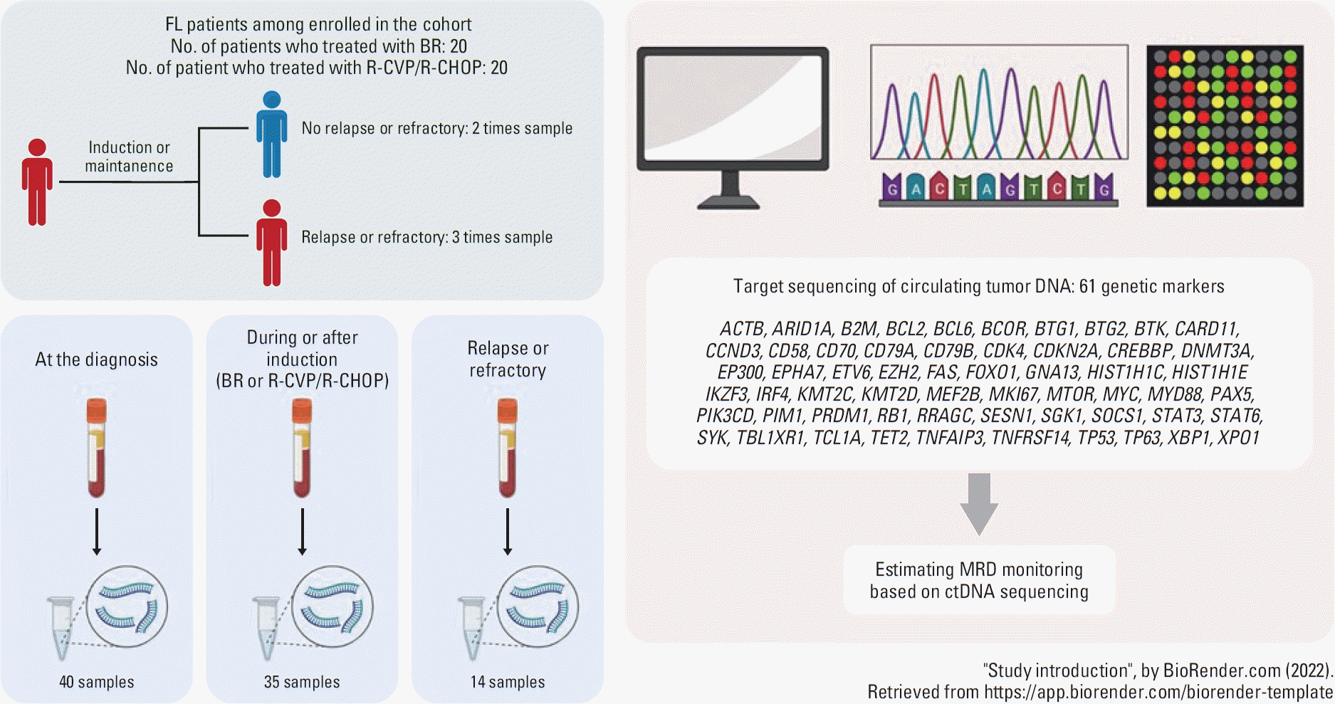
This study analyzed patients with newly diagnosed, grade 1-3A FL in a cohort of lymphoma patients who were prospectively enrolled in our prospective cohort study between April 2017 and February 2020 (NCT03117036). Plasma cellfree DNA (cfDNA) was collected at the time of enrollment, at response assessment, and at relapse after written informed consent was obtained (Fig. 1). Analysis was performed in patients who fulfilled the following criteria: (1) patients who received rituximab-containing immunochemotherapy and rituximab maintenance; (2) patients who had archived plasma cfDNA samples available for ctDNA profiling; (3) patients for whom baseline and follow-up data, including at relapse and survival, were available; (4) patients who were pathologically confirmed with FL grade 1-3A after a review of tumor tissue samples obtained at the time of diagnosis.

2. Case report forms and treatments

The case report form contained patient information and related clinical and laboratory data as follows: Ann Arbor stage, Follicular Lymphoma International Prognostic Index (FLIPI), Eastern Cooperative Oncology Group (ECOG) performance status (PS), B-symptoms, serum lactate dehydrogenase (LDH), β-2 microglobulin (B2M), number of nodal sites, presence of organomegaly, and bone marrow biopsy results [21]. Patients received three kinds of regimen as frontline treatment: BR; R-CHOP (rituximab, cyclophosphamide, vincristine, doxorubicin, and prednisolone); R-CVP (rituximab, cyclophosphamide, vincristine, and prednisolone). According to the Lugano response classification, the treatment response was evaluated by computed tomography (CT) and 18F-fluorodeoxyglucose positron emission tomography CT (PET-CT) [22]. Patients achieving complete (CR) or partial response (PR) to the frontline treatment received rituximab (375 mg/m2 intravenous infusion or 1,400 mg subcutaneous injection) every 2 months for 2 years as maintenance therapy. During rituximab maintenance, surveillance evaluations were performed to monitor disease relapse. The cut-off date for update of disease and survival status was May 31, 2022.

3. cfDNA extraction and library preparation

Whole blood samples were collected in cfDNA (BCT, Streck Inc., Omaha, NE) tubes. Plasma was prepared using three centrifugation steps with increasing centrifugal force. Cell-free nucleic acid was extracted from plasma using a QIAamp Circulating Nucleic Acid Kit (Qiagen, Santa Clarita, CA). Genomic DNA (gDNA) was isolated using a QIAamp DNA Mini Kit (Qiagen). DNA concentrations were quantified using an Infinite M200 Pro NanoQuant (Tecan, Mannedorf, Switzerland) and the fragment size distribution was measured using a 4200 TapeStation Instrument (Agilent Technologies, Santa Clara, CA). An AllPrep DNA/RNA Mini Kit (Qiagen) was used to extract gDNA from formalin-fixed paraffin-embedded tumor tissue. gDNA was quantified and fragmented in the same manner. Purified gDNA was sonicated (7 minutes, 0.5% duty, intensity of 0.1, and 50 cycles/ burst) into 150-200 bp fragments using a Covaris S2 (Covaris Inc., Woburn, MA). gDNA and plasma DNA libraries were created using a KAPA Hyper Prep Kit (Kapa Biosystems, Woburn, MA). For the library construction of plasma cfDNA, hybrid selection was performed using the customized panel. The panel comprised 61 genes and covered 171,988 bp across the human genome (Fig. 1) [23-27].

4. Sequencing data processing

All data were aligned to the hg19 reference using BWA-mem (v0.7.5). GATK v4.0.0 [28], and SAMTOOLS v1.6 [29] were used for base quality recalibration, cross-validation of the UID family, and sorting SAM and BAM files, respectively. After the sequencing data alignment step, discordant paired and off-target sequencing reads were removed. Picard (v2.9.4) was used to group reads into the same UMI families, and then a home-built Python (v2.7.10) script was used for error suppression. The error suppression method was based on a previous study [30].

5. Detection of ctDNA mutations

All bases were subjected to Phred quality filtering using a threshold Q of 30, and only positions where total depths were above 500× were considered for variant identification. To exclude germline mutations in analysis, non-reference alleles present at a frequency greater than 1% in the matched germline DNA were removed. After applying the error suppression method to the sequencing data, the following selection steps were used to eliminate remaining sequencing errors: (1) Variants not significantly greater than the error found in matched germline DNA (Binomial Bonferroni adjusted p-value < 0.01) were filtered out; (2) Variant candidates with a high strand bias (90% if supporting reads ≥ 20; Fisher exact test, p-value < 0.1 if supporting reads < 20) were removed; (3) If the z-statistic of the variant was not significantly higher than the background error obtained from gDNA (Bonferroni adjusted p-value < 0.05), it was excluded from the analysis. Allele frequency ≥ 0.15% and alternative allele count ≥ 5 were selected for candidate mutations, whereas a variant of ≥ 5% was selected for the case of indel. We employed VEP (v102) [31] to perform functional annotation and excluded synonymous mutations from the analysis. The subsequent analyses considered variants that were non-synonymous mutations, including splicing variants. The levels of ctDNA were quantified as genome equivalents (GEs) that were determined as the product of total cfDNA concentration and the maximal variant allele frequency (VAF) of somatic mutations [32]. Tumor mutation burden (TMB) was defined as the total sum of selected variants across the target panel regions after removing errors. The human GE, converted from VAF, was used for clinical interpretation of the detected variants. Furthermore, we classified the patients into high- and low-mutation burdens using the third quantile value of the TMB (< 10 or ≥ 10). For evaluation of concordance between mutation profiles of plasma ctDNA and primary tumor, we have used the archived mutation data from formalin-fixed paraffin-embedded tumor tissue samples where targeted genetic sequencing was performed using the panel, including 405 genes in practice as previously reported [33]. Thus, we compared the mutation profiles of ctDNA with that of primary tumor tissue in those patients with available tumor tissue sequencing data out of the total study population.
Based on ctDNA information, the M7-FLIPI score was estimated with a weighted summation of high-risk FLIPI, ECOG-PS > 1, and occurrence of at least one non-silent mutation among any seven genes (EZH2, ARID1A, MEF2B, EP300, FOXO1, CREBBP, and CARD11) through the calculator presented at the German Low-Grade Lymphoma Study Group official internet site (https://www.german-lymphoma-alliance.de). In addition, the low- and high-risk groups (< 0.8 vs. ≥ 0.8) were defined according to the same cut-off values in the previous study [24].

6. Statistical analysis

Descriptive statistics were determined as proportions and medians, and the intergroup comparisons for categorical variables were assessed by Fisher’s exact test. PFS was calculated as the date from diagnosis to disease progression or death related to any cause. Overall survival (OS) was defined as the period from the date of diagnosis to death or the last date of follow-up. Survival curves were described using Kaplan-Meier estimates and were compared between groups using the log-rank test. Statistical analyses were performed using IBM PASW ver. 25.0 software program (IBM SPSS Inc., Armonk, NY) and survival analysis package in R software (Survival 3.4-0).

Results

1. Characteristics and outcomes of patients

The characteristics of 40 patients are summarized in Table 1. The median age was 52 years (range, 29 to 81 years), and 65.0% (n=26) were less than 60 years old. There were more males (n=25, 62.5%) than females (n=15, 37.5%). Eighty-seven point five percent of patients showed good general condition on ECOG-PS 0 (n=35). Only five patients (12.5%) experienced B-symptoms at diagnosis. In laboratory analysis, 92.5% of patients (n=37) showed elevated LDH levels, and 47.4% (n=18) presented elevated B2M. Twenty-three patients (59.0%) presented with grade 1-2 FL, and 16 (41%) showed 3A FL. Advanced-stage FL was estimated at 82.5% (n=33), high-risk FLIPI showed 65% (n=26), and a high value of m7-FLIPI was documented at 30.0% (n=12). No significant difference in clinical characteristics was observed between the patients who received BR or R-CHOP/R-CVP. The overall response rate (CR and PR) was 92.5% (37/40) and CR rate was 65.0% (26/40). At a median follow-up of 49.2 months (95% confidence interval [CI], 47.0 to 51.4 months), relapse or progression occurred in 14 patients (n=14/40, 35.0%), and eight patients (n=8/14, 57.1%) died due to disease progression. Three of the 14 patients with disease progression were definitively diagnosed with transformed FL (S1 Fig.). The median PFS and OS did not reach the median value (S2 Fig.). Of 14 patients who experienced disease progression, six received the BR regimen, and eight were treated with R-CVP or R-CHOP. Although the follow-up period of patients treated with BR was relatively shorter than R-CVP/R-CHOP, there was no difference in PFS (NR [not reached] vs. 108.6 months, p=0.987) and OS (NR vs. 128.6 months, p=0.921) according to the type of immunochemotherapy (S2 Fig.).

2. Mutation profiles of ctDNA at diagnosis

To detect ctDNA mutations, we performed targeted deep sequencing in plasma cfDNA and white blood cell (WBC) gDNA from 40 patients. The target panel was designed as a pool of RNA baits covering 61 FL-related genes and targeting 171,988 bp of the human genome. The total reads generated from the plasma cfDNA and WBC gDNA samples were 54.5 and 30.7 million reads on average. After filtering unaligned reads and polymerase chain reaction duplicates from sequencing data, the mean of unique sequencing coverage depths of plasma cfDNA and WBC gDNA were 5283.3 and 6172.8 (S3 Table).
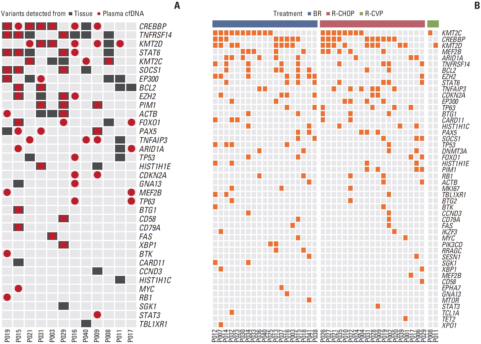
After removing putative false positives according to our statistical criteria, we obtained the list of somatic mutations from plasma cfDNA samples (see Materials and Methods). The mutations of ctDNA were detected in all baseline samples that were obtained at the time of diagnosis (100%, 40/40). The mutation profiles of ctDNA were compared with that of primary tumor tissue in 12 patients who had tissue samples available for analysis. The comparison showed 51.5% (34/66) sensitivity and 50.7% (34/67) positive predictive value of ctDNA of the somatic mutation library from tissue (Fig. 2A). The mutation profiles of cfDNA in 40 patients showed that the most-detected somatic mutation was KMT2C (n=20, 50%), CREBBP (n=18, 45%), KMT2D (n=18, 45%), MEF2B (n=10, 25%), and ARID1A (n=9, 23%) (Fig. 2B). According to the type of frontline treatment, there was no significant difference among mutation profiles. The frequency of M7-FLIPI genes was as follows: CREBBP (n=18, 45%), ARID1A (n=9, 23%), MEF2B (n=10, 25%), EZH2 (n=8, 20%), EP300 (n=6, 15%), CARD11 (n=5, 13%), and FOXO1 (n=4, 10%) (Fig. 2B).
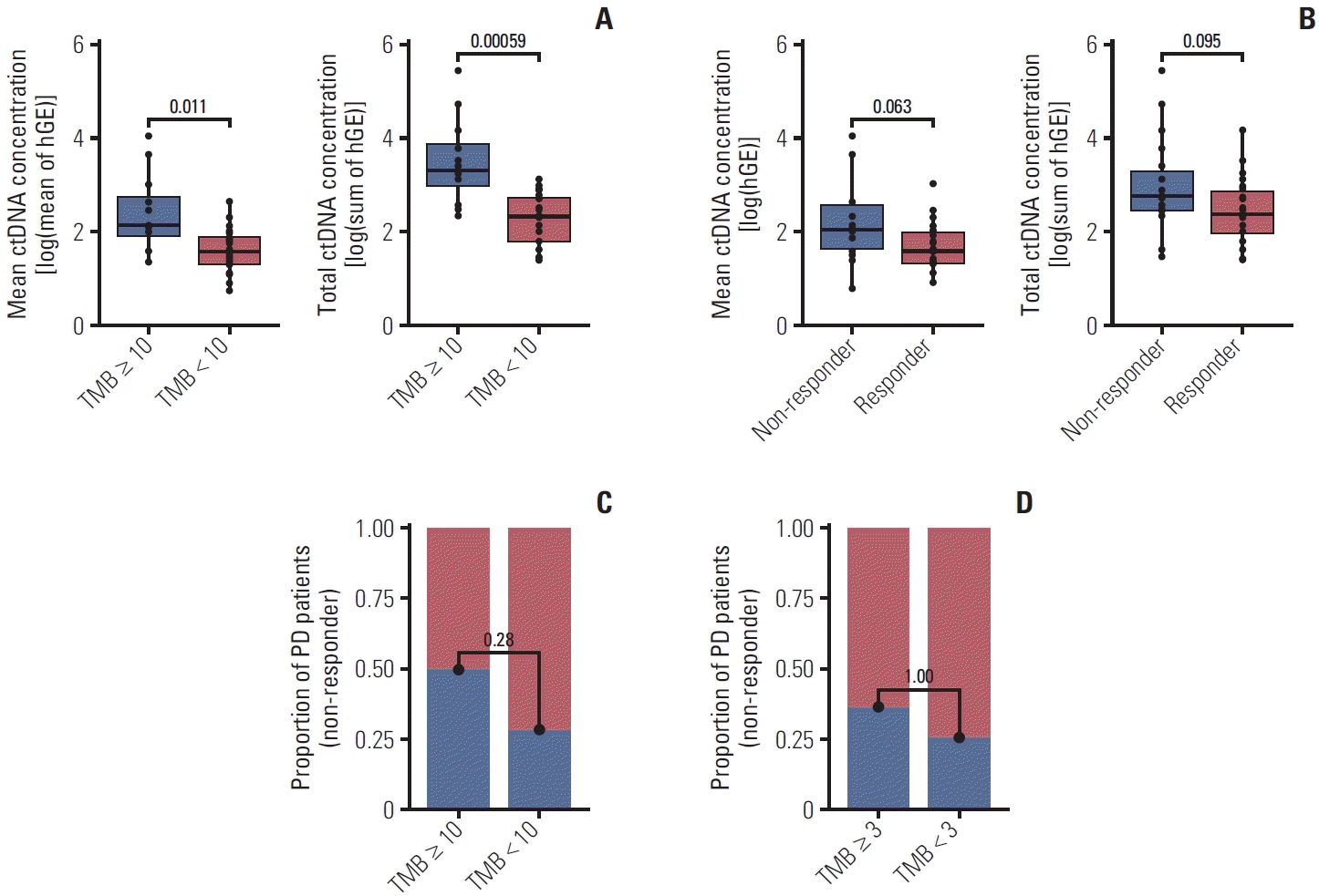
The median ctDNA concentration in the 40 patients was 2.50 human GEs per mL (hGE/mL) (range, 1.38 to 5.45 hGE/mL), and the mean total ctDNA concentration was estimated at 2.62 hGE/mL (2.62±0.88 hGE/mL). Thus, we dichotomized patients into low- and high-ctDNA groups based on the median (< 2.50 hGE/mL or ≥ 2.50 hGE/mL), mean values (< 2.62 hGE/mL or ≥ 2.62 hGE/mL) of ctDNA. Moreover, we classified the patients into high- and low-mutation burdens using the third quantile value of TMB (< 10 or ≥ 10) (Table 2). The ctDNA concentrations were significantly associated with TMB (p < 0.001) (Fig. 3A). However, the ctDNA concentrations were unrelated to the treatment response (Fig. 3B and C). Furthermore, the mutation burdens of seven genes in m7-FLIPI were not different between responders and nonresponders to frontline treatment (p > 0.999) (Fig. 3D).

3. Mutations of ctDNA and survival outcome

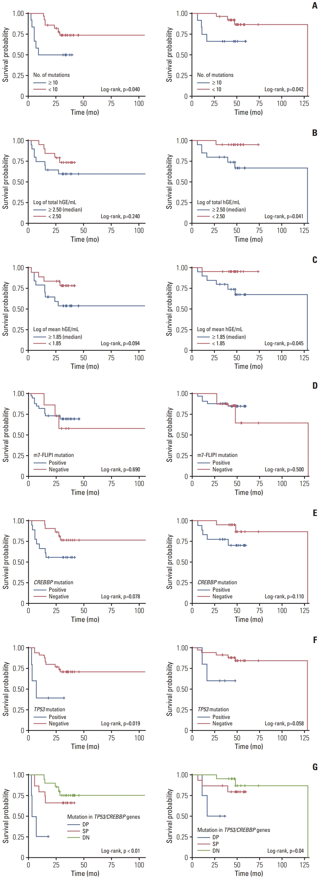
The patients with high TMB (≥ 10) showed shorter PFS (p=0.040) and OS (p=0.042) than patients with low TMB (< 10) (Fig. 4A). The patients with high median ctDNA concentration (≥ 2.50 hGE/mL) were associated with poor OS (p=0.041), but not with PFS (p=0.240) (Fig. 4B). Likewise, patients with high mean ctDNA concentration (≥ 2.62 hGE/mL) showed a poor OS (p=0.045), but not PFS (p=0.094) (Fig. 4C). However, the survival outcomes in patients who had the mutational profile on m7-FLIPI were not affected in terms of PFS (p=0.690) and OS (p=0.500) (Fig. 4D).
Among seven genes of M7-FLIPI (EZH2, ARID1A, MEF2B, EP300, FOXO1, CREBBP, and CARD11), only those with a CREBBP mutation showed a tendency to poor PFS (p=0.078) without an association with OS (p=0.110) (Fig. 4E). When we analyzed the association of mutations of each gene in our panel, TP53 mutation was significantly related with worse PFS (p=0.019), and it also showed a tendency to poor OS (p=0.058) (Fig. 4F). In addition, patients having both mutations of CREBBP and TP53 had a tendency to earlier disease progression than those patients without them, regardless of frontline treatment regimens (Fig. 4G). Thus, the mutations of CREBBP and TP53 were correlated with POD24 in the univariate (HR, 3.983; 95% CI, 1.655 to 9.586; p=0.002) and multivariate (HR, 2.433; 95% CI, 0.628 to 9.419; p=0.011) analysis (S4 Table). The CREBBP gene was found in 18 of the 40 patients (45%), and TP53 was found in five patients (13%). When we compared the mutation profiles between ctDNA and tumor tissue in 12 patients who had tissue samples available, CREBBP had a sensitivity of 33.0%, but due to the small sample size in this study, it is difficult to determine the tissue mutation concordance rate for TP53.

4. Longitudinal analysis of ctDNA

Longitudinal analysis of patients achieving CR showed a decrease in ctDNA concentration at the time of CR; mutations detected at diagnosis disappeared at CR in the P016, P020, and P031 (Fig. 5A). On the other hand, patients with relapse or progression showed an increase or reappearance of mutations (Fig. 5B). Of the 14 patients who exhibited disease progression throughout the entire follow-up period, samples were analyzed in 13 cases, except for a patient who had experienced disease progression recently. When we analyzed the dynamics of ctDNA mutations, a patient showed a reappearance of ctDNA mutations that were detected at diagnosis (P002). This patient eventually relapsed during rituximab maintenance. By contrast, in two patients (P011 and P014) who failed to respond to their frontline treatment, the mutations of ctDNA either remained at the same level or increased at the time of disease progression. The longitudinal clonal evolution mutation profiles of the remaining ten patients are shown in S5 Fig., with the exclusion of the three patients presented.

Discussion

In this study, we analyzed ctDNA mutations profiles using serially collected plasma cfDNA from newly diagnosed FL patients to explore the feasibility of ctDNA mutations as a biomarker for the management of patients. The sequencing of plasma cfDNA with baseline samples identified ctDNA mutations in all cases, and high baseline TMB (≥ 10) correlated with higher ctDNA concentration and poor PFS. Among various genes where mutations were detected, the presence of CREBBP and TP53 mutations were associated with the POD24, implying poor prognosis of FL. In a previous study, a TP53 mutation was reported to be associated with poor prognosis in FL patients although the frequency was around 10%- 20% [27]. The frequency of mutations in CREBBP is around 60%-70% and known to be related to the genomic evolution in tumor cells [24,26]. Thus, our findings suggest that the treatment strategy for FL patients could be modified according to the ctDNA mutation profiles including TP53 and CREBBP mutations at diagnosis. In addition, we evaluated the role of ctDNA mutations as a tool for disease monitoring. Although the number of analyzed patients was small, the persistence or reappearance of ctDNA mutations correlated with disease relapse or progression in PET-CT (Fig. 5). This result suggests there is a potential role for ctDNA mutations as a tool for disease prognosis and monitoring in FL patients.
To our knowledge, this is the first study to evaluate the feasibility of ctDNA mutations in newly diagnosed Asian FL patients who received immunochemotherapy and rituximab maintenance. All patients were from our prospective cohort, and serially collected plasma cfDNA samples were made available to us for analysis. Nevertheless, our results should be interpreted with caution due to several limitations. First, there were limitations in the sensitivity and specificity of detecting ctDNA mutations reflecting the mutation profiles of primary tumors in our study because the concordance of mutations between tumor tissue and ctDNA was confirmed in only half of the genes examined (Fig. 2). This low rate of concordance in our study might be associated with the following issues. First, we only compared the mutation profiles of ctDNA with mutations of primary tumor tissue in 12 patients out of 40 patients due to lack of available data for analysis because we only had 12 primary tumor tissue samples for sequencing. Considering that mutations in ctDNA were detected in all baseline samples at diagnosis (100%, 40/40), we might be able to provide more robust data if we could perform the comparison between blood ctDNA and tumor tissue in all patients. Moreover, we focused on patients who underwent rituximab maintenance after induction immunochemotherapy out of all patients with FL, which might also influence the relatively small number of tissue samples in our study population. Secondly, the limitations of our methodology could lead to this result because our panel included a limited number of genes. The targeted sequencing of tumor tissue was conducted in clinical practice using the panel with more than 400 genes at our hospital. We used the data for comparison with that of known ctDNA mutation profiles. However, the number of genes in our ctDNA analysis was much smaller than that of LymphomaSCAN because our study used only 61 genes to detect ctDNA mutations, including essential genes of M7-FLIPI. In our previous studies evaluating the role of ctDNA mutation profiles in various subtypes of NHL including diffuse large B-cell lymphoma and peripheral T-cell lymphomas, the restricted range of genes available for the analysis has been the limitation [34-37]. Furthermore, the analytical performance of our platform might have limitations in the detection of ctDNA mutations. Indeed, the concordance rate increased in our recent study for natural killer/T-cell lymphoma using the different platform with more than 160 genes [38]. Therefore, the feasibility of ctDNA analysis in the clinical application could be influenced by the performance of analytical platforms and the number of genes included in the panel.
In conclusion, the analysis of ctDNA mutations in plasma at diagnosis and during follow-up might help to both predict outcome and monitor disease status of patients with advanced FL considering its potential as a tool for predicting the occurrence of relapse as well as large-cell transformation earlier than it becomes clinically evident. However, there are technical limitations in the low sensitivity of ctDNA analysis results due to heterogeneous mutational presentations. The feasibility of ctDNA measurement should be further improved before it becomes an appropriate and clinically relevant test in FL patients.

Electronic Supplementary Material

Supplementary materials are available at Cancer Research and Treatment website (https://www.e-crt.org).

Notes

Ethical Statement

This study was approved by the Institutional Review Board of Samsung Medical Center (approval number: 2016-11-040-019). It was conducted in accordance with the ethical principles of the Declaration of Helsinki and the Korea Good Clinical Practice guidelines. All participants provided written informed consent before taking part in the study.

Author Contributions

Conceived and designed the analysis: Yoon SE, Shin SH, Kim WS, Kim SJ.

Collected the data: Yoon SE, Cho J, Kim WS, Kim SJ.

Contributed data or analysis tools: Yoon SE, Shin SH, Cho J, Kim SJ.

Performed the analysis: Yoon SE, Shin SH, Nam DK, Kim SJ.

Wrote the paper: Yoon SE, Shin SH, Kim SJ.

Conflicts of Interest

Conflict of interest relevant to this article was not reported.

ACKNOWLEDGMENTS

This research was supported by grants provided by National Research Foundation of Korea funded by the Korean government (NRF-2021R1A2C1007531; 2022R1F1A1064058).

References

1. Swerdlow SH, Campo E, Pileri SA, Harris NL, Stein H, Siebert R, et al. The 2016 revision of the World Health Organization classification of lymphoid neoplasms. Blood. 2016; 127:2375–90.
crossref
2. Teras LR, DeSantis CE, Cerhan JR, Morton LM, Jemal A, Flowers CR. 2016 US lymphoid malignancy statistics by World Health Organization subtypes. CA Cancer J Clin. 2016; 66:443–59.
crossref
3. Dreyling M, Ghielmini M, Rule S, Salles G, Vitolo U, Ladetto M, et al. Newly diagnosed and relapsed follicular lymphoma: ESMO Clinical Practice Guidelines for diagnosis, treatment and follow-up. Ann Oncol. 2016; 27:v83–90.
crossref
4. Hiddemann W, Kneba M, Dreyling M, Schmitz N, Lengfelder E, Schmits R, et al. Frontline therapy with rituximab added to the combination of cyclophosphamide, doxorubicin, vincristine, and prednisone (CHOP) significantly improves the outcome for patients with advanced-stage follicular lymphoma compared with therapy with CHOP alone: results of a prospective randomized study of the German Low-Grade Lymphoma Study Group. Blood. 2005; 106:3725–32.
crossref
5. Marcus R, Imrie K, Solal-Celigny P, Catalano JV, Dmoszynska A, Raposo JC, et al. Phase III study of R-CVP compared with cyclophosphamide, vincristine, and prednisone alone in patients with previously untreated advanced follicular lymphoma. J Clin Oncol. 2008; 26:4579–86.
crossref
6. Rummel MJ, Niederle N, Maschmeyer G, Banat GA, von Grunhagen U, Losem C, et al. Bendamustine plus rituximab versus CHOP plus rituximab as first-line treatment for patients with indolent and mantle-cell lymphomas: an openlabel, multicentre, randomised, phase 3 non-inferiority trial. Lancet. 2013; 381:1203–10.
7. Schulz H, Bohlius JF, Trelle S, Skoetz N, Reiser M, Kober T, et al. Immunochemotherapy with rituximab and overall survival in patients with indolent or mantle cell lymphoma: a systematic review and meta-analysis. J Natl Cancer Inst. 2007; 99:706–14.
crossref
8. Marcus R, Davies A, Ando K, Klapper W, Opat S, Owen C, et al. Obinutuzumab for the first-line treatment of follicular lymphoma. N Engl J Med. 2017; 377:1331–44.
crossref
9. Flinn IW, van der Jagt R, Kahl BS, Wood P, Hawkins TE, Macdonald D, et al. Randomized trial of bendamustine-rituximab or R-CHOP/R-CVP in first-line treatment of indolent NHL or MCL: the BRIGHT study. Blood. 2014; 123:2944–52.
crossref
10. Bachy E, Seymour JF, Feugier P, Offner F, Lopez-Guillermo A, Belada D, et al. Sustained progression-free survival benefit of rituximab maintenance in patients with follicular lymphoma: long-term results of the PRIMA study. J Clin Oncol. 2019; 37:2815–24.
crossref
11. Casulo C, Byrtek M, Dawson KL, Zhou X, Farber CM, Flowers CR, et al. Early relapse of follicular lymphoma after rituximab plus cyclophosphamide, doxorubicin, vincristine, and prednisone defines patients at high risk for death: an analysis from the National LymphoCare Study. J Clin Oncol. 2015; 33:2516–22.
crossref
12. Seymour JF, Marcus R, Davies A, Gallop-Evans E, Grigg A, Haynes A, et al. Association of early disease progression and very poor survival in the GALLIUM study in follicular lymphoma: benefit of obinutuzumab in reducing the rate of early progression. Haematologica. 2019; 104:1202–8.
crossref
13. Wagner-Johnston ND, Link BK, Byrtek M, Dawson KL, Hainsworth J, Flowers CR, et al. Outcomes of transformed follicular lymphoma in the modern era: a report from the National LymphoCare Study (NLCS). Blood. 2015; 126:851–7.
crossref
14. Freeman CL, Kridel R, Moccia AA, Savage KJ, Villa DR, Scott DW, et al. Early progression after bendamustine-rituximab is associated with high risk of transformation in advanced stage follicular lymphoma. Blood. 2019; 134:761–4.
crossref
15. Yoon SE, Cho J, Kim WS, Kim SJ. Impact of transformation on the survival of patients diagnosed with follicular lymphoma that progressed within 24 months. J Cancer. 2021; 12:2488–97.
crossref
16. Diaz LA Jr, Bardelli A. Liquid biopsies: genotyping circulating tumor DNA. J Clin Oncol. 2014; 32:579–86.
crossref
17. Camus V, Viennot M, Lequesne J, Viailly PJ, Bohers E, Bessi L, et al. Targeted genotyping of circulating tumor DNA for classical Hodgkin lymphoma monitoring: a prospective study. Haematologica. 2021; 106:154–62.
crossref
18. Lakhotia R, Melani C, Dunleavy K, Pittaluga S, Saba N, Lindenberg L, et al. Circulating tumor DNA predicts therapeutic outcome in mantle cell lymphoma. Blood Adv. 2022; 6:2667–80.
crossref
19. Herrera AF, Tracy S, Croft B, Opat S, Ray J, Lovejoy AF, et al. Risk profiling of patients with relapsed/refractory diffuse large B-cell lymphoma by measuring circulating tumor DNA. Blood Adv. 2022; 6:1651–60.
crossref
20. Freedman A, Jacobsen E. Follicular lymphoma: 2020 update on diagnosis and management. Am J Hematol. 2020; 95:316–27.
crossref
21. Solal-Celigny P, Roy P, Colombat P, White J, Armitage JO, Arranz-Saez R, et al. Follicular lymphoma international prognostic index. Blood. 2004; 104:1258–65.
crossref
22. Cheson BD, Fisher RI, Barrington SF, Cavalli F, Schwartz LH, Zucca E, et al. Recommendations for initial evaluation, staging, and response assessment of Hodgkin and non-Hodgkin lymphoma: the Lugano classification. J Clin Oncol. 2014; 32:3059–68.
crossref
23. Tobin JW, Keane C, Gunawardana J, Mollee P, Birch S, Hoang T, et al. Progression of disease within 24 months in follicular lymphoma is associated with reduced intratumoral immune infiltration. J Clin Oncol. 2019; 37:3300–9.
crossref
24. Pastore A, Jurinovic V, Kridel R, Hoster E, Staiger AM, Szczepanowski M, et al. Integration of gene mutations in risk prognostication for patients receiving first-line immunochemotherapy for follicular lymphoma: a retrospective analysis of a prospective clinical trial and validation in a population-based registry. Lancet Oncol. 2015; 16:1111–22.
crossref
25. Carbone A, Roulland S, Gloghini A, Younes A, von Keudell G, Lopez-Guillermo A, et al. Follicular lymphoma. Nat Rev Dis Primers. 2019; 5:83.
crossref
26. Green MR. Chromatin modifying gene mutations in follicular lymphoma. Blood. 2018; 131:595–604.
crossref
27. O’Shea D, O’Riain C, Taylor C, Waters R, Carlotti E, Macdougall F, et al. The presence of TP53 mutation at diagnosis of follicular lymphoma identifies a high-risk group of patients with shortened time to disease progression and poorer overall survival. Blood. 2008; 112:3126–9.
crossref
28. McKenna A, Hanna M, Banks E, Sivachenko A, Cibulskis K, Kernytsky A, et al. The Genome Analysis Toolkit: a MapReduce framework for analyzing next-generation DNA sequencing data. Genome Res. 2010; 20:1297–303.
crossref
29. Li H. A statistical framework for SNP calling, mutation discovery, association mapping and population genetical parameter estimation from sequencing data. Bioinformatics. 2011; 27:2987–93.
crossref
30. Newman AM, Lovejoy AF, Klass DM, Kurtz DM, Chabon JJ, Scherer F, et al. Integrated digital error suppression for improved detection of circulating tumor DNA. Nat Biotechnol. 2016; 34:547–55.
crossref
31. McLaren W, Gil L, Hunt SE, Riat HS, Ritchie GR, Thormann A, et al. The Ensembl variant effect predictor. Genome Biol. 2016; 17:122.
32. Avanzini S, Kurtz DM, Chabon JJ, Moding EJ, Hori SS, Gambhir SS, et al. A mathematical model of ctDNA shedding predicts tumor detection size. Sci Adv. 2020; 6:eabc4308.
crossref
33. Cho J, Yoon SE, Kim SJ, Ko YH, Kim WS. Comparison of tumor mutation burden of 300 various non-Hodgkin lymphomas using panel based massively parallel sequencing. BMC Cancer. 2021; 21:972.
crossref
34. Shin SH, Kim YJ, Lee D, Cho D, Ko YH, Cho J, et al. Analysis of circulating tumor DNA by targeted ultra-deep sequencing across various non-Hodgkin lymphoma subtypes. Leuk Lymphoma. 2019; 60:2237–46.
crossref
35. Yoon SE, Kim YJ, Shim JH, Park D, Cho J, Ko YH, et al. Plasma circulating tumor DNA in patients with primary central nervous system lymphoma. Cancer Res Treat. 2022; 54:597–612.
crossref
36. Hur JY, Kim YJ, Yoon SE, Son DS, Park WY, Kim SJ, et al. Plasma cell-free DNA is a prognostic biomarker for survival in patients with aggressive non-Hodgkin lymphomas. Ann Hematol. 2020; 99:1293–302.
crossref
37. Kim SJ, Kim YJ, Yoon SE, Ryu KJ, Park B, Park D, et al. Circulating tumor DNA-based genotyping and monitoring for predicting disease relapses of patients with peripheral T-cell lymphomas. Cancer Res Treat. 2023; 55:291–303.
crossref
38. Kim JJ, Kim HY, Choi Z, Hwang SY, Jeong H, Choi JR, et al. In-depth circulating tumor DNA sequencing for prognostication and monitoring in natural killer/T-cell lymphomas. Front Oncol. 2023; 13:1109715.
crossref

Fig. 1.
Overview of study design: design for sample collection. Target genes for mutation profiling. BR, bendamustine and rituximab; ctDNA, circulating tumor DNA; FL, follicular lymphoma; MRD, minimal residual disease; RCHOP, rituximab, cyclophosphamide, vincristine, doxorubicin, and prednisolone; RCVP, cyclophosphamide, vincristine, and prednisolone.
crt-2023-869f1.tif
Fig. 2.
Comparison of mutations according to sample types and treatments (A). Concordance of somatic mutations between tissue and plasma cell-free DNA (cfDNA) samples (n=12). (B) The landscape of somatic mutation detected in plasma cfDNA samples from 40 patients, with mutational profiles grouped according to their treatments: BR (bendamustine plus rituximab; n=19), R-CHOP (rituximab, cyclophosphamide, vincristine, doxorubicin, and prednisolone; n=19), and R-CVP (cyclophosphamide, vincristine, and prednisolone; n=2).
crt-2023-869f2.tif
Fig. 3.
Relationship between circulating tumor DNA (ctDNA) concentration, tumor mutation burden (TMB) (A), and treatment response (B). Comparison of response rates according to TMB high and low group across entire targeted 61 genes (C) and the region covered by only 7-FLIPI (Follicular Lymphoma International Prognostic Index) genes (D). hGE, human genome equivalents; PD, progression of disease.
crt-2023-869f3.tif
Fig. 4.
Progression-free survival (left panel) and overall survival (right panel) according to tumor mutation burden (A), total circulating tumor DNA concentration (B), mean ctDNA concentration (C), absence of mutation on 7-FLIPI (Follicular Lymphoma International Prognostic Index) genes (EZH2, ARID1A, MEF2B, EP300, FOXO1, CREBBP, and CARD11) (D) and TP53/CREBBP gene mutation (E-G). DN, double negative; DP, double negative; hGE, human genome equivalents; SP, single positive.
crt-2023-869f4.tif
Fig. 5.
Monitoring circulating tumor DNA (ctDNA) concentrations of patients in therapeutic response (A) and disease relapse (B) groups. The chemotherapeutic history is displayed above each monitoring plot. The patients have their positron emission tomography computed tomography (PET-CT) results at nearby time points and the cell-free DNA (cfDNA) sampling and response were classified using Deauville criteria (which is an internationally recommended scale in PET-CT assessment). BR, bendamustine and rituximab; R, rituximab; RB, rituximab, bendamustine; R-CHOP, rituximab, cyclophosphamide, vincristine, doxorubicin, and prednisolone; R-CVP, cyclophosphamide, vincristine, and prednisolone; hGE, human genome equivalent.
crt-2023-869f5.tif
Table 1.
Baseline patients’ characteristics (n=40)
Baseline value Total (n=40) BR (n=20) R-CHOP/R-CVP (n=20) p-value
Age (yr)
 < 60 26 (65.0) 14 (53.8) 12 (46.2) 0.333
 ≥ 60 14 (35.0) 5 (35.7) 9 (64.3)
Sex
 Male 25 (62.5) 11 (44.0) 14 (56.0) 0.745
 Female 15 (37.5) 8 (53.3) 7 (46.7)
ECOG PS
 0 35 (87.5) 16 (45.7) 19 (54.3) 0.654
 ≥ 1 5 (12.5) 3 (60.0) 2 (40.0)
B-symptom
 Absence 35 (87.5) 17 (45.9) 20 (54.1) 0.596
 Presence 5 (12.5) 2 (66.7) 1 (33.3)
Anemia (g/dL)
 ≤ 12 11 (27.5) 4 (36.4) 7 (63.6) 0.488
 > 12 29 (72.5) 15 (51.7) 14 (48.3)
LDH (≥ 200 U/L)
 Normal 3 (7.5) 3 (100) 0 0.098
 Elevated 37 (92.5) 16 (43.2) 21 (56.8)
B2M (n=38)
 Normal 20 (52.6) 11 (55.0) 9 (45.0) 0.746
 Elevated 18 (47.4) 8 (44.4) 10 (55.6)
Grade (n=39)
 1-2 23 (59.0) 14 (60.9) 9 (39.1) 0.105
 3a 16 (41.0) 5 (31.3) 11 (68.8)
Stage
 I/II 7 (17.5) 4 (57.1) 3 (42.9) 0.689
 III/IV 33 (82.5) 15 (45.5) 18 (54.5)
FLIPI
 0-2 14 (35.0) 9 (64.3) 5 (35.7) 0.186
 ≥ 3 26 (65.0) 10 (38.5) 16 (61.5)
m7-FLIPI
 Low (< 0.8) 28 (70.0) 16 (57.1) 12 (42.9) 0.089
 High (≥ 0.8) 12 (30.0) 3 (25.0) 9 (75.0)
Splenomegaly
 Absence 34 (85.0) 16 (47.1) 18 (52.9) > 0.99
 Presence 6 (15.0) 3 (50.0) 3 (50.0)
No. of extranodal
 0 16 (40.0) 6 (37.5) 10 (62.5) 0.349
 ≥ 1 24 (60.0) 13 (54.2) 11 (45.8)
BM involvement
 Absence 20 (50.0) 10 (50.0) 10 (50.0) > 0.99
 Presence 20 (50.0) 9 (45.0) 11 (55.0)

Values are presented as number (%). B2M, β-2 microglobulin; BM, bone marrow; BR, bendamustine plus rituximab; ECOG PS, Eastern Cooperative Oncology Group performance status; FLIPI, Follicular Lymphoma International Prognostic Index; LDH, serum lactate dehydrogenase; R-CHOP, rituximab, cyclophosphamide, vincristine, doxorubicin, and prednisolone; R-CVP, cyclophosphamide, vincristine, and prednisolone.

Table 2.
patient characteristics according to hGE and TMB
Baseline value hGE median
hGE mean
TMB
< 2.50 (n=20) ≥ 2.50 (n=20) p-value < 2.62 (n=22) ≥ 2.62 (n=18) p-value < 10 (n=28) ≥ 10 (n=12) p-value
Age (yr)
 < 60 16 (61.5) 10 (38.5) 0.096 18 (69.2) 8 (30.8) 0.021 20 (76.9) 6 (23.1) 0.281
 ≥ 60 4 (28.6) 10 (71.4) 4 (28.6) 10 (71.4) 8 (57.1) 6 (42.9)
Sex
 Male 11 (44.0) 14 (56.0) 0.514 12 (48.0) 13 (52.0) 0.332 20 (80.0) 5 (20.0) 0.091
 Female 9 (60.0) 6 (40.0) 10 (66.7) 5 (33.3) 8 (53.3) 7 (46.7)
ECOG PS
 0 17 (48.6) 18 (51.4) > 0.99 18 (51.4) 17 (48.6) 0.355 24 (68.6) 11 (31.4) > 0.99
 ≥ 1 3 (60.0) 2 (40.0) 4 (80.0) 1 (20.0) 4 (80.0) 1 (20.0)
B-symptom
 Presence 3 (100) 0 0.231 3 (100) 0 0.238 3 (100) 0 0.541
Anemia
 ≤ 12 g/dL 6 (54.5) 5 (45.5) > 0.99 6 (54.5) 5 (45.5) > 0.99 6 (54.5) 5 (45.5) 0.254
LDH
 Normal 2 (66.7) 1 (33.3) > 0.99 3 (100) 0 0.238 1 (33.3) 2 (66.7) 0.209
 Elevated 18 (48.6) 19 (51.4) 19 (51.4) 18 (48.6) 27 (73.0) 10 (27.0)
B2M (n=38)
 Normal 12 (60.0) 8 (40.0) 0.330 13 (65.0) 7 (35.0) 0.328 15 (75.0) 5 (25.0) 0.489
 Elevated 7 (38.9) 11 (61.1) 8 (44.4) 10 (55.6) 11 (61.1) 7 (38.9)
Grade (n=38)
 1-2 11 (47.8) 12 (52.2) > 0.99 12 (52.2) 11 (47.8) > 0.99 16 (69.6) 7 (30.4) > 0.99
 3a 8 (50.0) 8 (50.0) 9 (56.3) 7 (43.8) 11 (68.8) 5 (31.3)
Stage
 I/II 5 (71.4) 2 (28.6) 0.407 5 (71.4) 2 (28.6) 0.427 7 (100) 0 0.081
 III/IV 15 (45.5) 18 (54.5) 17 (51.5) 16 (48.5) 21 (63.6) 12 (36.4)
FLIPI
 0-2 10 (71.4) 4 (28.6) 0.096 11 (78.6) 3 (21.4) 0.046 12 (85.7) 2 (14.3) 0.157
 ≥ 3 10 (38.5) 16 (61.5) 11 (42.3) 15 (57.7) 16 (61.5) 10 (38.5)
Splenomegaly
 Presence 3 (50.0) 3 (50.0) > 0.99 4 (66.7) 2 (33.3) 0.673 3 (50.0) 3 (50.0) 0.341
BM involvement
 Presence 9 (45.0) 11 (55.0) 0.752 10 (50.0) 10 (50.0) 0.751 12 (60.0) 8 (40.0) 0.301
Chemotherapy
 BR 9 (47.4) 10 (52.6) > 0.99 11 (57.9) 8 (42.1) 0.761 12 (63.2) 7 (36.8) 0.494
 R-CHOP or R-CVP 11 (52.4) 10 (47.6) 11 (52.4) 10 (47.6) 16 (76.2) 5 (23.8)

Values are presented as number (%). B2M, β-2 microglobulin; BM, bone marrow; BR, bendamustine and rituximab; ECOG PS, Eastern Cooperative Oncology Group performance status; FLIPI, Follicular Lymphoma International Prognostic Index; hGE, human genome equivalents; LDH, serum lactate dehydrogenase; R-CHOP, rituximab, cyclophosphamide, vincristine, doxorubicin, and prednisolone; R-CVP, cyclophosphamide, vincristine, and prednisolone; TMB, tumor mutation burden.

TOOLS
Similar articles