Abstract
Purpose
This study investigated the incidence of secondary malignancy in multiple myeloma (MM) patients compared with that in the general population using a population-based database covering all residents in Korea.
Materials and Methods
Based on the national health insurance system in Korea, all people primarily diagnosed with MM between January 1, 2010 to December 31, 2018 were identified. A total of 9,985 MM patients aged ≥ 20 years in Korea were included.
Results
Among them, 237 (2.4%) developed secondary malignancies by 2018. The standardized incidence rates (SIRs) of all secondary malignancies in MM patients were 0.87 (95% confidence interval [CI], 0.76 to 0.98), with a higher incidence of hematologic malignancies than in the general population with an SIR of 3.80 (95% CI, 2.61 to 5.00). The incidence rates of both lymphoid malignancy (SIR, 3.56; 95% CI, 2.31 to 4.82) and myeloid malignancy (SIR, 3.78; 95% CI, 1.16 to 6.39) were higher in MM patients than in the general population. In contrast, a lower incidence of solid cancer was observed in MM patients than in the general population (SIR, 0.76, 95% CI, 0.65 to 0.86). There was no significant difference in survival in MM patients without secondary malignancies, with hematologic malignancy, and with solid cancer (p=0.413).
Conclusion
MM patients had a greater risk of secondary malignancies, especially hematologic malignancies, than the general population. Future studies with a focus on analyzing patients’ history, treatment details, and genetic information in various stages of MM patients are needed to better understand the mechanism behind this increased risk.
Multiple myeloma (MM) is a disorder of plasma cells that accounted for 0.9% of all cancer incident cases and 1.2% of cancer deaths in 2020 [1]. The treatment and prognosis of MM have shown remarkable progress compared to other types of cancer, especially in young patients [2,3]. The relative 5-year survival rate for patients with MM increased from 0.28 in the 1970s to 0.41 in the 21st century with the introduction of novel immunomodulatory agents and hematopoietic stem cell transplantation (HSCT) [4]. As survival has improved, the development of chronic diseases in MM patients, such as secondary primary malignancies (SPM), has emerged as a new clinical challenge.
The powerful anticancer effect of a medication is one of the most important factors to consider when making decisions for the treatment of malignancies. However, the potent efficacy of treatment modalities is a double-edged sword due to their simultaneous control of current malignancies and damage to other normal cellular systems. Especially for diseases that most patients get cured of and survive long, such as Hodgkin lymphoma and pediatric acute lymphoblastic leukemia, judicious choice of treatment for balancing cancer cure and adverse effect are one of the important topics. In standard-risk myeloma, the expected median overall survival is approximately 10 years with stem cell transplantation and various novel agents [5]. In diseases such as MM, in which the expected median survival, despite its incurable nature, is relatively longer compared to other aggressive malignancies, investigating later events of the treatment journey matters because of longer exposure to the various agents. The possible occurrence of SPM after or during MM treatment should be a concern for physicians. For this reason, meticulous analysis of the incidence rate and survival impact of SPM in patients with MM is imperative.
According to the review of the International Myeloma Working Group in 2017, the risk of SPM in MM patients has not increased as compared to the incidence rate from the population cancer registry [6]. When observing stratified cancer types, hematologic second malignancies, including myelodysplastic syndromes, acute myeloid leukemia, and nonHodgkin lymphoma, generally increased in MM patients, whereas the risk of solid secondary cancer has shown inconsistent results [6]. However, most studies have been conducted in Western countries, with a few studies conducted in Asian countries. In addition, many studies are not recent and include MM patients diagnosed as far back as 2010 [6]. However, the few recent reviews that have been conducted demonstrated similar findings as previous reviews: a similar decreased risk of secondary solid cancer and an increased risk of hematologic SPM have been observed in MM patients [5,7].
Despite the lower incidence of MM in Asian countries as compared to that in Western countries, an increased pattern of MM incidence has been more prominent in Asian countries, and the highest increment has been observed in men aged 50 years or more in Korea [8]. Recently, a few studies on SPM have been reported in Taiwan [9,10], but the data on the Asian population remains limited. Therefore, this study investigated the incidence of SPM in MM patients as compared to that in the general population in Korea using a population-based database covering all residents in Korea.
In Korea, a single and mandatory national health insurance system (NHIS) provides coverage for health care utilization for the prevention, diagnosis, treatment, prescription, and rehabilitation of diseases for the population. We used the NHIS - National Health Information Database (NHID) in Korea to identify all MM incident cases between January 1, 2010 to December 31, 2018. Healthcare utilization and sociodemographic information (such as sex, birth year, residential area, income percentile, and occupation) were obtained from the NHIS-NHID [11].
To define MM cases, the International Classification of Diseases 10 (ICD-10) code C90.0, which indicates MM and malignant plasma cell neoplasms, was applied in combination with the code for rare and intractable diseases code to represent cancer. The rare and intractable diseases code is a special code in the NHIS to reduce the economic burden for diseases with high medical expenses by reducing copayment; thus, the accuracy of cancer identification in combination with ICD-10 and rare and intractable diseases code is high [12]. The ICD-10 has been applied since 1995. Therefore, cancer identification, including MM, was not affected by the revision of the disease code. To identify people with a primary diagnosis of MM, the year 2009 was set as the wash-out period, and individuals who had utilized healthcare due to any cancer diagnosis or had received anticancer treatments before the date of the first diagnosis of MM were excluded. To exclude malignant plasma cell neoplasms other than MM, we excluded patients with codes other than C90.0. The codes for exclusion included plasma cell leukemia (C90.1), extramedullary plasmacytoma (C90.2), and solitary plasmacytoma (C90.3). In addition, patients with C90.1 within two months of the date they were first diagnosed with MM were also excluded [13]. Treatment for MM with autologous HSCT was defined by the codes used for surgical procedures the NHIS-NHID. If MM patients had a surgical code for autologous HSCT after their first diagnosis of MM, they were defined as individuals who had received autologous HSCT (SCT group); otherwise, they were defined as individuals who did not receive autologous HSCT (non-SCT group).
The incidence of SPM after a diagnosis of MM was defined by the ICD-10 codes for new malignancy (Any C codes) as the primary diagnostic code 180 days after their diagnosis of MM and more than one hospitalization and outpatient visit within 1 year after their first date of a diagnosis of a SPM. The malignancies within 180 days of a diagnosis of MM were considered co-prevalent with MM. SPMs were classified as hematologic malignancies [ICD-10 codes of C81-C96, except for MM (C90.0)] and solid malignancies (ICD-10 codes of C00-C80). The types of solid malignancies were further classified as gastrointestinal cancer (C15-C20), head and neck cancer (C01-C13, C30-C32, and C73), hepatobiliary cancer (C22-C25), breast cancer (C50), genitourinary cancer (C61-C67), gynecological cancer (C53), musculoskeletal and soft tissue cancer (C41, C44, and C49), thoracic cancer (C34, C37, and C78), or central nervous system cancer (C71). The types of hematologic malignancies were classified into either lymphoid malignancy (other hematologic malignancies other than myeloid cancer) or myeloid malignancy (C92). Lymphoid malignancies included Hodgkin lymphoma (C81), follicular lymphoma (C82), non-follicular lymphoma (C83), mature T/natural killer (NK)-cell lymphomas (C84), other and unspecified types of non-Hodgkin lymphoma (C85), other specified types of T/NK-cell lymphoma (C86), lymphoid leukemia (C91), leukemia of unspecified cell type (C95), and other and unspecified malignant neoplasms of lymphoid, hematopoietic and related tissue (C96). The myeloid malignancy (C92) includes acute myeloblastic leukemia (C92.0), chronic myeloid leukemia (C92.2, C92.3), myeloid sarcoma (C92.4), acute promyelocytic leukemia (C92.5), acute myelomonocytic leukemia (C92.6), acute myeloid leukemia with 11q23-abnormality (C92.7), other myeloid leukemia (C92.8), acute myeloid leukemia with multilineage dysplasia (C92.9), and myeloid leukemia unspecified. The time to onset of the SPM was calculated from the MM index date and the first date of the diagnosis of a SPM. Myeloproliferative neoplasms without C-code by ICD-10 were not included.
Demographic and clinical characteristics of all MM patients with and without the development of SPM after a diagnosis of MM were compared using chi-square statistics. The considered characteristics included sex, age group (20-49, 50-59, 60-69, and ≥ 70 years), year of the diagnosis of MM, quartile of income, Charlson comorbidity index (CCI) score, and comorbidities associated with MM. The CCI score was identified based on codes of primary and secondary diagnoses using the ICD-10 diagnostic coding system proposed by Quan et al. [14,15], which had occurred 12 months before the date of MM diagnosis, as described in a previous study [15]. The weight for the CCI was an original weight ranging from 1-6 [16]. Having primary and secondary diagnoses of renal injury or renal failure, anemia, bone fracture, or recurrent bacterial infections based on ICD-10 code within 12 months prior to the date of a diagnosis of MM was defined as comorbidities associated with MM.
The SPM incidence rate in MM patients was presented as per 100,000 person-years. The follow-up person-years were calculated from the date of the diagnosis of MM to the date of the diagnosis of a SPM defined as above, date of death, or December 31, 2018, whichever came first. The SPM incidence rate of MM patients between 2010 and 2018 was compared with that of the Korea Central Cancer Registry during the same period through indirect standardization adjusted for sex and the 5-year age group. The Korea Central Cancer Registry is a population-based national cancer registry covering more than 96% of all cancers developed in Korea. The standardized incidence rate (SIR) for all SPMs, hematologic malignancy, solid cancer, and site-specific cancers was presented using the following equation, and 95% confidence intervals (CIs) for the SIRs were estimated based on the Poisson distribution. Through standardization, adjustment for age and sex distribution between patients with MM and general population was performed.
A comparison of cancer incidence rate with the general population was performed after stratification by sex and autologous HSCT status. SPM incidence rate by the year in which MM was diagnosed (2010-2013 and 2014-2018) was considered. In addition, the survival probability of all patients without SPM development, and MM patients with secondary solid cancer and hematologic malignancies was compared by the Kaplan-Meier curve and log-rank test. All analyses were performed separately for the two sexes. SAS software ver. 9.4 (SAS Institute Inc., Cary, NC) was used for all statistical analyses.
A total of 9,985 MM patients aged ≥ 20 years diagnosed between 2010 and 2018 in Korea were included. Of them, 237 (2.4%) developed SPM by December 31, 2018 (Table 1). Among patients with MM with SPM development, the proportion of males, earlier diagnosis, and highest income quartile were higher compared to patients with MM without SPM development. The proportion of males was 65.4% and 53.0% in patients with MM with and without SPM development (p < 0.001). Patients diagnosed in the earlier period and with the highest income quartile level accounted for a higher proportion of MM patients with SPM (p < 0.001 and p=0.004). The proportion of age group, CCI score, and presence of coexisting myeloma-defining events, including renal failure, anemia, and bone fracture, were not significantly different in MM patients with or without SPM.
Table 2 shows the SPM incidence rate and SIR of malignancy in MM patients compared with those in the general Korean population. With follow-up of 22,890.3 years (mean follow-up of 2.3 years), the crude SPM incidence rate was 1,035.4 (95% CI, 904.3 to 1,166.5). SPM comprised 39 hematologic malignancies (incidence rate of 170.4; 95% CI, 117.0 to 223.8) and 198 solid cancers (incidence rate of 860.5; 95% CI, 745.0 to 985.0). Gastrointestinal cancer (205.3/100,000 person-years), followed by hepatobiliary cancer (139.8/100,000 person-years), lymphoid malignancy (135.4/100,000 person-years), thoracic cancer (135.4/100,000 person-years), and genitourinary cancer (118.0/100,000 person-years) were the five most common incident cancers in MM patients.
The SIRs of all SPM in MM patients were 0.87 (95% CI, 0.76 to 0.98; p=0.020), showing lower cancer incidence in MM patients compared with those of the general population (Table 2). However, MM patients showed a higher incidence of hematologic malignancies than that in the general population with an SIR of 3.80 (95% CI, 2.61 to 5.00; p < 0.001). The incidence rate of both lymphoid malignancy (SIR, 3.56; 95% CI, 2.31 to 4.82; p < 0.001) and myeloid malignancy (SIR, 3.78; 95% CI, 1.16 to 6.39; p=0.038) were higher in MM patients than the general population. In contrast, a lower incidence of solid cancer was observed in MM patients than in the general population (SIR, 0.76; 95% CI, 0.65 to 0.86; p < 0.001). The SIR of site-specific solid cancer showed a lower incidence for most types of cancer in MM patients than in the general population.
When stratified by sex or autologous HSCT treatment, the SPM incidence pattern in patients with MM compared with the general population was comparable. Stratified by sex, the SIRs of hematologic SPM, especially lymphoid malignancies, also were higher in patients with MM than the general population in both men and women with SIR of 3.26 (95% CI, 1.75 to 4.77; p=0.003) and 3.74 (95% CI, 1.71 to 5.78; p=0.008), respectively (S1 Table). In myeloid malignancies, SIR was 4.19 (95% CI, 0.83 to 7.53; p=0.062) in men and 2.56 (95% CI, 0.00 to 6.12; p=0.389) in women were not statistically significant due to very small number of cases. Most solid tumors SIRs were lower in both sex groups. Similarly, patients with MM without receiving autologous HSCT in men was higher for hematologic SPM (SIR, 3.28; 95% CI, 1.91 to 4.66; p=0.001 for lymphoid; SIR, 4.33; 95% CI, 1.12 to 7.53; p=0.042 for myeloid). However, in female patients with MM who received autologous HSCT, the SIR of hematologic SPM was not statistically significant due to a very small number of cases (S2 Table).
The change of incidence rate of SPM by the year in which MM was diagnosed (2010-2013 and 2014-2018) is shown in Table 3. Despite of similar overall incidence rate (1,076.7 per 100,000 person-year in 2010-2013 and 985.7 per 100,000 person-years in 2014-2018), the incidence rate of most sites of solid cancer was higher in MM patients diagnosed in 2010-2013 than those diagnosed in 2014-2018. Otherwise, the incidence rate of hematologic malignancies, especially lymphoid malignancy, was much higher in patients with MM diagnosed in 2014-2018 compared with those diagnosed in 2010-2013, despite relatively shorter duration of chance for cancer development.
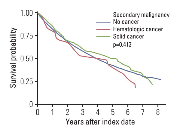
When comparing survival probability in patients with MM according to SPM development (Fig. 1), the median survival time in patients without SPM, with hematologic SPM, and solid SPM was 3.92 years, 4.60 years, and 5.25 years. The 8-year survival rate was estimated to be 27.2%, 16.6%, and 21.5% in patients with MM without SPM, with hematologic SPM, and solid SPM, and there were no significant differences in survival rates between the three groups (p=0.413).
In this research, we analyzed a total of 9,985 Korean MM patients diagnosed between 2010 and 2018 and found 237 SPM cases (2.4%) based on the NHIS-NHID. To the best of our knowledge, this is the largest Asian population-based study on SPM following the diagnosis of MM. SIRs of secondary hematologic malignancies were higher in patients with MM than in the general population in both lymphoid and myeloid malignancies. Among solid tumors, SIRs of secondary gastrointestinal tract malignancies, head and neck cancers, and breast cancer were significantly lower in MM patients. In sum, the total SIR of SPM compared with the general population was lower in MM patients. The survival difference between patients with SPM and without SPM was not observed.
The abovementioned findings of our study are consistent results with previous reports. In large number of datasets researches, using California Cancer Registry data, Rosenberg et al. [17] reported among a total of 16,331 MM patients, a 1.3% increased risk of hematologic SPM for 10 years after diagnosis, but not of solid tumors in MM patients who underwent autologous HSCT. Among previous studies analyzed Asian patients, Tzeng et al. [9] investigated 3,970 MM patients in Taiwan and reported an 11-fold greater incidence of hematologic malignancies and a 2-fold lower incidence of solid tumors in MM patients compared to those in the 15,880 compared other cancer patients without MM. More recently, Liu et al. [10] reported 1.33% of the cumulative SPM incidence in 14 years of follow-up data from 4,327 MM patients using the Taiwan National Cancer Registry and National Health Insurance Research databases.
In our result, SIR of SPM showed similar patterns in patients who did not receive autologous HSCT and patients who received autologous HSCT (S2 Table). Several studies have indicated that long-term exposure to alkylators such as oral melphalan in patients with MM might be a relevant risk factor for SPM, and autologous HSCT with alkylating agents could be a risk factor for SPM in patients with lymphoma [5,18,19]. However, the incidence of SPM after autologous HSCT in MM reportedly did not increase in previous studies [6]. These findings support that the risk of SPM from once or twice exposure to high-dose intravenous melphalan for autologous HSCT in patients with MM should not be an obstacle in making clinical decisions.
Among patients with the development of SPM, the proportion of males, highest income quartile, and patients diagnosed with MM earlier in the study period was higher. Longer time lags and follow-up periods after a diagnosis of MM could be more opportunities for the occurrence of SPM. However, in hematologic SPM, patients who were diagnosed with MM between the years 2014 and 2018 had a higher incidence rate than those who were diagnosed at the earlier period. This finding could be explained by various factors. MM has shown a trend of increasing incidence rate globally and in the Korean National registry data analysis, especially in men older than 50 years [8,20]. Extended survival duration and complexity of treatment as numerous novel agents’ development could provide more opportunities of arising SPM. As more novel drugs and anti-tumor mechanisms have been discovered, more complicated disturbances of the normal immune milieu and damages to the DNA repair system, and to the tumor microenvironment and malignant altered genes are being reported, which could be possible causes of SPM [5]. Furthermore, more cumulation of anti-myeloma drugs and longer extension of total treatment duration including immune-modulating drug (IMiD) maintenance therapy could facilitate the growth of SPM.
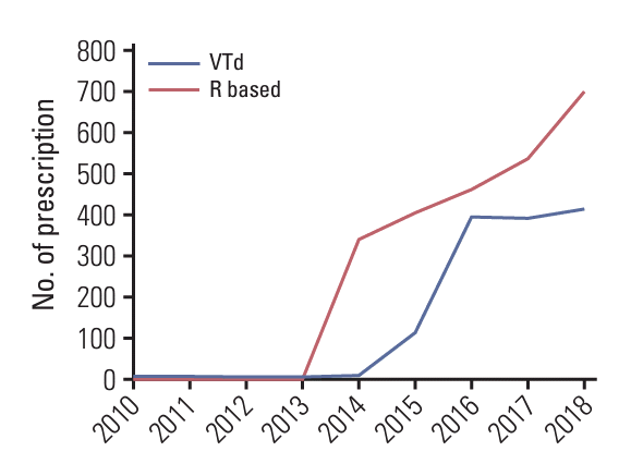
Among IMiDs, lenalidomide has been indicated consistently as a possible contributor to SPM development in several large-scale randomized studies and meta-analyses [5,20-24]. In our research, in the same context, lenalidomide might be an attributable factor for SPM because lenalidomide had been introduced to the clinical practice with being covered by NHIS in Korea since the year 2014. Since 2014, the exposure to lenalidomide has dramatically increased in Korea (Fig. 2). Before 2014, the number of prescriptions of lenalidomide was less than 10 but steeply increased to 338 prescriptions in 2014 and continuously increased. Patients with the highest upper quartile income presented more development of SPM. Although the efficacy of lenalidomide for relapsed or refractory MM was ascertained in 2007 [22], before the year 2014, except in clinical trials, many patients with lower income might have had difficulties affording lenalidomide-based treatment, which eventually might have led to less exposure to lenalidomide.
As in Fig. 1, we could not identify the differences in survival between patients with MM with SPM and patients with MM without SPM. A recent study from the American Society of Clinical Oncology (ASCO) CancerLinq MM real-world data registry reported that longer median overall survival was observed in patients with MM subsequently diagnosed with an SPM [25]. This result is consistent with our findings. In the study by Cooper et al., secondary malignancy site-specific survival in patients with MM compared with patients with MM without secondary malignancy was presented. According to the result, improved survival was observed in patients with MM who developed non-melanoma cutaneous cancers, breast cancer, prostate cancer, non-Hodgkin lymphoma, diffuse large B cell lymphoma, and melanoma as secondary cancer. Survival was not different in patients with MM with colorectal cancer, acute lymphoblastic leukemia, or chronic lymphocytic leukemia compared with patients with MM without secondary cancer. Survival was worse in patients with MM diagnosed with acute myeloid leukemia and lung cancer compared with patients with MM without secondary cancer [25]. Considering the shorter survival time of patients with MM, those who survived longer could develop secondary malignancy; thus, the survival probability would not be different.
Increased incidence of SPM without survival disadvantages has been consistently observed in many previous studies [8,15,16,18,20,22]. Although lenalidomide therapy is associated with SPM development in MM, survival outcomes of patients who had SPM were not inferior to those of patients who did not. The survival gain from lenalidomide in various stages of MM has been clearly demonstrated in numerous phase III randomized control trials [22,26-29]. Although lenalidomide is one of the most used novel agents for MM, the survival benefit is greater than the risk of SPM-related death as the survival benefit of autologous HSCT outweighs the risk of death from SPM [17,19].
This study has several limitations. Due to the nature of national registry data, the association between SPM development and exact anti-myeloma treatment intensity and schedule could not be evaluated. However, our data could indicate the annual trend of SPM according to the practice pattern change such as the introduction of lenalidomide. Patient selection bias could exist because we excluded patients who presented with malignancies other than MM within 6 months after the diagnosis of MM to sort out true SPM from pre-existing but undetectable malignancies. As such, patients who died within 6 months after the diagnosis of MM, implying that the disease progressed rapidly and the patients were fragile, could not be included in the survival analysis. However, we excluded plasma cell leukemia from the analysis to generalize patients and applied the CCI score to tell the fragility. Data on familial history, environmental factors and lifestyle associated with carcinogenesis, and genetic information could not be collected. Due to the small number of SPM by each cancer type, the cancers were grouped into a wide range for SIR estimation and as hematologic and solid cancer for survival probability. Because MM has wide heterogenetic molecular features and dynamic evolution of mutation through the developmental stages [16], future studies are needed to discover genetic alterations causing SPMs by combining large-scale registry data for each type of cancer and detailed genetic information.
In conclusion, MM patients had a higher risk of SPM, especially hematologic malignancies. However, the survival differences were not observed between patients with SPM and those without. Even though the survival benefit of recently developed treatment has been proven in various stages of MM, as the treatment indication of novel agents (including lenalidomide) has been extended to high-risk smoldering myeloma and maintenance therapy after autologous HSCT, the cumulative dose and duration of exposure will increase more. The potential risk of SPM in anti-myeloma treatment should be one of the major concerns for physicians. Consensus or guidelines for surveillance may be needed for SPM, especially lymphoid malignancies. Future studies are warranted converging patients’ history, treatment details, and genetic information in various stages of MM.
Electronic Supplementary Material
Supplementary materials are available at Cancer Research and Treatment website (https://www.e-crt.org).
Notes
Ethical Statement
This study was approved by the Institutional Review Board of the National Cancer Center, Korea (approval No. NCC2019-0238). The requirement for informed consent was waived because individual identities were already anonymized in the NHIS-NHID, and the NHIS-NHID was utilized after the IRB approval.
References
1. Sung H, Ferlay J, Siegel RL, Laversanne M, Soerjomataram I, Jemal A, et al. Global cancer statistics 2020: GLOBOCAN estimates of incidence and mortality worldwide for 36 cancers in 185 countries. CA Cancer J Clin. 2021; 71:209–49.
2. Turesson I, Velez R, Kristinsson SY, Landgren O. Patterns of improved survival in patients with multiple myeloma in the twenty-first century: a population-based study. J Clin Oncol. 2010; 28:830–4.
3. Thomas A, Mailankody S, Korde N, Kristinsson SY, Turesson I, Landgren O. Second malignancies after multiple myeloma: from 1960s to 2010s. Blood. 2012; 119:2731–7.
4. Thorsteinsdottir S, Dickman PW, Landgren O, Blimark C, Hultcrantz M, Turesson I, et al. Dramatically improved survival in multiple myeloma patients in the recent decade: results from a Swedish population-based study. Haematologica. 2018; 103:e412–5.
5. Maclachlan K, Diamond B, Maura F, Hillengass J, Turesson I, Landgren CO, et al. Second malignancies in multiple myeloma: emerging patterns and future directions. Best Pract Res Clin Haematol. 2020; 33:101144.
6. Musto P, Anderson KC, Attal M, Richardson PG, Badros A, Hou J, et al. Second primary malignancies in multiple myeloma: an overview and IMWG consensus. Ann Oncol. 2017; 28:228–45.
7. Poh C, Keegan T, Rosenberg AS. Second primary malignancies in multiple myeloma: a review. Blood Rev. 2021; 46:100757.
8. Huang J, Chan SC, Lok V, Zhang L, Lucero-Prisno DE 3rd, Xu W, et al. The epidemiological landscape of multiple myeloma: a global cancer registry estimate of disease burden, risk factors, and temporal trends. Lancet Haematol. 2022; 9:e670–7.
9. Tzeng HE, Lin CL, Tsai CH, Tang CH, Hwang WL, Cheng YW, et al. Time trend of multiple myeloma and associated secondary primary malignancies in Asian patients: a Taiwan population-based study. PLoS One. 2013; 8:e68041.
10. Liu Y, Hou HA, Qiu H, Tang CH. Is the risk of second primary malignancy increased in multiple myeloma in the novel therapy era? A population-based, retrospective cohort study in Taiwan. Sci Rep. 2020; 10:14393.
11. Lee J, Lee JS, Park SH, Shin SA, Kim K. Cohort profile: The National Health Insurance Service-National Sample Cohort (NHIS-NSC), South Korea. Int J Epidemiol. 2017; 46:e15.
12. Yang MS, Park M, Back JH, Lee GH, Shin JH, Kim K, et al. Validation of cancer diagnosis based on the National Health Insurance Service Database versus the National Cancer Registry Database in Korea. Cancer Res Treat. 2022; 54:352–61.
13. Tang CH, Liu HY, Hou HA, Qiu H, Huang KC, Siggins S, et al. Epidemiology of multiple myeloma in Taiwan, a population based study. Cancer Epidemiol. 2018; 55:136–41.
14. Quan H, Sundararajan V, Halfon P, Fong A, Burnand B, Luthi JC, et al. Coding algorithms for defining comorbidities in ICD-9-CM and ICD-10 administrative data. Med Care. 2005; 43:1130–9.
15. Kim KH. Comparative study on three algorithms of the ICD-10 Charlson comorbidity index with myocardial infarction patients. J Prev Med Public Health. 2010; 43:42–9.
16. Charlson ME, Pompei P, Ales KL, MacKenzie CR. A new method of classifying prognostic comorbidity in longitudinal studies: development and validation. J Chronic Dis. 1987; 40:373–83.
17. Rosenberg AS, Brunson A, Tuscano J, Jonas BA, Hoeg R, Wun T, et al. Effect of autologous hematopoietic stem cell transplant on the development of second primary malignancies in multiple myeloma patients. Blood Cancer J. 2021; 11:5.
18. Miller JS, Arthur DC, Litz CE, Neglia JP, Miller WJ, Weisdorf DJ. Myelodysplastic syndrome after autologous bone marrow transplantation: an additional late complication of curative cancer therapy. Blood. 1994; 83:3780–6.
19. Palumbo A, Bringhen S, Kumar SK, Lupparelli G, Usmani S, Waage A, et al. Second primary malignancies with lenalidomide therapy for newly diagnosed myeloma: a meta-analysis of individual patient data. Lancet Oncol. 2014; 15:333–42.
20. Lee H, Park HJ, Park EH, Ju HY, Oh CM, Kong HJ, et al. Nationwide statistical analysis of lymphoid malignancies in Korea. Cancer Res Treat. 2018; 50:222–38.
21. Saleem K, Franz J, Klem ML, Yabes JG, Boyiadzis M, Jones JR, et al. Second primary malignancies in patients with haematological cancers treated with lenalidomide: a systematic review and meta-analysis. Lancet Haematol. 2022; 9:e906–18.
22. Dimopoulos M, Spencer A, Attal M, Prince HM, Harousseau JL, Dmoszynska A, et al. Lenalidomide plus dexamethasone for relapsed or refractory multiple myeloma. N Engl J Med. 2007; 357:2123–32.
23. Wang J, Lv C, Zhou M, Xu JY, Chen B, Wan Y. Second primary malignancy risk in multiple myeloma from 1975 to 2018. Cancers (Basel). 2022; 14:4919.
24. Jones JR, Cairns DA, Gregory WM, Collett C, Pawlyn C, Sigsworth R, et al. Second malignancies in the context of lenalidomide treatment: an analysis of 2732 myeloma patients enrolled to the Myeloma XI trial. Blood Cancer J. 2016; 6:e506.
25. Cooper JD, Thornton JA, Gibson SJ, Pham K, Sunderland K, DeStefano CB. Survival of patients with multiple myeloma diagnosed with second primary malignancies: an ASCO Cancerlinq analysis. Blood. 2022; 140(Suppl 1):10039–40.
26. Lonial S, Jacobus S, Fonseca R, Weiss M, Kumar S, Orlowski RZ, et al. Randomized trial of lenalidomide versus observation in smoldering multiple myeloma. J Clin Oncol. 2020; 38:1126–37.
27. Cavo M, Gay F, Beksac M, Pantani L, Petrucci MT, Dimopoulos MA, et al. Autologous haematopoietic stem-cell transplantation versus bortezomib-melphalan-prednisone, with or without bortezomib-lenalidomide-dexamethasone consolidation therapy, and lenalidomide maintenance for newly diagnosed multiple myeloma (EMN02/HO95): a multicentre, randomised, open-label, phase 3 study. Lancet Haematol. 2020; 7:e456–68.
Table 1.
Table 2.
Cancer site | Incident rate (95% CI)b) | Observed N | Expected N | SIR | p-value |
---|---|---|---|---|---|
All | 1,035.4 (904.3-1,166.5) | 237 | 272.9 | 0.87 (0.76-0.98) | 0.020 |
Hematologic malignancy | 170.4 (117.0-223.8) | 39 | 10.8 | 3.80 (2.61-5.00) | < 0.001 |
Lymphoid malignancy | 135.4 (87.8-183.0) | 31 | 8.7 | 3.56 (2.31-4.82) | < 0.001 |
Myeloid malignancy | 34.9 (10.7-59.1) | 8 | 2.1 | 3.78 (1.16-6.39) | 0.038 |
Solid cancer | 865.0 (745.0-985.0) | 198 | 262.1 | 0.76 (0.65-0.86) | < 0.001 |
Gastrointestinal cancer | 205.3 (146.7-263.9) | 47 | 85.3 | 0.55 (0.39-0.71) | < 0.001 |
Head and neck cancer | 48.1 (19.7-76.5) | 11 | 24.1 | 0.54 (0.25-0.83) | 0.002 |
Hepatobiliary cancer | 139.8 (91.4-188.2) | 32 | 40.6 | 0.79 (0.51-1.06) | 0.129 |
Breast cancer | 13.1 (0.0-27.9) | 3 | 13.2 | 0.23 (0.00-0.49) | < 0.001 |
Genitourinary | 118.0 (73.5-162.5) | 27 | 32.9 | 0.82 (0.51-1.13) | 0.258 |
Gynecology | 8.7 (0.0-20.8) | 2 | 2.5 | 0.80 (0.00-1.91) | 0.729 |
Musculoskeletal and soft tissue | 52.4 (22.8-82.0) | 12 | 9.7 | 1.24 (0.54-1.95) | 0.498 |
Thorax | 135.4 (87.8-183.0) | 31 | 40.6 | 0.76 (0.50-1.03) | 0.086 |
CNS | 4.4 (0.0-13.0) | 1 | 17.6 | 0.06 (0.00-0.17) | < 0.001 |
CI, confidence interval; CNS, central nervous system; SIR, standardized incidence ratio. A total of 30 cases of other solid cancer that could not be classified into specific group were not shown in table. Total follow-up period was 22,890.3 person-years.