Journal List > Obstet Gynecol Sci > v.67(4) > 1516087836

Adamyan, Pivazyan, Obosyan, Krylova, and Isaeva: Preimplantation genetic testing for aneuploidy in patients of different age: a systematic review and meta-analysis

Abstract

This study aimed to summarize the current knowledge on the benefits of In vitro fertilization/intracytoplasmic sperm injection with preimplantation genetic testing for aneuploidy (PGT-A) and to discuss the role of PGT-A in patients of different ages undergoing assisted reproduction. A systematic review was conducted in accordance with the Preferred Reporting Items for Systematic Reviews and Meta-Analyses 2020 checklist. Registration number: CRD42022354697. Studies were identified by searching the PubMed, Cochrane Library, Google Scholar, Scopus, Embase, and ClinicalTrials databases. Seven meta-analyses were performed with additional stratification of age and prognosis of the women studied. Clinical pregnancy rate per embryo transfer in patients aged >35 years was higher in the PGT-A group (P=0.0002) than in controls. Live birth rate (LBR) per embryo transfer in women 35 years old or younger (P=0.002) was higher in the PGT-A group. The LBR per patient in women aged >35 years was higher in the PGT-A group (P=0.004). The effects of PGT-A on LBR in patients with poor prognosis showed a statistically significant increase (P=0.003). There was no significant difference in the rate between the two groups. PGT-A is effective and can be recommended for patients aged >35 years undergoing assisted reproduction to improve their reproductive outcomes. Moreover, our study showed the possible benefits of PGT-A in patients with a poor prognosis. Overall, our findings suggest that PGT-A is a valuable tool for improving the reproductive outcomes of assisted reproductive procedures in older women and those with a history of pregnancy complications.

Introduction

Assisted reproductive technologies (ART) allow for the treatment of most infertile couples with the aim of securing a healthy birth. The success of in vitro fertilization (IVF) cycles depends on various factors and is generally evaluated by implantation efficiency, clinical pregnancy, and live birth. These results are influenced by the ovarian response to stimulation, oocyte quality, embryo culture, transfer selections, and the age of the patient [1].
It is well established that embryonic aneuploidy is prevalent in IVF cycles, especially in women of advanced maternal age (AMA) [2]. Most embryos with an abnormal number of chromosomes are not compatible with life [3], and these fatal genetic defects are responsible for implantation failure and early miscarriage after the transfer of a morphologically good-quality embryo. This prevalence increases with age; the estimated aneuploidy rate increases from 25% for oocytes from women under 35 years old to more than 75% for oocytes from women aged >40 years [4]. On these and many other bases, one of the most important challenges for the embryologist is to discern which embryo is the most appropriate to transfer.
Preimplantation genetic testing (PGT) is a procedure used to identify genetic abnormalities in embryos created by IVF and can be used as a tool to select embryos of good quality for embryo transfer (ET), which, in theory, should improve implantation rates, decrease miscarriage rates (MR), and reduce the time to achieve a successful pregnancy. However, studies that directly compare the outcomes of cycles with and without PGT are scarce. Moreover, there are doubts about the benefits of using PGT for certain pathologies. There are many data points on the benefits of using PGT in a distinctive cohort of patients characterized by AMA, recurrent implantation failure (RIF) [5], recurrent pregnancy loss (RPL), severe male infertility, or elective single ET [6,7]. However, there are many controversies regarding this topic; thus, it needs to be clarified.
Therefore, to fully validate the advantages of PGT for aneuploidy in terms of an increased chance of successful pregnancy and live birth, and to provide an update on the efficacy of PGT for aneuploidy (PGT-A) in clinical outcomes, we aimed to conduct a systematic review in which we summarized all published studies.

Materials and methods

This systematic review was registered with the International Prospective Systematic Review Registry of the National Institute of Health Research (PROSPERO). Protocol and registration number: PROSPERO 2022 CRD42022354697. This systematic review was conducted in accordance with the Preferred Reporting Items for Systematic Reviews and Meta-Analyses (PRISMA) 2020 guidelines for reporting systematic reviews [8]. Institutional Review Board approval was not required because the present study was a review.

1. Study selection

An electronic database search was conducted using PubMed, the Cochrane Library, ClinicalTrials.gov, Scopus, Embase, and Google Scholar. The authors used a combination of the following terms: “preimplantation genetic testing”, “PGT-A”, “pregnancy”, and “live birth”. The last screening date was October 1, 2022.
No restrictions or search filters (publication status, type of article, or language of publication) were applied to verify all potentially relevant studies.
The search strategy for the PubMed electronic database was as follows: using the advanced search builder in PubMed, the following combinations of keywords were used: (preimplantation genetic testing) AND (PGT-A) AND (pregnancy) AND (live birth), and no filters or limits were used.
In addition to PubMed, the Cochrane Library electronic database was searched. The search combinations were as follows: (preimplantation genetic testing) AND (PGT-A) AND (pregnancy); no filters or limits were used.
The electronic databases Scopus and Google Scholar were searched using the following words: ((preimplantation AND genetic AND testing) AND (PGT-A) AND (live AND birth) AND (pregnancy) AND (embryo AND biopsy)).
The search was also conducted in the ClinicalTrials.gov electronic database using an advanced search combination of “preimplantation genetic testing” and “pregnancy”.
Additionally, the search was conducted using MeSH terms in PubMed ((((«Preimplantation Diagnosis”[MeSH]) AND “Pregnancy”[MeSH]) AND “Pregnancy Rate”[MeSH]) AND “Live Birth»[MeSH]) and in the Cochrane Library (MeSH descriptor: [Preimplantation Diagnosis] explode all trees).
The search was conducted independently by three investigators (L.O., E.K., and S.I.) and the search results were saved to a reference manager (Zotero version 6.0.8; Corporation for Digital Scholarship, Fairfax, VA, USA). After searching, all articles were reviewed based on their titles and abstracts. All studies were selected, and each potentially relevant study was obtained in full text and independently assessed for inclusion by the authors. Additionally, a manual search of the references of the articles was performed to identify additional studies of interest. Any disagreements regarding the inclusion or exclusion of the preselected studies and other disagreements during the review process were resolved with the help of a fourth author (L.P.).

2. Eligibility criteria and main outcomes

The inclusion criteria for the present systematic review were: women of reproductive age who underwent assisted reproduction, array comparative genomic hybridization (aCGH) or next-generation sequencing (NGS)-based PGT-A. Fresh embryo transfer was considered in groups without PGT-A intervention.
Studies containing information on other molecular methods used to assess chromosomal content were excluded. The use of aCGH or NGS is recommended by the European Society of Human Reproduction and Embryology (ESHRE) [9].
Randomized and non-randomized clinical trials published in English were included. Papers in languages other than English, case reports, preclinical studies, reviews, opinion articles, and studies published as abstracts were excluded.
The primary analysis aimed to assess the risk ratios of the clinical pregnancy rate (CPR) and live birth rate (LBR). CPR was defined as the number of clinical pregnancies expressed per 100 initiated, aspirated, or embryo transfer cycles. LBR was defined as the number of deliveries resulting in at least one live birth and is expressed per 100 cycle attempts [3].
Secondary analyses evaluated MR, implantation (IR), ongoing pregnancy/live birth, and spontaneous abortion rates. MR is defined as the spontaneous loss of intrauterine pregnancy before 22 weeks of gestational age [3].
The implantation rate was calculated as the number of gestational sacs visualized by transvaginal ultrasonography (number of implanted embryos) divided by the total number of embryos transferred. The ongoing pregnancy/live birth rate was defined as the number of ongoing pregnancies after the presence of a fetal pole with fetal heart tones and/or live births, divided by the total number of embryos transferred.
We grouped these results as “per embryo transfer” and “per patient”. “LBR per embryo transfer” refers to the LBR calculated based on the number of live births per embryo transfer procedure. “LBR per patient” refers to the LBR calculated based on the number of live births per individual patient undergoing fertility treatment. “CPR per embryo transfer” refers to the percentage of ET procedures that result in a clinical pregnancy. “CPR per patient” refers to the overall percentage of patients who achieve a clinical pregnancy following ET. “MR per embryo transfer” refers to the percentage of embryos that resulted in miscarriage following a transfer procedure. “MR per patient” refers to the likelihood of miscarriage in individual patients undergoing fertility treatment.

3. Quality assessment

A risk of bias assessment was performed for each of the included studies using the Cochrane Handbook for Systematic Reviews of Interventions [10]. Three review authors independently evaluated the quality of the selected studies. Any discrepancies between the reviewers were resolved through discussion or consultation with the fourth review author (L.P.).
Following the Cochrane Handbook for Systematic Reviews of Interventions, the risk of bias (RoB) 2 tool [11] was used to assess the risk of bias in randomized controlled studies, and risk of bias in non-randomised studies-of interventions (ROBINS-I) [12] was used for non-randomized studies (prospective controlled, prospective cohort, retrospective studies, and other types of studies). Additionally, these tools were used to assess the risk of bias arising from reporting biases resulting from missing synthesis results.

4. Statistical analysis

For quantitative synthesis, a meta-analysis (forest plot) was performed using the RevMan 5.4. (Cochrane Collaboration, London, UK) (recommended by the Cochrane Society). According to the Cochrane Handbook for Systematic Reviews of Interventions, an I2 value of 0 indicates no observed heterogeneity, whereas I2 values from 30% to 60% represent moderate heterogeneity, I2 values from 50% to 90% represent substantial heterogeneity, and I2 values from 75% to 100% represent considerable heterogeneity. Meta-analyses with heterogeneity greater than 75% were excluded.

Results

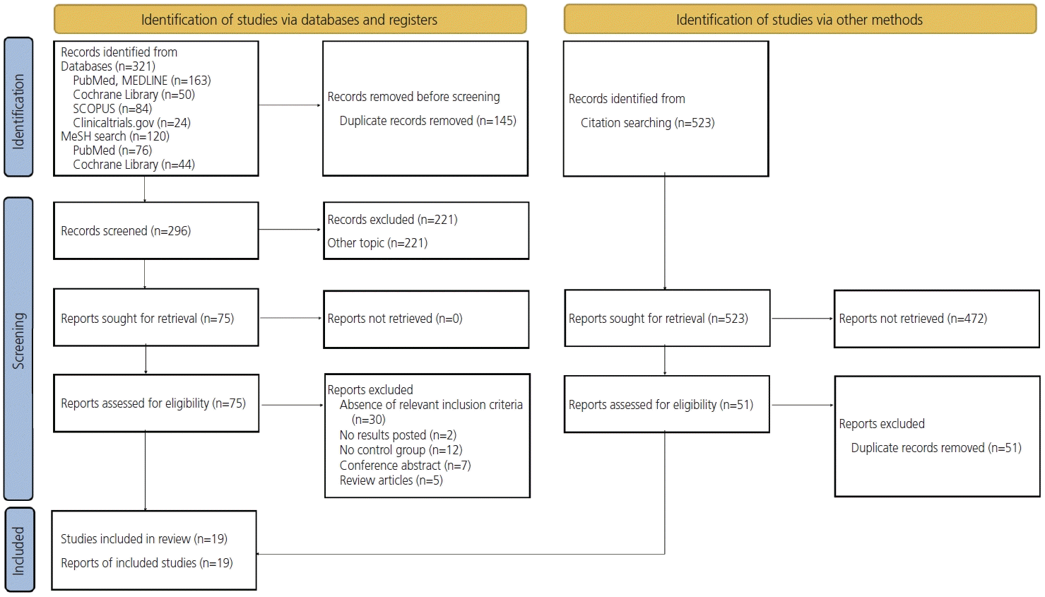
The entire search strategy and results are presented in the flow diagram (Fig. 1). The initial search yielded a total of 321 articles. After a MeSH search, 120 reports were identified: 76 from PubMed and 44 from the Cochrane Library. After removing duplicates and searching the titles and abstracts of the articles, 296 publications were selected. Therefore, 75 reports remained for full-text screening and analysis based on our inclusion criteria.
Fifty-six articles did not meet the inclusion criteria for various reasons, as detailed in Fig. 1, and were excluded from the study. The most common reasons for exclusion were that the article concerned a conference abstract [13-19], absence of relevant inclusion criteria [20-49], absence of published study results [50,51], absence of a control group [52-63], and some studies [64-68] were review articles.
Additionally, 51 articles were found in the references of the 19 articles included in the qualitative analyses, all of which met the eligibility criteria. However, none of these studies were included in the systematic review because they were duplicates found earlier. Therefore, 19 studies [69-87] were retained for qualitative synthesis.
Five publications were randomized studies and 14 were nonrandomized studies. The process of inclusion and exclusion is detailed in the PRISMA flow diagram shown in Fig. 1. The characteristics of the included studies are provided in Table 1. The outcomes of the studies included in the meta-analysis are presented in Table 2.
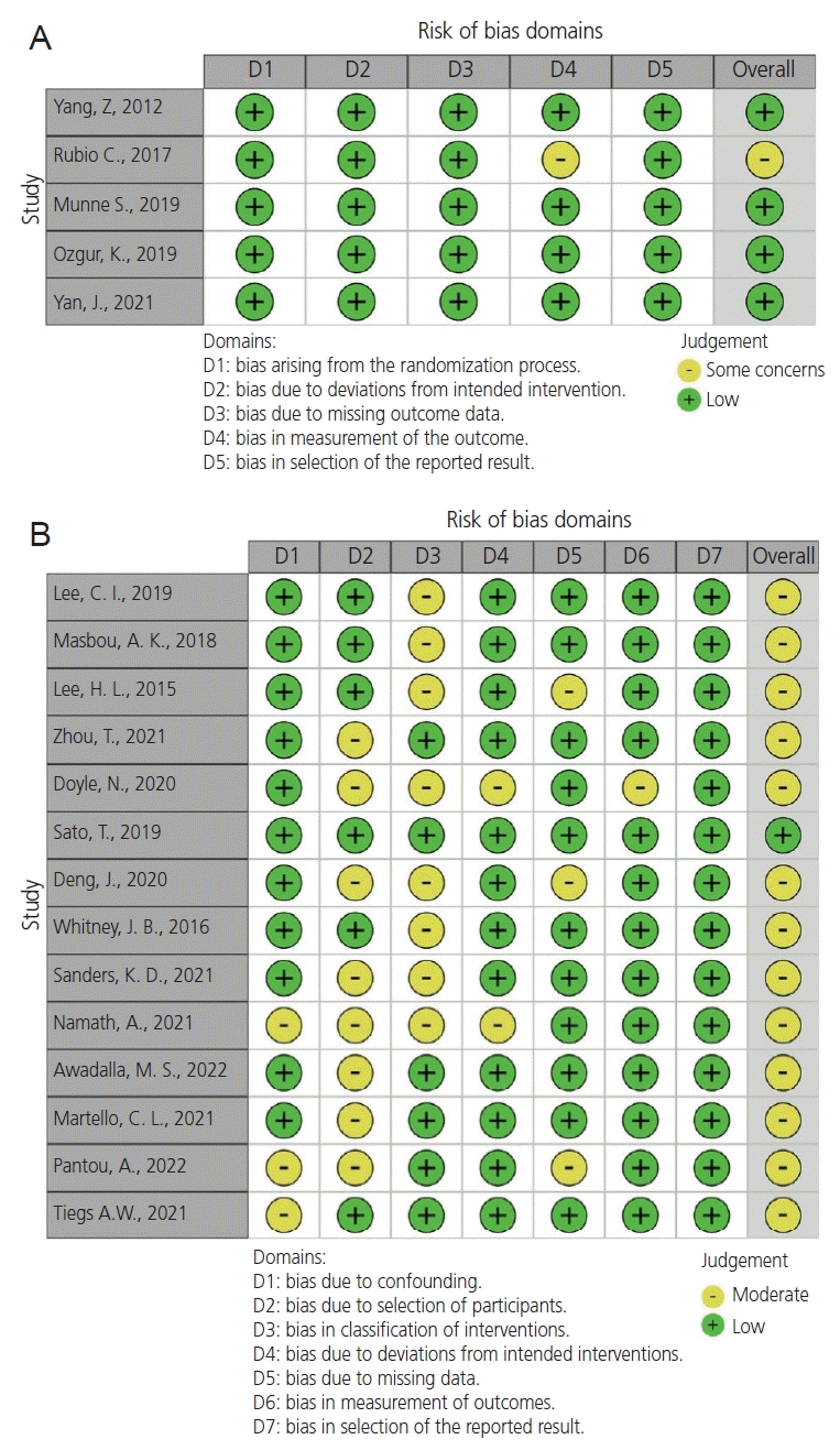
According to the Cochrane Handbook, three reviewers (L.O., E.K., and S.I.) assessed the risk of bias in each of the included studies using RoB 2 for randomized control trials and ROBINS-I for non-randomized trials. Disagreements were resolved through discussion with a fourth author (L.P.)
Visualization tools were created using ROBVIS App (National Insitute of Health Research, Newcastle upon Tyne, UK) [88]. This application created “traffic light” graphs of domain-level judgments for each result and weighted bar graphs of the distribution of risk-of-bias judgments within each bias domain.
Based on these tools, randomized controlled trials had a low risk of bias and nonrandomized trials had a moderate risk of bias (Fig. 2).

1. Clinical pregnancy rates per embryo transfer

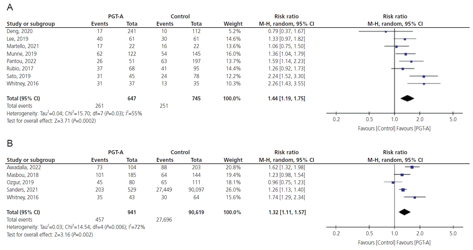
Eight studies reported results on CPR per ET cycle in women aged 35 years or older (risk ratio [RR], 1.44; 95% confidence interval [CI], 1.19-1.75; P=0.0002). Heterogeneity in this comparison was 55% (Fig. 3A). In this group, PGT-A improved CPR.

2. Live birth rates per embryo transfer

Five studies reported results on LBR per ET cycle in women 35 years old or younger (RR, 1.32; 95% CI, 1.11-1.57; P=0.002). The live birth rate per ET improved after PGT-A compared to controls. Heterogeneity in this comparison was 72% (Fig. 3B).

3. Live birth rates per patient

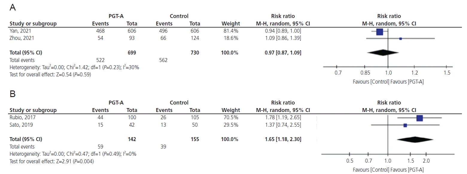
Two studies reported results on LBR per patient in women 38 years of age or younger (RR, 0.97; 95% CI, 0.87-1.09; P=0.59). No significant differences were found between the two groups. Heterogeneity in this comparison was 30% (Fig. 4A).
Two studies reported results on LBR per patient in the over-35-year-old age group; in this comparison, PGT-A improved live birth rates (RR, 1.65; 95% CI, 1.18-2.30; P=0.004). The heterogeneity of this comparison was 0% (Fig. 4B).
Among patients aged <35 years, PGT-A resulted in a higher LBR per ET than in those who did not undergo PGT-A. However, no significant differences were observed in the number of live births per patient in this age group. In patients aged >35 years, PGT-A improved live birth rates compared to those without PGT-A. Overall, PGT-A appears to have a more positive effect on live birth outcomes in older patients.

4. Effect of PGT-A on the live birth rate in patients with a poor prognosis

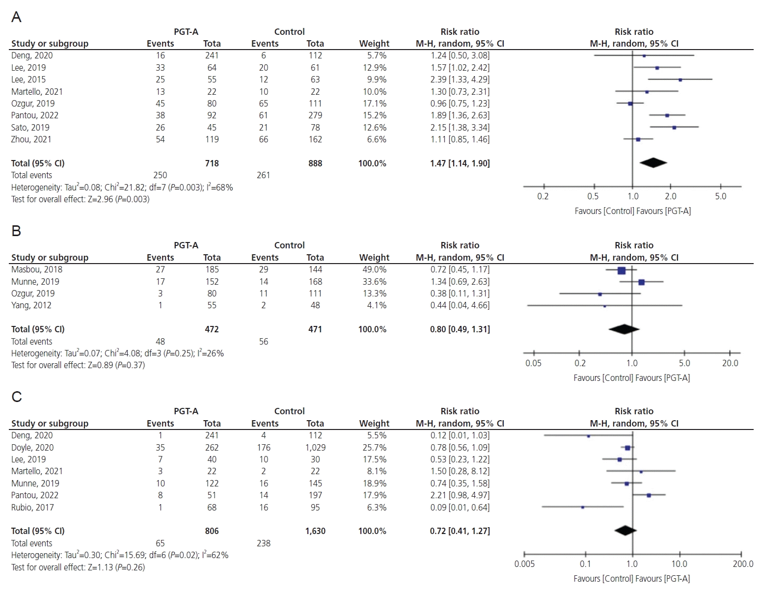
This meta-analysis of eight studies compared the LBR in patients with a history of previous miscarriage, RPL, or RIF (RR, 1.47; 95% CI, 1.14-1.90;  P=0.003). The heterogeneity of this comparison was 68%. Thus, PGT-A improved LBR in this cohort of patients (Fig. 5A).

5. Miscarriage rates per embryo transfer cycle

Four studies reported results on MR per cycle of ET in women 35 years old or younger (RR, 0.80; 95% CI, 0.49-1.31; P=0.37). Consequently, no significant difference was observed between the PGT-A and control groups. Heterogeneity in this comparison was 26% (Fig. 5B).
Seven studies reported results on MR in the over-35-year-old age group. No significant differences between the two groups (RR, 0.72; 95% CI, 0.41-1.27; P=0.26). Heterogeneity in this comparison was 62% (Fig. 5C).

Conclusion

1. Our results

In this meta-analysis, we evaluated the effectiveness of PGT-A in IVF/intracytoplasmic sperm injection (ICSI) cycles in patients of different ages and found that PGT improved the efficiency of ART, increasing clinical pregnancy and LBR, especially in women of AMA and with a poor prognosis. However, no benefits were demonstrated when applied to younger women. The advantages of these groups of patients can be explained by the higher rates of aneuploidy, which is rational because it is well established that embryonic aneuploidy is the main genetic factor influencing human reproductive success, not patient age alone. Poor oocyte quality in these patients can be explained by cytoplasmic errors, particularly in mitochondrial function [89,90]. According to our results in the specific population under 35 years of age, PGT-A did not reduce the MR as expected, which can be attributed to different factors. MR increase with age, and although this population is still relatively young, some age-related factors that are not related to chromosomal abnormalities may contribute to miscarriages. For example, pregnancy complications such as gestational diabetes or preeclampsia are more common in older mothers [91-93].
It is also important to highlight that the PGT-A is not a perfect test, and there can be technical limitations that lead to false results. For example, mosaic embryos (with both normal and abnormal cells) may be incorrectly classified as abnormal and not transferred, or vice versa. Additionally, some chromosomal abnormalities, particularly those that affect only a small portion of the chromosome, may not be detected by PGT-A [77,94,95].  Additionally, studies that have looked at the effectiveness of PGT-A in reducing MR in this age group may have had small sample sizes, which may limit the generalizability of the results.
In a study conducted by Anderson et al. [96] in 2020, the authors suggested that age did not appear to be a factor when considering embryo implantation and live birth rates between treatment groups.
However, according to published data, aneuploid embryos account for at least 10% of human pregnancies and the incidence can exceed 50% in women over 35 years of age [97].
However, these findings remain controversial. One of the included studies, the Single Embryo Transfer of Euploid Embryo (STAR) study trial [79], was highly debated. For example, the published reanalysis of this study revealed significant shortcomings in its statistical analyses [98]. Thus, the STAR study revealed that PGT-A did not beneficially affect IVF outcomes. Moreover, based on the Preimplantation Genetic Diagnosis International Society (PGDIS) 2019 analysis of this trial, not even reaching statistical significance (P=0.053), the authors did not hesitate in reporting that “a significant increase in ongoing pregnancy rate” was observed [99].

2. Stage of embryo biopsy and its influence on embryo development

One of the factors that can affect the results is that the stages of embryo biopsy in the enrolled studies were different; some of them involved biopsy in the cleavage stage, while others involved biopsy in the blastocyst stage. However, the role of biopsy duration remains controversial. According to the ESHRE recommendations on the most appropriate day, blastocyst biopsy is performed on days 5-7 post-insemination, according to the rate of development, once the inner cell mass is clearly visible [100].
Some authors have suggested that blastomere biopsy can lead to potential embryonic damage, higher levels of abnormality, and mosaicism. In a randomized controlled trial conducted by Scott et al. [101] in 2013, the effect of pre-implantation genetic testing for monogenic diseases biopsy on developing embryos was assessed, and the results demonstrated a relative 39% reduction in IR in the cleavage-stage biopsy group compared to controls without a reduction in the trophectoderm (TE) biopsy group. Only the D5 biopsy group showed a statistically significant increase in the LBR per ET. In a recent study, Sarkar et al. [102] assessed whether embryo biopsy for PGT-A affected the birth weight or preterm birth rate. The authors reported that trophectoderm biopsy for PGT-A did not increase the risk of small for gestation age, low birth weight, or preterm birth in IVF pregnancies [102]. 

3. Comprehensive chromosome screening (CCS)

The types of PGT methods used for complete chromosome screening (aCGH and NGS) in the included studies differed, which may have affected the results. NGS is the newest technique used for incorporation into second-generation PGT. Various studies that validated the precision of the NGS approach for embryonic CCS have demonstrated 100% consistency in the diagnosis of aCGH [103,104]. NGS and aCGH results were compared. In a retrospective cohort study, Friedenthal et al. [105] compared the IR, ongoing pregnancy/LBR, biochemical pregnancy rate, and spontaneous abortion between NGS and aCGH groups. Preimplantation genetic screening using NGS significantly improves IR and LBR compared with PGT using aCGH in single-thawed euploid embryo transfer cycles, which might be attributed to the advantages of NGS in detecting small chromosomal deletions, duplications, and mosaicism [105].

4. Embryo mosaicism

Embryo mosaicism occurring during mitotic division of the embryo, giving rise to chromosomally different cell lines, is one of the main sources of error when performing PGT-A [106-111]. Several studies have shown that mosaic embryos theoretically have a reduced IR and an increased risk of miscarriage, pregnancy complications, and clinically affected live births [112].
According to the PGDIS 2021 statement [112], embryos with a mosaicism rate lower than 20% can be considered euploid (and transferable), whereas embryos with more than 80% abnormal cells are classified as aneuploid. The remaining (20-80%) can be classified as mosaics. However, establishing the thresholds between which embryos can be considered transferable remains controversial. In a criticism of the earlier PGDIS 2019 position statement [99], the authors declared that accurate percentages of aneuploid DNA could not be calculated because of the inability to determine how many cells were damaged during biopsy, contributing to the fractional loss of DNA content and sample contamination [113].
A single trophectoderm biopsy of on average 5-6 cells, as is currently the practice in PGT-A at the blastocyst stage, cannot mathematically represent the whole embryo. Gleicher et al. [114] established two mathematical models to assess the probabilities of false-negative and false-positive results of an average 6-cell biopsy from approximately 300-cell TE. Both models revealed that even under the best-case scenario, assuming an even distribution of mosaicism in TE (because mosaicism is usually clonal and a highly unlikely scenario), a biopsy of at least 27 TE cells would be required to achieve minimal diagnostic predictability from a single TEB [114]. The data did not support an equal distribution of mosaicism throughout the trophectoderm and suggesting that mosaicism levels may be highly dependent on the biopsy [115]. Therefore, the trophectoderm alone cannot reliably represent the inner cell mass for biological reasons. Given the nature of the biology of mosaicism genesis and propagation, any biopsy piece analyzed as mosaic may not accurately reflect the surrounding trophectoderm or the rest of the embryo [116].
It is important to assess the efficacy of PGT using the “per patient” indicator to avoid excluding patients with poorer prognosis whose embryos may never reach ET [99]. Preliminary data suggested a 50% aneuploidy rate at the blastocyst stage (and even higher rates at cleavage stages) and with further gradual self-correction downstream [117]. According to PGDIS 2019, given the current knowledge base, discarding embryos based on a single TE biopsy appears shortsighted, and represents another misunderstanding of embryo biology [99].
However, the transfer of blastocysts in which mosaic aneuploidies have been found should only be considered following expert advice and appropriate genetic counseling for patients. It is recommended that clinicians inform patients that there is currently no evidence-based method available to determine which embryos with mosaic results have the best chance of resulting in a successful pregnancy or which may have the lowest risk of an undesired outcome [118,119]. The question of the correlation between transfer of (under PGDIS definition) “mosaic” embryos and reduced implantation and/or increased rates of miscarriage needs further investigation, and the current available data clearly dispute these propositions [99].
Patient counseling should include a discussion of various possible explanations for the mosaic results of the PGT-A and potential outcomes. In clinical medicine, the responsibility of establishing validated evidence in support of a proposed treatment and/or test rests with the proponents of the treatments or tests, mandating that such evidence exists before such treatments or tests are integrated into routine clinical practice [100].  
Embryo mosaicism is another limitation of this study. The transfer threshold differed among the enrolled studies, which could also affect the results of these studies.

5. Fresh and frozen embryos were transferred

Both fresh and frozen embryos were used in the studies included in our meta-analysis. There are still many concerns regarding the effect of cryopreservation on the health of children born and the outcome data after frozen ET. In their systematic review, Maheshwari et al. [120] analyzed obstetric and perinatal outcomes after fresh or thawed frozen ET and found that frozen-thawed ET was associated with better perinatal outcomes than fresh IVF embryos. Based on these findings, we assume that the differences in the included studies may be confounding factors affecting the results.

6. Male factor

We did not consider the influence of male factor on infertility; however, some aneuploidies may be derived from sperm. Men with an abnormal karyotype and Y chromosome deletions tend to produce sperm with an unbalanced set of chromosomes. Several other factors such as varicocele, chemotherapy, age, and lifestyle can also negatively influence meiotic division during spermatogenesis [121]. Petousis et al. [122] demonstrated that the rate of abnormal spermatozoa after fluorescence in situ hybridization examination was significantly higher in male patients with infertility (55.8% vs. 15.0%) and that teratozoospermia was strongly correlated with the incidence of chromosome 17 aneuploidy. Recent studies examining the effect of advanced paternal age on sperm aneuploidy rates have found that men over 50 years of age have more DNA-damaged spermatozoa, a lower rate of blastocyst development, a higher overall rate of aneuploidy, and a higher rate of trisomy [123,124].

7. Cost-effectiveness

The PGT-A strategy becomes more cost-effective with age. Somigliana et al. [125] stated that it is not economically advantageous to use PGT in women aged <36 years of age. Sensitivity analyses that vary the cost of ET, the cost of genetic tests, the magnitude of the adverse effect of PGT-A on LBR, and overall LBR alter the efficacy thresholds to some extent but generally support the notion that PGT-A may be cost-effective in some specific subgroups [125-127].
Our systematic review and meta-analysis evaluated the effectiveness of PGT-A in IVF/ICSI cycles in patients of different ages and included 19 clinical trials that evaluated approximately 100,000 IVF cycles in quantitative synthesis. Furthermore, well-defined eligibility criteria that prioritized only studies using aCGH or NGS were used. Meta-analyses with moderate or low heterogeneity were included.
Nevertheless, there were limitations to our systematic review and meta-analysis. First, there was a lack of clinical studies with a low risk of bias. Thus, our meta-analysis included studies with both moderate and low risk. Additionally, we could not perform subgroup analysis in cases of high heterogeneity because of the small number of relevant clinical studies. The reasons for the high heterogeneity may include the inclusion of randomized and non-randomized studies, studies with low and moderate risk of bias, patients with poor prognosis with different pathologies, and different days of embryo biopsy in these studies (Table 1).
Second, our search strategy included studies published only in English; conference abstracts were excluded, limiting our electronic search. Additionally, not every study transferred mosaic embryos, and this was not mentioned by all of the authors. Owing to the known inaccuracy of PGT-A testing and the possible natural resolution of mosaicism, some authors have suggested that mosaic embryos should be considered normal and transferred [99]. Currently, this is not standard practice.
Moreover, the outcome “per embryo transfer” is controversial regarding PGT-A studies. For example, Rubio et al. [78] did not use a single ET. However, in the study published by Wilkinson [128], participants refused to perform ET in cases of poor prognosis.  
The largest trial included in our systematic review and meta-analysis by Sanders et al. [76] was rebutted by Roberts et al. [129]. These authors argue that the comparator group must consist of treatments that could have had PGT-A if the option were available. Their analysis obtained estimates of the effect of PGT-A, which suggested an overall modest reduction in treatment success rates. The treatment effect of PGT-A was different, with an overall odds ratio for a live birth event of 0.82 (0.68-1.00) using >one transferrable embryo control and 0.80 (0.64-0.99) using >five embryo-created controls.
The next limitation is that PGT-A and NGS use frozen ET and should not be compared with fresh ET controls. Finally, it is more relevant to assess the effectiveness of PGT-A on cumulative LBR. However, there was an insufficient number of studies to perform meta-analysis.
Implications for future research may include modern techniques for non-invasive PGT. This method may play an enormous role in future fertility treatment, as damage to the embryo and the associated risks are negligible. Therefore, their use in routine practice should be investigated. In addition, although there are some doubts regarding time-lapse techniques, they should be further evaluated for evidence-based evaluation and decreased controversy. However, we need to consider not only embryos, but also gametes for better pregnancy rates. Thus, it is essential to develop gamete rejuvenation techniques to improve IVF outcomes in couples of advanced parental age.
Based on our systematic review and meta-analysis, we evaluated the effectiveness of PGT-A in IVF/ICSI cycles in patients of different ages and found that PGT improved the efficiency of ART, increasing clinical pregnancy and LBR, especially in women of AMA and those with a poor prognosis; however, no benefits were demonstrated when applied to younger women. Nevertheless, further research is needed to fully understand the effectiveness of PGT-A and to answer all questions regarding the importance of the validation, accuracy, and safety of PGT-A.

Notes

Conflict of interest

The authors declare no competing interests.

Ethical approval

No ethical approval was needed to run this systematic review and meta-analysis.

Patient consent

No patient consent was needed to run this systematic review and meta-analysis.

Funding information

No funding was provided.

References

1. Heijnen EM, Macklon NS, Fauser BC. What is the most relevant standard of success in assisted reproduction? The next step to improving outcomes of IVF: consider the whole treatment. Hum Reprod. 2004; 19:1936–8.
2. Franasiak JM, Forman EJ, Hong KH, Werner MD, Upham KM, Treff NR, et al. The nature of aneuploidy with increasing age of the female partner: a review of 15,169 consecutive trophectoderm biopsies evaluated with comprehensive chromosomal screening. Fertil Steril. 2014; 101:656–63.e1.
crossref
3. Zegers-Hochschild F, Adamson GD, Dyer S, Racowsky C, de Mouzon J, Sokol R, et al. The International glossary on infertility and fertility care, 2017. Fertil Steril. 2017; 108:393–406.
crossref
4. Fragouli E, Alfarawati S, Daphnis DD, Goodall NN, Mania A, Griffiths T, et al. Cytogenetic analysis of human blastocysts with the use of FISH, CGH and aCGH: scientific data and technical evaluation. Hum Reprod. 2011; 26:480–90.
5. Pirtea P, De Ziegler D, Tao X, Sun L, Zhan Y, Ayoubi JM, et al. Rate of true recurrent implantation failure is low: results of three successive frozen euploid single embryo transfers.  Fertil Steril. 2021; 115:45–53.
crossref
6. Goossens V, Harton G, Moutou C, Traeger-Synodinos J, Van Rij M, Harper JC. ESHRE PGD consortium data collection IX: cycles from January to December 2006 with pregnancy follow-up to October 2007. Hum Reprod. 2009; 24:1786–810.
crossref
7. Twisk M, Mastenbroek S, van Wely M, Heineman MJ, Van der Veen F, Repping S. Preimplantation genetic screening for abnormal number of chromosomes (aneuploidies) in in vitro fertilisation or intracytoplasmic sperm injection. Cochrane Database Syst Rev. 2006; (1):CD005291.
crossref
8. Page MJ, McKenzie JE, Bossuyt PM, Boutron I, Hoffmann TC, Mulrow CD, et al. The PRISMA 2020 statement: an updated guideline for reporting systematic reviews. Syst Rev. 2021; 10:89.
9. Coonen E, Rubio C, Christopikou D, Dimitriadou E, Gontar J, Goossens V, et al. ESHRE PGT consortium good practice recommendations for the detection of structural and numerical chromosomal aberrations. Hum Reprod Open. 2020; 2020:hoaa017.
10. Higgins JPT, Thomas J, Chandler J, Cumpston M, Li T, Page MJ, et al. Cochrane Handbook for Systematic Reviews of Interventions version 6.1 [Internet]. Chichester: John Wiley & Sons; c2019 [cited 2021 Jan 4]. Available from: https://training.cochrane.org/handbook/archive/v6.1.
11. Sterne JAC, Savović J, Page MJ, Elbers RG, Blencowe NS, Boutron I, et al. RoB 2: a revised tool for assessing risk of bias in randomised trials. BMJ . 2019; 366:l4898.
crossref
12. Sterne JA, Hernán MA, Reeves BC, Savović J, Berkman ND, Viswanathan M, et al. ROBINS-I: a tool for assessing risk of bias in non-randomised studies of interventions. BMJ. 2016; 355:i4919.
crossref
13. Mantravadi K, Mathew S, Soorve S, Rao DG, Karunakaran S. Does pre-implantation genetic testing for aneuploidy optimise the reproductive outcomes in-women with idiopathic recurrent pregnancy loss? Fertil Steril. 2020; 114:e437.
crossref
14. McCulloh DH, Grifo JA. Preimplantation genetic testing (pgt) success in the united states (2014-2017): multiple outcome measures indicate superiority of pgt over no pgt. Fertil Steril. 2020; 114:e413. –4.
crossref
15. Aharon D, Gounko D, Lee JA, Mukherjee T, Copperman AB, Sekhon L. Preimplantation genetic testing for aneuploidy in donor oocyte ivf cycles: a matched, sibling oocyte cohort study. Fertil Steril. 2020; 114:e275.
16. Munné S, Kaplan B, Frattarelli JL, Gysler M, Child T, Nakhuda G, et al. Preimplantation genetic testing for aneuploidy: a pragmatic, multicenter randomized clinical trial of single frozen euploid embryo transfer versus selection by morphology alone. Reprod Biomed Onlin. 2019; 38:e9.
crossref
17. Yang Z, Kuang Y, Meng Y, Zhang X, Su L, Lyu Q, et al. Selecting single euploid blastocysts for transfer with NGS significantly improves IVF treatment outcomes: a randomized study. Reprod Biomed Online. 2019; 38:e20. –1.
crossref
18. Canon CM, Aharon D, Gounko D, Lee JA, Roth RM, Slifkin R, et al. Should patients with only two embryos eligbile for biopsy after a single controlled ovarian hyperstimulation cycle utilize pgt-a prior to transfer selection? Cycles without PGT-A. Fertil Steril. 2021; 116:e246.
crossref
19. Daneshmand S, Richter KS, Gu R, Miller D, Tober D, Lin MP, et al. Value of preimplantation genetic testing for aneuploidy (pgt-a) in the context of in vitro fertilization (ivf) using donor oocytes. Fertil Steril. 2021; 116:e401.
crossref
20. Scott RT Jr, Upham KM, Forman EJ, Hong KH, Scott KL, Taylor D, et al. Blastocyst biopsy with comprehensive chromosome screening and fresh embryo transfer significantly increases in vitro fertilization implantation and delivery rates: a randomized controlled trial. Fertil Steril. 2013; 100:697–703.
21. Comparison of the live birth rate of PGT versus expectant management in patients with RPL [Internet]. Shanghai: Shanghai First Maternity and Infant Hospital; c2022 [cited 2022 Jul 14]. Available from: https://clinicaltrials.gov/study/NCT05457335.
22. Sui YL, Lei CX, Ye JF, Fu J, Zhang S, Li L, et al. In vitro fertilization with single-nucleotide polymorphism microarray-based preimplantation genetic testing for aneuploidy significantly improves clinical outcomes in infertile women with recurrent pregnancy loss: a randomized controlled trial. Reprod Dev Med. 2020; 4:32–41.
crossref
23. Haviland MJ, Murphy LA, Modest AM, Fox MP, Wise LA, Nillni YI, et al. Comparison of pregnancy outcomes following preimplantation genetic testing for aneuploidy using a matched propensity score design. Hum Reprod. 2020; 35:2356–64.
crossref
24. Sacchi L, Albani E, Cesana A, Smeraldi A, Parini V, Fabiani M, et al. Preimplantation genetic testing for aneuploidy improves clinical, gestational, and neonatal outcomes in advanced maternal age patients without compromising cumulative live-birth rate. J Assist Reprod Genet. 2019; 36:2493–504.
crossref
25. Roeca C, Johnson R, Carlson N, Polotsky AJ. Preimplantation genetic testing and chances of a healthy live birth amongst recipients of fresh donor oocytes in the United States. J Assist Reprod Genet. 2020; 37:2283–92.
crossref
26. Ying LY, Sanchez MD, Baron J, Ying Y. Preimplantation genetic testing and frozen embryo transfer synergistically decrease very pre-term birth in patients undergoing in vitro fertilization with elective single embryo transfer. J Assist Reprod Genet. 2021; 38:2333–39.
27. Bhatt SJ, Marchetto NM, Roy J, Morelli SS, McGovern PG. Pregnancy outcomes following in vitro fertilization frozen embryo transfer (IVF-FET) with or without preimplantation genetic testing for aneuploidy (PGT-A) in women with recurrent pregnancy loss (RPL): a SARTCORS study. Hum Reprod . 2021; 35:2339–44.
crossref
28. Murphy LA, Seidler EA, Vaughan DA, Resetkova N, Penzias AS, Toth TL, et al. To test or not to test? A framework for counselling patients on preimplantation genetic testing for aneuploidy (PGT-A). Hum Reprod. 2019; 34:268–75.
crossref
29. Kushnir VA, Darmon SK, Albertini DF, Barad DH, Gleicher N. Effectiveness of in  vitro fertilization with preimplantation genetic screening: a reanalysis of United States assisted reproductive technology data 2011-2012.  Fertil Steril. 2016; 106:75–9.
30. Sarkar P, Jindal S, New EP, Sprague RG, Tanner J, Imudia AN. The role of preimplantation genetic testing for aneuploidy in a good prognosis IVF population across different age groups. Syst Biol Reprod Med. 2021; 67:366–73.
crossref
31. Mastenbroek S, Twisk M, van Echten-Arends J, Sikkema-Raddatz B, Korevaar JC, Verhoeve HR, et al. In vitro fertilization with preimplantation genetic screening. N Engl J Med. 2007; 357:9–17.
32. Mejia RB, Capper EA, Summers KM, Mancuso AC, Sparks AE, Van Voorhis BJ. Cumulative live birth rate in women aged ≤37 years after in vitro fertilization with or without preimplantation genetic testing for aneuploidy: a Society for Assisted Reproductive Technology clinic outcome reporting system retrospective analysis. F S Rep. 2022; 3:184–91.
33. Gianaroli L, Magli MC, Munné S, Fiorentino A, Montanaro N, Ferraretti AP. Will preimplantation genetic diagnosis assist patients with a poor prognosis to achieve pregnancy? Hum Reprod. 1997; 12:1762–7.
crossref
34. Staessen C, Platteau P, Van Assche E, Michiels A, Tournaye H, Camus M, et al. Comparison of blastocyst transfer with or without preimplantation genetic diagnosis for aneuploidy screening in couples with advanced maternal age: a prospective randomized controlled trial. Hum Reprod. 2004; 19:2849–58.
crossref
35. Dang TT, Phung TM, Le H, Nguyen TB, Nguyen TS, Nguyen TL, et al. Preimplantation genetic testing of aneuploidy by next generation sequencing: association of maternal age and chromosomal abnormalities of blastocyst. Open Access Maced J Med Sci. 2019; 7:4427–31.
crossref
36. Hardarson T, Hanson C, Lundin K, Hillensjö T, Nilsson L, Stevic J, et al. Preimplantation genetic screening in women of advanced maternal age caused a decrease in clinical pregnancy rate: a randomized controlled trial. Hum Reprod. 2008; 23:2806–12.
37. Staessen C, Verpoest W, Donoso P, Haentjens P, Van der Elst J, Liebaers I, et al. Preimplantation genetic screening does not improve delivery rate in women under the age of 36 following single-embryo transfer. Hum Reprod. 2008; 23:2818–25.
crossref
38. Ikuma S, Sato T, Sugiura-Ogasawara M, Nagayoshi M, Tanaka A, Takeda S. Preimplantation genetic diagnosis and natural conception: a comparison of live birth rates in patients with recurrent pregnancy loss associated with translocation.  PLoS One . 2015; 10:e0129958.
crossref
39. Werlin L, Rodi I, DeCherney A, Marello E, Hill D, Munné S. Preimplantation genetic diagnosis as both a therapeutic and diagnostic tool in assisted reproductive technology. Fertil Steril. 2003; 80:467–8.
40. Mersereau JE, Pergament E, Zhang X, Milad MP. Preimplantation genetic screening to improve in vitro fertilization pregnancy rates: a prospective randomized controlled trial.  Fertil Steril. 2008; 90:1287–9.
41. Meyer LR, Klipstein S, Hazlett WD, Nasta T, Mangan P, Karande VC. A prospective randomized controlled trial of preimplantation genetic screening in the “good prognosis” patient.  Fertil Steril. 2009; 91:1731–8.
42. Schoolcraft WB, Katz-Jaffe MG, Stevens J, Rawlins M, Munne S. Preimplantation aneuploidy testing for infertile patients of advanced maternal age: a randomized prospective trial.  Fertil Steril. 2009; 92:157–62.
43. Debrock S, Melotte C, Spiessens C, Peeraer K, Vanneste E, Meeuwis L, et al. Preimplantation genetic screening for aneuploidy of embryos after in vitro fertilization in women aged at least 35 years: a prospective randomized trial. Fertil Steril. 2010; 93:364–73.
crossref
44. Rubio C, Bellver J, Rodrigo L, Bosch E, Mercader A, Vidal C, et al. Preimplantation genetic screening using fluorescence in situ hybridization in patients with repetitive implantation failure and advanced maternal age: two randomized trials. Fertil Steril. 2013; 99:1400–7.
45. Gianaroli L, Magli MC, Ferraretti AP, Munné S. Preimplantation diagnosis for aneuploidies in patients undergoing in vitro fertilization with a poor prognosis: identification of the categories for which it should be proposed.  Fertil Steril. 1999; 72:837–44.
crossref
46. Blockeel C, Schutyser V, De Vos A, Verpoest W, De Vos M, Staessen C, et al. Prospectively randomized controlled trial of PGS in IVF/ICSI patients with poor implantation. Reprod Biomed Online. 2008; 17:848–54.
47. Preimplantation genetic testing for aneuploidy (PGT-A) in women over 36 years of age [Internet]. Margate: Genomic Prediction Inc.; c2020 [cited 2023 Nov 18]. Available from: https://clinicaltrials.gov/show/NCT04167748.
48. Mahesan AM, Chang PT, Ronn R, Paul ABM, Meriano J, Casper RF. Preimplantation genetic testing for aneuploidy in patients with low embryo numbers: benefit or harm? J Assist Reprod Genet. 2022; 39:2027–33.
crossref
49. Sadecki E, Rust L, Walker DL, Fredrickson JR, Krenik A, Kim T, et al. Comparison of live birth rates after IVF-embryo transfer with and without preimplantation genetic testing for aneuploidies. Reprod Biomed Online. 2021; 43:995–1001.
crossref
50. Preimplantation genetic diagnosis for the indication of advanced reproductive age [Internet]. Bethesda: ClinicalTrials.gov.; c2010 [cited 2022 Nov 4]. Available from: https://clinicaltrials.gov/ct2/show/NCT00646893.
51. Preimplantation genetic diagnosis for the indication of advanced reproductive age [Internet]. Beverly Hills: Reprogenetics; c2010 [cited 2017 Jul 11]. Available from: https://clinicaltrials.gov/ct2/show/NCT00646893.
52. Mazzilli R, Cimadomo D, Vaiarelli A, Capalbo A, Dovere L, Alviggi E, et al. Effect of the male factor on the clinical outcome of intracytoplasmic sperm injection combined with preimplantation aneuploidy testing: observational longitudinal cohort study of 1,219 consecutive cycles. Fertil Steril. 2017; 108:961–72.e3.
crossref
53. Del Carmen Nogales M, Cruz M, de Frutos S, Martínez EM, Gaytán M, Ariza M, et al. Association between clinical and IVF laboratory parameters and miscarriage after single euploid embryo transfers. Reprod Biol Endocrinol. 2021; 19:186.
crossref
54. Gorodeckaja J, Neumann S, McCollin A, Ottolini CS, Wang J, Ahuja K, et al. High implantation and clinical pregnancy rates with single vitrified-warmed blastocyst transfer and optional aneuploidy testing for all patients. Hum Fertil (Camb). 2020; 23:256–67.
55. Viñals Gonzalez X, Odia R, Naja R, Serhal P, Saab W, Seshadri S, et al. Euploid blastocysts implant irrespective of their morphology after NGS-(PGT-A) testing in advanced maternal age patients. J Assist Reprod Genet. 2019; 36:1623–9.
crossref
56. Zhao H, Tao W, Li M, Liu H, Wu K, Ma S. Comparison of two protocols of blastocyst biopsy submitted to preimplantation genetic testing for aneuploidies: a randomized controlled tria. Arch Gynecol Obstet. 2019; 299:1487–93.
crossref
57. Live birth rate in patients with unexplained recurrent pregnancy loss [Internet]. Beijing: Peking University Third Hospital; c2020 [cited 2020 Nov 9]. Available from: https://clinicaltrials.gov/study/NCT04621773.
58. Predictive value of embryonic testing (PROV-ET) [Internet]. Lake Mary: Reproductive Medicine Associates of New Jersey; c2022 [cited 2022 Jan 14]. Available from: https://clinicaltrials.gov/ct2/show/NCT03604107.
59. PGT-A in screening of embryos in the treatment of unexplained recurrent miscarriage [Internet]. Beijing: Peking University Third Hospital; c2020 [cited 2020 Nov 25]. Available from: https://clinicaltrials.gov/ct2/show/NCT04643938.
60. Tong J, Niu Y, Wan A, Zhang T. Next-generation sequencing (NGS)-based preimplantation genetic testing for aneuploidy (PGT-A) of trophectoderm biopsy for recurrent implantation failure (RIF) patients: a retrospective study. Reprod Sci. 2021; 28:1923–9.
crossref
61. Li N, Guan Y, Ren B, Zhang Y, Du Y, Kong H, et al. Effect of blastocyst morphology and developmental rate on euploidy and live birth rates in preimplantation genetic testing for aneuploidy cycles with single-embryo transfer. Front Endocrinol (Lausanne). 2022; 13:858042.
crossref
62. Simon AL, Kiehl M, Fischer E, Proctor JG, Bush MR, Givens C, et al. Pregnancy outcomes from more than 1,800 in  vitro fertilization cycles with the use of 24-chromosome single-nucleotide polymorphism-based preimplantation genetic testing for aneuploidy. Fertil Steril. 2018; 110:113–21.
63. Rubino P, Tapia L, Ruiz de Assin Alonso R, Mazmanian K, Guan L, Dearden L, et al. Trophectoderm biopsy protocols can affect clinical outcomes: time to focus on the blastocyst biopsy technique. Fertil Steril. 2020; 113:981–9.
crossref
64. Homer HA. Preimplantation genetic testing for aneuploidy (PGT-A): the biology, the technology and the clinical outcomes.   Aust N Z J Obstet Gynaecol. 2019; 59:317–24.
crossref
65. Jansen RP, Bowman MC, de Boer KA, Leigh DA, Lieberman DB, McArthur SJ. What next for preimplantation genetic screening (PGS)? Experience with blastocyst biopsy and testing for aneuploidy. Hum Reprod. 2008; 23:1476–8.
crossref
66. Griffin DK. Why PGT-A, most likely, improves IVF success.  Reprod Biomed Online. 2022; 45:633–7.
crossref
67. Cornelisse S, Zagers M, Kostova E, Fleischer K, van Wely M, Mastenbroek S. Preimplantation genetic testing for aneuploidies (abnormal number of chromosomes) in in vitro fertilisation. Cochrane Database Syst Rev. 2020; 9:CD005291.
68. L’Heveder A, Jones BP, Naja R, Serhal P, Nagi JB. Preimplantation genetic testing for aneuploidy: current perspectives.  Semin Reprod Med. 2021; 39:1–12.
69. Lee CI, Wu CH, Pai YP, Chang YJ, Chen CI, Lee TH, et al. Performance of preimplantation genetic testing for aneuploidy in IVF cycles for patients with advanced maternal age, repeat implantation failure, and idiopathic recurrent miscarriage.   Taiwan J Obstet Gynecol. 2019; 58:239–43.
crossref
70. Masbou AK, Friedenthal JB, McCulloh DH, McCaffrey C, Fino ME, Grifo JA, et al. A comparison of pregnancy outcomes in patients undergoing donor egg single embryo transfers with and without preimplantation genetic testing. Reprod Sci. 2019; 26:1661–5.
crossref
71. Yang Z, Liu J, Collins GS, Salem SA, Liu X, Lyle SS, et al. Selection of single blastocysts for fresh transfer via standard morphology assessment alone and with array CGH for good prognosis IVF patients: results from a randomized pilot study. Mol Cytogenet. 2012; 5:24.
crossref
72. Lee HL, McCulloh DH, Hodes-Wertz B, Adler A, McCaffrey C, Grifo JA. In vitro fertilization with preimplantation genetic screening improves implantation and live birth in women age 40 through 43. J Assist Reprod Genet. 2015; 32:435–44.
73. Yan J, Qin Y, Zhao H, Sun Y, Gong F, Li R, et al. Live birth with or without preimplantation genetic testing for aneuploidy. N Engl J Med. 2021; 385:2047–58.
crossref
74. Deng J, Hong HY, Zhao Q, Nadgauda A, Ashrafian S, Behr B, et al. Preimplantation genetic testing for aneuploidy in poor ovarian responders with four or fewer oocytes retrieved. J Assist Reprod Genet. 2020; 37:1147–54.
crossref
75. Zhou T, Zhu Y, Zhang J, Li H, Jiang W, Zhang Q, et al. Effects of PGT-A on pregnancy outcomes for young women having one previous miscarriage with genetically abnormal products of conception. Reprod Sci. 2021; 28:3265–71.
crossref
76. Sanders KD, Silvestri G, Gordon T, Griffin DK. Analysis of IVF live birth outcomes with and without preimplantation genetic testing for aneuploidy (PGT-A): UK Human Fertilisation and Embryology Authority data collection 2016-2018. J Assist Reprod Genet. 2021; 38:3277–85.
crossref
77. Tiegs AW, Tao X, Zhan Y, Whitehead C, Kim J, Hanson B, et al. A multicenter, prospective, blinded, nonselection study evaluating the predictive value of an aneuploid diagnosis using a targeted next-generation sequencing-based preimplantation genetic testing for aneuploidy assay and impact of biopsy. Fertil Steril. 2021; 115:627–37.
78. Rubio C, Bellver J, Rodrigo L, Castillón G, Guillén A, Vidal C, et al. In vitro fertilization with preimplantation genetic diagnosis for aneuploidies in advanced maternal age: a randomized, controlled study. Fertil Steril. 2017; 107:1122–9.
79. Munné S, Kaplan B, Frattarelli JL, Child T, Nakhuda G, Shamma FN, et al. Preimplantation genetic testing for aneuploidy versus morphology as selection criteria for single frozen-thawed embryo transfer in good-prognosis patients: a multicenter randomized clinical trial. Fertil Steril. 2019; 112:1071–9.e7.
crossref
80. Ozgur K, Berkkanoglu M, Bulut H, Yoruk GDA, Candurmaz NN, Coetzee K. Single best euploid versus single best unknown-ploidy blastocyst frozen embryo transfers: a randomized controlled trial.  J Assist Reprod Genet . 2019; 36:629–36.
crossref
81. Sato T, Sugiura-Ogasawara M, Ozawa F, Yamamoto T, Kato T, Kurahashi H, et al. Preimplantation genetic testing for aneuploidy: a comparison of live birth rates in patients with recurrent pregnancy loss due to embryonic aneuploidy or recurrent implantation failure. Hum Reprod. 2019; 34:2340–8.
crossref
82. Doyle N, Gainty M, Eubanks A, Doyle J, Hayes H, Tucker M, et al. Donor oocyte recipients do not benefit from preimplantation genetic testing for aneuploidy to improve pregnancy outcomes. Hum Reprod. 2020; 35:2548–55.
83. Whitney JB, Schiewe MC, Anderson RE. Single center validation of routine blastocyst biopsy implementation.  J Assist Reprod Genet . 2016; 33:1507–13.
crossref
84. Namath A, Jahandideh S, Devine K, O’Brien JE, Stillman RJ. Gestational carrier pregnancy outcomes from frozen embryo transfer depending on the number of embryos transferred and preimplantation genetic testing: a retrospective analysis. Fertil Steril. 2021; 115:1471–7.
crossref
85. Awadalla MS, Agarwal R, Ho JR, McGinnis LK, Ahmady A. Effect of trophectoderm biopsy for PGT-A on live birth rate per embryo in good prognosis patients. Arch Gynecol Obstet. 2022; 306:1321–7.
crossref
86. Martello CL, Kulmann MIR, Donatti LM, Bos-Mikich A, Frantz N. Preimplantation genetic testing for aneuploidies does not increase success rates in fresh oocyte donation cycles: a paired cohort study. J Assist Reprod Genet. 2021; 38:2909–14.
crossref
87. Pantou A, Mitrakos A, Kokkali G, Petroutsou K, Tounta G, Lazaros L, et al. The impact of preimplantation genetic testing for aneuploidies (PGT-A) on clinical outcomes in high risk patients. J Assist Reprod Genet. 2022; 39:1341–9.
88. McGuinness LA, Higgins JPT. Risk-of-bias VISualization (robvis): an R package and shiny web app for visualizing risk-of-bias assessments. Res Synth Methods. 2021; 12:55–61.
crossref
89. Zhao J, Li Y. Adenosine triphosphate content in human unfertilized oocytes, undivided zygotes and embryos unsuitable for transfer or cryopreservation.   J Int Med Res. 2012; 40:734–9.
crossref
90. Konstantinidis M, Alfarawati S, Hurd D, Paolucci M, Shovelton J, Fragouli E, et al. Simultaneous assessment of aneuploidy, polymorphisms, and mitochondrial DNA content in human polar bodies and embryos with the use of a novel microarray platform. Fertil Steril. 2014; 102:1385–92.
crossref
91. Wang X, Shi W, Zhao S, Gong D, Li S, Hu C, et al. Whole exome sequencing in unexplained recurrent miscarriage families identified novel pathogenic genetic causes of euploid miscarriage. Hum Reprod. 2023; 38:1003–18.
crossref
92. Lin J, Huang J, Zhu Q, Kuang Y, Cai R, Wang Y. Effect of maternal age on pregnancy or neonatal outcomes among 4,958 infertile women using a freeze-all strategy. Front Med (Lausanne). 2020; 6:316.
93. Lu XM, Liu YB, Zhang DD, Cao X, Zhang TC, Liu M, et al. Effect of advanced paternal age on reproductive outcomes in IVF cycles of non-male-factor infertility: a retrospective cohort study. Asian J Androl. 2023; 25:245–51.
crossref
94. Popovic M, Dhaenens L, Boel A, Menten B, Heindryckx B. Chromosomal mosaicism in human blastocysts: the ultimate diagnostic dilemma. Hum Reprod Update. 2020; 26:313–34.
crossref
95. Liu YL, Yu TN, Wang PH, Tzeng CR, Chen CH, Chen CH. Could PGT-A pick up true abnormalities that have clinical relevance? Retrospective analysis of 1043 embryos. Taiwan J Obstet Gynecol. 2020; 59:496–501.
crossref
96. Anderson RE, Whitney JB, Schiewe MC. Clinical benefits of preimplantation genetic testing for aneuploidy (PGT-A) for all in vitro fertilization treatment cycles. Eur J Med Gene. 2020; 63:103731.
crossref
97. Nagaoka SI, Hassold TJ, Hunt PA. Human aneuploidy: mechanisms and new insights into an age-old problem.  Nat Rev Genet. 2012; 13:493–504.
98. Orvieto R, Gleicher N. Preimplantation genetic testing for aneuploidy (PGT-A)-finally revealed.  J Assist Reprod Genet. 2020; 37:669–72.
crossref
99. Gleicher N, Albertini DF, Barad DH, Homer H, Modi D, Murtinger M, et al. The 2019 PGDIS position statement on transfer of mosaic embryos within a context of new information on PGT-A. Reprod Biol Endocrinol. 2020; 18:57.
crossref
100. Kokkali G, Coticchio G, Bronet F, Celebi C, Cimadomo D, Goossens V, et al. ESHRE PGT consortium and SIG embryology good practice recommendations for polar body and embryo biopsy for PGT. Hum Reprod Open. 2020; 2020:hoaa020.
101. Scott RT Jr, Upham KM, Forman EJ, Zhao T, Treff NR. Cleavage-stage biopsy significantly impairs human embryonic implantation potential while blastocyst biopsy does not: a randomized and paired clinical trial.   Fertil Steril. 2013; 100:624–30.
crossref
102. Sarkar P, New EP, Jindal S, Tanner JP, Imudia AN. The effect of trophectoderm biopsy for preimplantation genetic testing on fetal birth weight and preterm delivery. Minerva Obstet Gynecol. 2023; Jan. 16. [Epub]. https://doi.org/10.23736/S2724-606X.22.05196-X.
103. Fiorentino F, Biricik A, Bono S, Spizzichino L, Cotroneo E, Cottone G, et al. Development and validation of a next-generation sequencing-based protocol for 24-chromosome aneuploidy screening of embryos. Fertil Steril. 2014; 101:1375–82.
104. Kung A, Munné S, Bankowski B, Coates A, Wells D. Validation of next-generation sequencing for comprehensive chromosome screening of embryos. Reprod Biomed Online. 2015; 31:760–9.
crossref
105. Friedenthal J, Maxwell SM, Munné S, Kramer Y, McCulloh DH, McCaffrey C, et al. Next generation sequencing for preimplantation genetic screening improves pregnancy outcomes compared with array comparative genomic hybridization in single thawed euploid embryo transfer cycles. Fertil Steril. 2018; 109:627–32.
crossref
106. Munné S, Sandalinas M, Escudero T, Márquez C, Cohen J. Chromosome mosaicism in cleavage-stage human embryos: evidence of a maternal age effect. Reprod Biomed Online. 2002; 4:223–32.
crossref
107. Los Los FJ, Van Opstal D, van den Berg C. The development of cytogenetically normal, abnormal and mosaic embryos: a theoretical model. Hum Reprod Update. 2004; 10:79–94.
108. Baart EB, Van Opstal D, Los FJ, Fauser BC, Martini E. Fluorescence in situ hybridization analysis of two blastomeres from day 3 frozen-thawed embryos followed by analysis of the remaining embryo on day 5.   Hum Reprod . 2004; 19:685–93.
crossref
109. Baart EB, Martini E, van den Berg I, Macklon NS, Galjaard RJ, Fauser BC, et al. Preimplantation genetic screening reveals a high incidence of aneuploidy and mosaicism in embryos from young women undergoing IVF.  Hum Reprod . 2006; 21:223–33.
crossref
110. Daphnis DD, Fragouli E, Economou K, Jerkovic S, Craft IL, Delhanty JD, et al. Analysis of the evolution of chromosome abnormalities in human embryos from day 3 to 5 using CGH and FISH. Mol Hum Reprod. 2008; 14:117–25.
111. Frumkin T, Malcov M, Yaron Y, Ben-Yosef D. Elucidating the origin of chromosomal aberrations in IVF embryos by preimplantation genetic analysis. Mol Cell Endocrinol. 2008; 282:112–9.
112. Leigh D, Cram DS, Rechitsky S, Handyside A, Wells D, Munne S, et al. PGDIS position statement on the transfer of mosaic embryos 2021. Reprod Biomed Online. 2022; 45:19–25.
113. Gleicher N, Mochizuki L, Barad DH, Patrizio P, Orvieto R. A review of the 2021/2022 PGDIS position statement on the transfer of mosaic embryos.   J Assist Reprod Genet. 2023; 40:817–26.
114. Gleicher N, Metzger J, Croft G, Kushnir VA, Albertini DF, Barad DH. A single trophectoderm biopsy at blastocyst stage is mathematically unable to determine embryo ploidy accurately enough for clinical use. Reprod Biol Endocrinol. 2017; 15:33.
115. Popovic M, Dheedene A, Christodoulou C, Taelman J, Dhaenens L, Van Nieuwerburgh F, et al. Chromosomal mosaicism in human blastocysts: the ultimate challenge of preimplantation genetic testing? Hum Reprod. 2018; 33:1342–54.
116. Cram DS, Leigh D, Handyside A, Rechitsky L, Xu K, Harton G, et al. PGDIS position statement on the transfer of mosaic embryos 2019. Reprod Biomed Online. 2019; 39 Suppl 1:e1–4.
crossref
117. Rito T, Naftaly J, Gleicher N, Brivanlou AH. Self-correction of aneuploidy in human blastocysts and self-organizing gastruloids. Fertil Steril. 2019; 112:e127.
118. Committee P. Clinical management of mosaic results from preimplantation genetic testing for aneuploidy (PGT-A) of blastocysts: a committee opinion. Fertil Steril. 2020; 114:246–54.
crossref
119. De Rycke M, Capalbo A, Coonen E, Coticchio G, Fiorentino F, Goossens V, et al. ESHRE survey results and good practice recommendations on managing chromosomal mosaicism. Hum Reprod Open. 2022; 2022:hoac044.
120. Maheshwari A, Pandey S, Shetty A, Hamilton M, Bhattacharya S. Obstetric and perinatal outcomes in singleton pregnancies resulting from the transfer of frozen thawed versus fresh embryos generated through in vitro fertilization treatment: a systematic review and meta-analysis. Fertil Steril. 2012; 98:368–77.e1-9.
121. Colaco S, Sakkas D. Paternal factors contributing to embryo quality. J Assist Reprod Genet. 2018; 35:1953–68.
crossref
122. Petousis S, Prapas Y, Papatheodorou A, Margioula-Siarkou C, Papatzikas G, Panagiotidis Y, et al. Fluorescence in situ hybridisation sperm examination is significantly impaired in all categories of male infertility. Andrologia. 2018; 50:e12847.
123. García-Ferreyra J, Luna D, Villegas L, Romero R, Zavala P, Hilario R, et al. High aneuploidy rates observed in embryos derived from donated oocytes are related to male aging and high percentages of sperm DNA fragmentation. Clin Med Insights Reprod Health. 2015; 9:21–7.
crossref
124. García-Ferreyra J, Hilario R, Dueñas J. High percentages of embryos with 21, 18 or 13 trisomy are related to advanced paternal age in donor egg cycles. JBRA Assist Reprod. 2018; 22:26–34.
125. Somigliana E, Busnelli A, Paffoni A, Vigano P, Riccaboni A, Rubio C, et al. Cost-effectiveness of preimplantation genetic testing for aneuploidies.  Fertil Steril. 2019; 111:1169–76.
crossref
126. Bakkensen JB, Flannagan KSJ, Mumford SL, Hutchinson AP, Cheung EO, Moreno PI, et al. A SART data cost-effectiveness analysis of planned oocyte cryopreservation versus in vitro fertilization with preimplantation genetic testing for aneuploidy considering ideal family size. Fertil Steril. 2022; 118:875–84.
127. Lee E, Costello MF, Botha WC, Illingworth P, Chambers GM. A cost-effectiveness analysis of preimplantation genetic testing for aneuploidy (PGT-A) for up to three complete assisted reproductive technology cycles in women of advanced maternal age.  Aust N Z J Obstet Gynecol. 2019; 59:573–9.
128. Wilkinson J. Neither relevant nor randomized: the use of “per embryo transfer” in the analysis of preimplantation genetic testing for aneuploidy trials.  Fertil Steril. 2023; 119:910–2.
crossref
129. Roberts SA, Wilkinson J, Vail A, Brison DR. Does PGT-A improve assisted reproduction treatment success rates: what can the UK Register data tell us? J Assist Reprod Genet. 2022; 39:2547–54.
crossref

Fig. 1.
Flow diagram of the literature search and study selection process according to the PRISMA guidelines. PRISMA, Preferred Reporting Items for Systematic Reviews and Meta-Analyses.
ogs-24028f1.tif
Fig. 2.
Traffic light plots. (A) RoB2.0 tool for randomized controlled trials; (B) ROBINS-I tool for nonrandomized studies of interventions. RoB2, risk of bias-2; ROBINS-I, risk of bias in nonrandomised studies-of Interventions.
ogs-24028f2.tif
Fig. 3.
(A) Forest plot regarding the clinical pregnancy rate in IVF patients aged >35 years (per embryo transfer cycle). (B) Forest plot of the live birth rate in IVF patients aged <35 years (per embryo transfer cycle). PGT-A, preimplantation genetic testing for aneuploidy; CI, confidence interval; IVF, in vitro fertilization.
ogs-24028f3.tif
Fig. 4.
(A) Forest plot regarding the live birth rate in IVF patients aged <38 years (per patient). (B) Forest plot of the live birth rate in IVF patients aged >35 years (per patient). PGT-A, preimplantation genetic testing for aneuploidy; CI, confidence interval; IVF, in vitro fertilization.
ogs-24028f4.tif
Fig. 5.
(A) Effect of PGT-A on the live birth rate in patients with a poor prognosis (per embryo transfer cycle). (B) Miscarriage rate in IVF patients aged <35 years old (per embryo transfer cycle). (C) Miscarriage rate in IVF patients aged >35 years (per embryo transfer cycle). PGT-A, preimplantation genetic testing for aneuploidy; CI, confidence interval; IVF, in vitro fertilization.
ogs-24028f5.tif
Table 1.
Principal characteristics of the studies included in this network meta-analysis
Study Patient’s age (yr) Fresh/frozen cycles Biopsy day/embryo stage ET day/embryo stage Ploidy analysis technique PGT-A group (No. of patients/ET cycles) Control group (No. of patients/ET cycles) Outcome measures
Lee et al. [69] (2019) ≥38 Frozen D5, 6/blastocyst D5, 6/blastocyst aCGH 61 61 IR, CPR, MR, and LBR
Masbou et al. [70] (2019) 26-30 Fresh/frozen D5, 6/blastocyst D5/blastocyst aCGH/NGS 185 ·112 fresh IR, OPR/LBR, and SAR
·144 frozen
Yang et al. [71] (2012) <35 Fresh/frozen D5/blastocyst D6/blastocyst aCGH 55 48 CPR, OPR, and MR
Lee et al. [72] (2015) 40-43 Fresh/frozen D5, 6/blastocyst D5/blastocyst aCGH 49 ·127 fresh IR and LBR
·28 frozen
Yan et al. [73] (2021) 20-37 Frozen D5/blastocyst D5/blastocyst NGS 606 606 LBR
Deng et al. [74] (2020) ·<38 Fresh/frozen D5/blastocyst D3, 5/blastocyst NGS 241 112 CPR, LBR, and MR
·38-40
·>40
Zhou et al. [75] (2021) <38 Fresh/frozen D5/blastocyst D5/blastocyst NGS 124 93 CPR, LBR, and MR
Sanders et al. [76] (2021) ·<35 Fresh/frozen D5/blastocyst D5/blastocyst NGS 2,464 187,546 LBR
·35-37
·38, 39
·40-42
·43, 44
·>44
Tiegs et al. [77] (2021) 18-44 Frozen D5/blastocyst D5/blastocyst NGS 484 1,208 IR
Rubio et al. [78] (2017) 38-41 Fresh D3/cleavage D5, 6/blastocyst aCGH 100 105 CPR, OPR, MR, and CLB
Munné et al. [79] (2019) 25-40 Frozen D5, 6/blastocyst D5, 6/blastocyst NGS 330 331 CPR, OPR, and MR
Ozgur et al. [80] (2019) 35 Frozen D5/blastocyst D5/blastocyst NGS 109 111 CPR, OPR, and MR
Sato et al. [81] (2019) 35-42 Frozen D5/blastocyst D5/blastocyst aCGH ·41 (RPL) ·38 (RPL) CPR and LBR
·42 (RIF) ·50 (RIF)
Doyle et al. [82] (2020) ·21-32 OD Fresh/frozen ·D3/cleavage D5, 6/blastocyst aCGH/NGS 262 1,029 LBR and MR
·40-45 ·D5/blastocyst
Whitney et al. [83] (2016) ·≤34 Fresh/frozen D5, 6/blastocyst D5, 6/blastocyst aCGH 134 153 IR, CPR, and LBR
·35-37
·38-40
·41, 42
·≥43
Namath et al. [84] (2021) NP Frozen D5/blastocyst D5, 6/blastocyst NGS 194 389 LBR
Awadalla et al. [85] (2022) 28-44 Fresh/frozen D5/blastocyst D5-7/blastocyst NGS 92 140 LBR
Martello et al. [86] (2021) ·21-32 OD Frozen D5/blastocyst D5, 6/blastocyst aCGH/NGS 22 22 IR, CPR, MR, and LBR
·41, 42
Pantou et al. [87] (2022) 28-50 Fresh/frozen D5/blastocyst D6/blastocyst aCGH 176/92 279/279 IR, CPR, LBR, and MR

ET, embryo transfer; PGT-A, preimplantation genetic testing for aneuploidy; D5, days 5; aCGH, array comparative genomic hybridization; IR, implantation rate; CPR, clinical pregnancy rate; MR, miscarriage rates; LBR, live birth rate; NGS, next-generation sequencing; OPR, ongoing pregnancy rates; SAR, spontaneous abortion rate; D6, days 6; D3, days 3; CLB, cumulative live-birth rates; RPL, recurrent pregnancy loss; RIF, recurrent implantation failure; OD, oocyte donation; NP, not provided.

Table 2.
Outcomes of the included literature
Study Design Outcome
Randomized control trials
 Yang et al. [71] (2012) Randomized pilot study Clinical pregnancy rate
 ·Morphology+aCGH 39.0 (70.9); P=0.017
 ·Morphology alone 22.0 (45.8); P=0.017
Ongoing pregnancy rat (≥20 weeks GA)
 ·Morphology+aCGH 38.0 (69.1); P=0.009
 ·Morphology alone 20.0 (41.7); P=0.009
Miscarriage rate
 ·Morphology+aCGH 1.0 (2.6); P=0.597
 ·Morphology alone 2.0 (9.1); P=0.597
 Rubio et al. [78] (2017) Multicenter, prospective, and randomized clinical trial Clinical pregancy rate per ET
 ·PGD-A 54.4 (37/68)
 ·Control 43.1 (41/105); P=NS
Live birth rate
 ·PGD-A 31.9 (44/138)
 ·Control 18.6 (26/140); P=0.003
Miscarriage rate
 ·PGD-A 2.7 (1)
 ·Control 39.0 (16); P=0.0007
 Munné et al. [79] (2019) Randomized controlled trial Miscarriage rate
 ·PGT-A 9.9 (27/274)
 ·Control 9.6 (30/313); P=0.89
Ongoing pregnancy rate
 ·PGT-A 50.0 (137/274)
 ·Control 45.7 (143/313); P=0.317
 Ozgur et al. [80] (2019) Randomized controlled trial Clinical pregnancy
 ·PGT-A: euploid subgroup 61.3 (49/80)
 ·Morphology group 68.5 (76/111); P=0.301
Miscarriage
 ·PGT-A: euploid subgroup 6.1 (3/80)
 ·Morphology group 14.5 (11/111); P=0.148
Live birth
 ·PGT-A: euploid subgroup 56.3 (45/80)
 ·Morphology group 58.6 (65/111); P=0.750
 Yan et al. [73] (2021) Multicenter, randomized, and controlled trial Cumulative live birth rate
 ·PGT-A 77.2 (468); P<0.001
 ·Conventional-IVF 81.8 (496); P<0.001
Cumulative clinical pregnancy
 ·PGT-A 83.3 (505)
 ·Conventional IVF 91.7 (556)
Cumulative pregnancy loss
 ·PGT-A 8.7 (46/526)
 ·Conventional-IVF 12.6 (72/571)
Non-randomized trials
 Lee et al. [69] (2019) Retrospective study Pregnancy rate
 ·PGT-A 65.6 (40/61); P=0.067
 ·Control 49.2 (30/61); P=0.067
Live birth rate
 ·PGT-A 54.1 (33/61); P=0.018
 ·Control 32.8 (20/61); P=0.018
Miscarriage rate
 ·PGT-A 17.5 (7/40); P=0.126
 ·Control 33.3 (10/30); P=0.126
Implantation rate
 ·PGT-A 56.1 (55/98); P<0.001
 ·Control 27.3 (38/139); P<0.001
The maternal age
 ·PGT-A 39.6±1.7; P=0.003
 ·Control 38.8±1.1; P=0.003
 Masbou et al. [70] (2019) Retrospective cohort study Ongoing pregnancy rate/live birth rate
 ·FET with PGT-A 54.6 (101/185); P>0.05
 ·FET without PGT-A 45.1 (64/144); P>0.05
 ·Fresh without PGT-A 55.4 (62/112); P>0.05
Implantation rate
 ·FET with PGT-A 63.2 (117/185); P>0.05
 ·FET without PGT-A 56.3 (81/144); P>0.05
 ·Fresh without PGT-A 70.5 (79/112); P>0.05
Spontaneous abortion rate
 ·FET with PGT-A 14.5; P>0.05
 ·FET without PGT-A 19.8; P>0.05
 Lee et al. [72] (2015) Retrospective cohort study Live birth rate
 ·PGS FET 45.5 (25/55)
 ·No-PGS FET 19.0 (12/63)
 ·No-PGS fresh 15.8 (48/303); P=0.0028 (FET vs. euploid FET)
Implantation rate
 ·PGS FET 50.9 (28/55)
 ·No-PGS FET 25.4 (16/63)
 ·No-PGS fresh 23.8 (72/303); P=0.0072 (FET vs. euploid FET)
 Deng et al. [74] (2020) Retrospective cohort study Clinical pregnancy rate per retrieval
 ·PGT-A 7.1 (17/241); P=0.526
 ·Non PGT-A 8.9 (10/112); P=0.526
Miscarriage rate per pregnancy
 ·PGT-A 5.9 (1/17); P=0.047
 ·Non PGT-A 40.0 (4/10); P=0.047
Live birth rate per retrieval
 ·PGT-A 6.6 (16/241); P=0.814
 ·Non PGT-A 5.4 (6/112); P=0.814
 Zhou et al. [75] (2021) Retrospective study Clinical pregancy rate per ET
 ·PGT-A 67.23 (80/119); P=0.01
 ·Control 51.85 (84/162); P=0.01
Live birth rate per ET
 ·PGT-A 45.38 (54/119); P=0.44
 ·Control 40.74 (66/162); P=0.44
Miscarriage rate per CP
 ·PGT-A 16.25 (13/80); P=0.73
 ·Control 14.29 (12/84); P=0.73
 Sanders et al. [76] (2021) Retrospective cohort analysis Live birth per ET
 ·PGT-A 38.4 (203/529); P<0.001
 ·Non PGT-A 30.5 (27,449/90,097); P<0.001
Live birth PTC
 ·PGT-A 38.5 (203/527); P=0.026
 ·Non PGT-A 33.9 (27,449/80,097); P=0.026
 Tiegs et al. [77] (2021) Multicenter, prospective, blinded, and nonselection study Sustained implantation rate
 ·PGT-A 47.9 (232/484); P=0.17
 ·Control 45.8 (553/1,208); P=0.17
 Sato et al. [81] (2019) A multicenter, prospective study In patients with a history of RPL
 Live births/patients
  ·PGT-A 26.8 (11/41)
  ·Non-PGT-A 21.1 (8/38); P=0.60
 Live births/embryo transfers
  ·PGT-A 52.4 (11/21)
  ·Non-PGT-A 21.6 (8/37); P=0.028
 Clinical pregnancies/embryo transfers
  ·PGT-A 66.7 (14/21)
  ·Non-PGT-A 29.7 (11/37); P=0.008
 Miscarriages/clinical pregnancies
  ·PGT-A 14.3 (2/14)
  ·Non-PGT-A 20.0 (2/10); P=0.68 (0.06-6.51)
In patients with a history of RIF
 Live births/embryo transfers
  ·PGT-A 62.5 (15/24)
  ·Non-PGT-A 31.7 (13/41); P=0.016
 Live births/patients
  ·PGT-A 35.7 (15/42)
  ·Non-PGT-A 26.0 (13/50); P=0.26
 Clinical pregnancies/embryo transfers
  ·PGT-A 70.8 (17/24)
  ·Non-PGT-A 31.7 (13/41); P=0.003
 Miscarriages/clinical pregnancies
  ·PGT-A 11.8 (2/17)
  ·Non-PGT-A 0.0 (0/13); P=0.999
 Doyle et al. [82] (2020) Retrospective paired cohort study Live birth
 First embryo transfer results
  ·PGT-A 53.8
  ·No PGT-A 55.8; P=0.44
 All embryo transfer outcomes
  ·PGT-A 48.4
  ·No PGT-A 47.2; P=0.7
 Total pregnancy loss
  First embryo transfer results
   ·PGT-A 13.0
   ·No PGT-A 15.9; P=0.29
  All embryo transfer outcomes
   ·PGT-A 13.4
   ·No PGT-A 17.1; P=0.16
 Whitney et al. [83] (2016) Retrospective cohort study Per transfer arm PGS versus no-PGS in <34, 35-37, 38-40, 41-42, and 43+aged groups
CPR per ET 88.4 (38/43) vs. 51.6 (33/64); P≤0.01
 ·85.4 (35/41) vs. 62.5 (20/32); P≤0.05; 83.8 (31/37) vs. 37.1 (13/35); P≤0.01
 ·66.7 (8/12) vs. 6.7 (1/15); P≤0.01; 100.0 (1/1) vs. 0.0 (0/7); P=0.11
Live birth per ET in ≤34, 35-37, 38-40, 41-42, and 43+aged groups
 ·81.4 (35/43) vs. 46.9 (30/64); P≤0.01; 73.1 (30/41) vs. 53.1 (17/32); P=0.08; 81.1 (30/37) vs. 28.6 (10/35); P≤0.01; 66.7 (8/12) vs. 6.7 (1/15); P≤0.01; 100.0 (1/1) vs. 0.0 (0/7); P=0.111
Implantation per ET in 34, 35-37, 38-40, 41-42, and 43+age groups
 ·84.6 (44/52) vs. 39.5 (49/124); P≤0.01; 78.6 (44/56) vs. 36.6 (26/71); P≤0.01
 ·81.4 (35/43) vs. 23.6 (17/72); P≤0.01; 2.2 (13/18) vs. 2.6 (1/38); P≤0.01
 ·100.0 (1/1) vs. 0.0 (0/19); P≤0.05
Live birth/cycle
 ·76.1 vs. 46.2; P≤0.01; 69.8 vs. 48.6; P=0.07
 ·63.8 vs. 27.8; P≤0.01; 28.6 vs. 6.3; P=0.124
 ·12.5% vs. 0.0; P=1.0
Overall spontaneous abortion rate
 ·PGS 4.4
 ·Non-PGS 12.9; P≤0.05
 Namath et al. [84] (2021) A retrospective cohort study Live birth rate
 ·PGT-A 41.2 (80/194)
 ·Non-PGT-A 43.7 (157/389); P=0.9
 Awadalla et al. [85] (2022) In retrospective cohort study Live birth rate per ET
 ·PGT-A 70.0 (73/104)
 ·Non-PGT-A 43.0 (88/203); P<0.01
 Martello et al. [86] (2021) The paired cohort retrospective study Pregnancy rate
 ·PGT-A 77.3 (17/22); P=1.0000
 ·Control 72.7 (16/22); P=1.0000
Live birth rate
 ·PGT-A 59.1 (13/22); P=0.4646
 ·Control 45.5 (10/22); P=0.4646
Miscarriage rate
 ·PGT-A 13.6 (3/22); P=1.0000
 ·Control 9.1 (2/22); P=1.0000
Implantation rate
 ·PGT-A 72.0 (18/22); P=0.4040
 ·Control 60.0 (18/22); P=0.4040
 Pantou et al. [87] (2022) Retrospective cohort study In patients with AMA
 Pregnancy rate/ET
  ·PGT-A (26/51)
  ·Control (63/197)
 Live birth rate/ET
  ·PGT-A (18/51); P=0.116
  ·Control (48/197); P=0.116
 Miscarriage rate/ET
  ·PGT-A (8/51)
  ·Control (14/197)
 Implantation rate/ET
  ·PGT-A (27/51); P=0.427
  ·Control (92/197); P=0.427
In patients with RM
 Pregnancy rate/ET
  ·PGT-A (11/18)
  ·Control (26/40)
 Live birth rate/ET
  ·PGT-A (9/18)
  ·Control (5/40)
 Miscarriage rate/ET
  ·PGT-A (2/18)
  ·Control (21/40)
 Implantation rate/ET
  ·PGT-A (11/18)
  ·Control (28/40)
In patients with RIF
 Pregnancy rate/ET
  ·PGT-A (14/23)
  ·Control (12/42)
 Live birth rate/ET
  ·PGT-A (11/23)
  ·Control (8/42)
 Miscarriage rate/ET
  ·PGT-A (3/23)
  ·Control (3/42)
 Implantation rate/ET
  ·PGT-A (16/23)
  ·Control (14/42)

aCGH, array comparative genomic hybridization; GA, gestational age; ET, embryo transfer; PGD-A, preimplantation genetic testing for aneuploidy; NS, not significant; PGT-A, preimplantation genetic testing for aneuploidy; IVF, in vitro fertilization; FET, frozen embryo transfer; PGS, preimplantation genetic screening; CP, clinical pregnancy; PTC, per treatment cycle; RPL, recurrent pregnancy loss; RIF, recurrent implantation failure; CPR, clinical pregnancy rate; AMA, advanced maternal age; RM, recurrent miscarriages.

TOOLS
Similar articles