Journal List > J Korean Neurosurg Soc > v.67(4) > 1516087750

Jang, Gwak, Kwon, Shin, and Yoo: Clinical Safety and Efficiency of the H-Port for Treatment of Leptomeningeal Metastasis

Abstract

Objective

To evaluate the usefulness of a cranial implantable chemoport, the H-port, as an alternative to the Ommaya reservoir for intraventricular chemotherapy/cerebrospinal fluid (CSF) access in patients with leptomeningeal metastasis (LM).

Methods

One hundred fifty-two consecutive patients with a diagnosis of LM and who underwent H-port installation between 2015 and 2021 were evaluated. Adverse events associated with installation and intraventricular chemotherapy, and the rate of increased intracranial pressure (ICP) control via the port were evaluated for safety and efficacy. These indices were compared with published data of Ommaya (n=89), from our institution.

Results

Time-to-install and installation-related complications of intracranial hemorrhage (n=2) and catheter malposition (n=5) were not significantly different between the two groups. Intraventricular chemotherapy-related complications of CSF leakage occurred more frequently in the Ommaya than in the H-port group (13/89 vs. 3/152, respectively, p<0.001). Intracranial hemorrhage during chemotherapy occurred only in the Ommaya group (n=4). The CSF infection rate was not statistically different between groups (14/152 vs. 12/89, respectively). The ICP control rate according to reservoir type revealed a significantly higher ICP control rate with the H-port (40/67), compared with the Ommaya result (12/58, p<0.001). Analyzing the ICP control rate based on the CSF drainage method, continuous extraventricular drainage (implemented only with the H-port), found a significantly higher ICP control rate than with intermittent CSF drainage (33/40 vs. 6/56, respectively, p<0.0001).

Conclusion

The H-port for intraventricular chemotherapy in patients with LM was superior for ICP control; it had equal or lower complication rates than the Ommaya reservoir.

INTRODUCTION

Leptomeningeal metastasis (LM) of systemic cancer is a terminal-stage complication of cancers. The natural history of this condition is dismal because patient survival is <2 months without proper management [3,24]. Repeated intrathecal or intraventricular chemotherapy can prolong patients’ survival for 4–9 months [5,9,11,21]. The Ommaya reservoir provides sterile access to ventricular cerebrospinal fluid (CSF) and has long been used to aspirate CSF and deliver chemotherapeutic agents in patients with LM [19,21,23]. However, complications including infection, catheter malfunction, CSF leakage, and intracranial hemorrhage/intraventricular hemorrhage (ICH/IVH) are associated with Ommaya reservoir use in patients who undergo LM treatment using intraventricular chemotherapy [7,17,19]. Previously, we reported improvement in complications associated with the Ommaya reservoir when we used the Chemoport® for treatment of LM. It has a non-collapsible chamber sealed with a titanium cage on a plastic cast by engraving the skull. The easily palpable chamber reduces the ICH and wound dehiscence associated with the chemotherapeutic agent reflux that occurs when the collapsible Ommaya reservoir is compressed [10]. We also found another use for the solid-chamber Chemoport for treatment of increased intracranial pressure (ICP) resulting from disturbed CSF flow. This increased ICP occurs in 40% to 70% of patients with LM during the course of therapy [6]. It necessitates frequent CSF drainage or ventriculoperitoneal shunt use for control [13,14,21,23,27]. The Ommaya reservoir cannot be used for continuous extraventricular drainage (EVD) of CSF due to the lack of a fixed needling system. But, in the case of Chemoport, EVD for continuous CSF drainage can be applied because the designated hooked needle with plastic wings can be fixed without special support and can resist pulling that occurs during non-intentional movement. However, the plastic-caged Chemoport cannot reliably be fixed tightly into an engraved skull, and the relatively high vertical dimension causes cosmetic problems and wound tension. To solve these problems, we adopted the H-port as an intraventricular access port for patients with LM. The H-port is a commercially available domestic product for intravenous access to chemotherapy. It has a solid chamber and a titaniumcaged base with three holes for fixation. The advantages of the secure installation supported by microplate screw fixation of the port base to the skull are recognized by neurosurgeons; its relatively low height above the scalp is welcomed by patients. Later, we invented the V-port system. The base of the port is fit to a burr hole, which reduces the operation time and incision size [2]. But, although the Korean Ministry of Food and Drug Safety has approved it for medical use, the V-port is not commercially available. In summary, commercially available H-port, compared to Ommaya reservoir, has advantage of non-collapsible chamber reducing chemotherapy-related complications and allowing continuous EVD with a designate hook needle for better ICP control.
In this study, we evaluated the safety and efficacy of the H-port used for treatment of patients with LM. We retrospectively analyzed clinical results for 152 consecutive patients with LM who received H-port installation. We compared the safety and efficacy results with those of the Ommaya intraventricular reservoir (n=89).

MATERIALS AND METHODS

Informed consent for H-port insertion was obtained from all individual participants included in this study and this retrospective study was approved by the Institutional Review Board of National Cancer Center of Korea (IRB Nos. NCC2014-0135, NCC2023-0130). It was conducted according to the Declaration of Helsinki guidelines for biomedical research.

Patient selection

We selected 155 patients who underwent H-port insertion at our institution between 2015 and 2021. H-port insertion was performed for intraventricular chemotherapy for LM in 152 patients who had a diagnosis of LM based on MRI or CSF cytology evidence. One hundred fifty-two patients were enrolled in the study after we excluded three patients who had a catheter placed into a cystic mass in the brain parenchyma. All patients and guardians agreed to H-port insertion after sufficient explanation of the possible risks of the procedure. Published data for 89 patients who underwent installation of the Ommaya reservoir for LM treatment were used for comparison [10].

Surgical technique of the H-port installation

The patients underwent stereotactic or navigation-guided installation of the H-port (INSUNG Medical Co., Wonju, Korea) as a route for intraventricular chemotherapy at Kocher’s point. The composition and dimensions of the H-port are similar to those previously reported for the V-port, as they share the same chamber made of polysulfone and epoxy insulated with a titanium cage (Fig. 1A). The difference between the V-port and H-port is that the direction of the chamber outlet connector is perpendicular to the V-port, but is a horizontal side outlet of the H-port (Fig. 1B). The total width of the H-port is 19 mm, and the total height is 9.5 mm. The outer leaflet surrounding the titanium cage has three small holes used to secure the H-port to the skull using mini-screws (3 mm). The dimension of side outlet type Ommaya reservoir is illustrated for comparison (Fig. 1C).
The basic technique for intraventricular Chemoport placement was similar to that previously reported for the V-port [2]. Briefly, after marking the entry point using stereotactic frame coordinates or frameless navigation, an elongated semi-circular incision was made on the anterior one-third, centered at Kocher’s point. After burr hole trephination on Kocher’s point, the skull was engraved with the shape of the H-port base (Fig. 1D). A ventricular catheter was then inserted at a planned trajectory and depth. After confirmation of ventricular access by visualizing CSF flow, the catheter was connected to the side outlet of the port. The H-port in position is secured to engraved skull with mini-plate screw and a periosteal apron is closed (Fig. 1E). Postoperative brain computed tomography (CT) imaging was performed immediately to detect malposition of the catheter or installation-associated intraventricular and/or intracerebral hemorrhage. The surgical procedure of this H-port is almost similar to that of the Ommaya reservoir, except for the process of fixing it to the skull using screws. And the size of the surgical wound tended to be slightly larger due to the need for skull space for engraving in the H-port. When using a stereotactic frame, local anesthesia was considered first, and when using navigation guidance, general anesthesia was considered first, but the type of anesthesia was decided by comprehensively considering patient preference.

Intraventricular chemotherapy

All patients were transferred to their respective oncologists after port installation. Intraventricular chemotherapy was performed by the oncologist as early as 3 days after installation. The intraventricular chemotherapy regimen administered was methotrexate mono-therapy or intrathecal triple therapy, according to the oncologist’s preference [11]. Before injection of the chemotherapeutic drugs, correct needle placement was confirmed based on aspiration of clear CSF; the opening pressure was recorded as a corresponding ICP in a head flat position. In cases of symptomatic increased ICP, drainage of CSF was tried using intermittent aspiration or continuous EVD using the designated hooked needle (Fig. 2), according to the physician’s decision.

Safety and efficacy evaluation

Among the adverse events recorded, ICH (intraventricular or intracerebral hemorrhage) and catheter malposition were classified as installation-related complications. Leakage of CSF, infection, and wound dehiscence during the course of therapy were recorded as intraventricular injection-related complications. Increased ICP was defined if the intraoperative ICP was more than 15 cm H2O. To assess efficacy, the number of rounds of intraventricular chemotherapy, the use of the H-port for continuous EVD, and the success of the ICP control rate were recorded.

Statistical methods

The incidence of various complications and the rate of ICP control were compared between the H-port and Ommaya reservoir using chi-square tests or Fisher’s exact tests, as appropriate. The significance of differences in therapeutic factors that possibly affected the outcome in terms of increased ICP control were evaluated using logistic regression analysis. A p-value <0.05 was considered to be significant.

RESULTS

Characteristics of patients

The clinical characteristics of the 152 patients are summarized in Table 1. Among 152 patients, 103 were female and 49 were male. The mean age was 56 years (range, 5–84). The most common primary cancer, lung cancer, was diagnosed in 79 patients (52.0%), followed by breast cancer in 47 patients (31.0%), gastric cancer in eight patients (5.0%), ovarian cancer in four patients (3.0%), and other malignancies (e.g., central nervous system lymphoma) in 14 patients (9.0%). The median pretreatment Karnofsky performance scale score was 80 (range, 40–100). The most common clinical manifestation of LM was headache with nausea and vomiting (49.0%), followed by cauda equina symptoms (27.0%), altered mentality (16.0%), and cranial neuropathy (15.0%). Increased ICP was recorded if the intraoperative ICP was more than 15 cm H2O, regardless of symptoms; it was recorded in 71 patients (47.0%). These pretreatment characteristics of patients were not significantly different between the H-port and Ommaya groups, except for the greater proportion of lung cancer as a primary cancer in the H-port group and more headache as a presenting symptom in the Ommaya group.

Reservoir installation-related factors

The average H-port installation operation time was 64 minutes (standard deviation [SD], ±38 minutes), which was not significantly different from that of the Ommaya group (54 minutes; SD, ±13 minutes). Although skull engraving for H-port installation took another mean value of 10 minutes, the entire operative procedure was completed in about 1 hour. Of the 152 patients, 55 underwent local anesthesia and 97 underwent general anesthesia.
Intraventricular reservoir malfunction was noted in five patients (two patients in the H-port and three patients in the Ommaya group) (Table 2). In one patient with an H-port installation, postoperative CT images revealed that the ventricular catheter tip was located in the contralateral brain parenchyma. The operator corrected the catheter position during the immediate re-operation. Another patient from the H-port group had a problem with a ‘unidirectional’ catheter malfunction; normal saline could be injected slowly, but aspiration was resisted due to a relatively small ventricle size. At the request of the physician, the operator re-positioned the catheter at a shorter distance in the ventricle so that the catheter was not in contact with the ventricle wall. The same ‘unidirectional’ catheter malfunction occurred in three patients in the Ommaya group. For these patients, intraventricular chemotherapy was allowed to proceed with caution. The drug was injected slowly after a careful review to ensure that the catheter tip position was inside the ventricle.
IVH was found on immediate postoperative CT images in two patients in the H-port group. A subsequent brain CT image indicated that in one patient, a small amount of IVH in the ipsilateral ventricle was absorbed 5 days after surgery. But the proximal catheter was found to be obstructed with debris during the revision operation, due to a malfunction. Another patient with an H-port had ICH along the catheter and a considerable amount of IVH on immediate postoperative CT images. As an increased ICP was noticed during the operation (>50 cm H2O), continuous EVD was applied for 3 weeks, until he died of disseminated intravascular coagulopathy. The incidence values for intraventricular reservoir installation-related complications were not significantly different between the H-port and Ommaya reservoir groups (4/152 vs. 3/89, respectively, p>0.05).

Intraventricular chemotherapy-related complications

Patients with the H-port received a median six times of intraventricular chemotherapy (SD, ±9.8; range, 0–47); this result was not significantly different from the median value of five rounds of intraventricular chemotherapy in the Ommaya group (SD, ±0.71; range, 1–49). Complications that occurred during the course of therapy were defined as intraventricular chemotherapy-related complications; the results are presented in Table 2 according to reservoir type. In 16 patients (13 patients from the Ommaya group and three patients from the H-port group), CSF leaked through the wound edge during the course of intraventricular chemotherapy from 2 to 19 weeks postoperatively, and a median time of 3 weeks. The incidence of CSF leakage was significantly higher in the Ommaya group than in the H-port group (p<0.001). In the Ommaya group, all except two patients had increased ICP in the range of 22 to 54 cm H2O at the time of leakage. Thus, leakage of CSF mixed with methotrexate into the subcutaneous space, secondary to increased ICP, was thought to be one of the reasons. All patients were managed using CSF aspiration, suturing, antibiotics, and compressive dressing during the first attempt. In five of 11 patients with uncontrolled ICP, the Ommaya reservoir was removed due to subsequent CSF infection. The two patients with normal ICP had a relatively small amount of leakage and were managed successfully using the above conservative management. Two patients in the H-port group received the installation 3 and 4 days after whole-brain RT, and the semi-circular incisions were relatively small to increase the tension of thinned scalp edge. In one patient, CSF leakage was noticed soon after concomitant systemic chemotherapy. Wound revision using z-plasty decreased scalp tension and solved the problem. Another patient was managed using simple revision. CSF aspiration for sampling was difficult in the other patient in the H-port group who had intermittent CSF leakage after intraventricular MTX administration. Wound revision revealed a partial disconnection between the port and ventricular catheter.
CSF infection occurred at a median of 4 weeks (range, 1–43) after installation and for a median of four times (range, 1–20) after intraventricular chemotherapy in 26 patients (14 patients in the H-port group and 12 patients in the Ommaya group). In five patients in the Ommaya group, infection was preceded by the CSF leakage described above. All except two patients were managed by removal of the reservoir/port, in addition to antibiotics given via the intravenous round for at least 2 weeks. One patient with normal ICP was managed with antibiotics only (the pathogen was methicillin-susceptible staphylococcus aureus). The other patient with increased ICP suffered from CSF otorrhea and died 6 days later, despite daily CSF aspiration.
On 7 to 15 days after port installation, four patients from the Ommaya group had a diagnosis of ICH, which had not been found on immediate postoperative brain CT images. In these patients, the removed CSF was bloody after one to three times of intraventricular chemotherapy. In two patients, aggravating factors were evident as they required low molecular weight heparin subcutaneous injections from postoperative days 3 and 8 for deep vein thrombosis. One patient experienced massive IVH without ICH after two times of intraventricular chemotherapy. This patient was managed using an EVD catheter after removal of the reservoir until the IVH was no longer visible on follow-up CT scans. In the other patient, postoperative 1-week brain CT images revealed ICH along the ventricular catheter and IVH. The family declined another operation, due to the dismal condition of the patient. The third patient, who had no evident aggravating factors, except increased ICP up to 70 cm H2O had scalp swelling after the second intraventricular chemotherapy. Brain CT images on postoperative day 15 revealed massive IVH with faint ICH along the catheter tract. The family did not want any aggressive treatment and the patient died 1 month later. In the fourth patient with increased ICP, there was failure-to-aspirate CSF from the Ommaya reservoir at postoperative 3 days. Scalp swelling appeared around the reservoir, and a brain CT at 8 postoperative days revealed ICH under the burr hole site and along the catheter tract. Despite CSF drainage via a lumbar puncture, the patient died of pneumonia at postoperative 3 weeks.
The difference in the incidence of intraventricular chemotherapy-related complications between the Ommaya group and the H-port group was statistically significant (17/152 vs. 29/96, respectively, p<0.001).

ICP control

In addition to patients with increased ICP at the time of installation, 18 patients in the H-port group and 15 patients in the Ommaya group developed increased ICP during the course of treatment. Thus, a total of 149 patients (89 patients in the H-port group and 60 patients in the Ommaya group) were found to have increased ICP. Among these, the ICP was controlled (≤15 cm H2O) in 76 patients (62 in the H-port and 14 in the Ommaya group, respectively) by the end of the study period. For the analysis of increased ICP control according to reservoir type, we excluded 22 patients in the H-port and two patients in the Ommaya group who received ventriculoperitoneal or lumboperitoneal shunt surgery for control of the increased ICP. According to the type of reservoir, ICP was controlled in 40 of 67 patients (60.0%) in the H-port and 12 of 58 patients (21.0%) in the Ommaya group with or without the additional interventions of intermittent CSF aspiration or EVD (Table 3). The rate of increased ICP control was significantly higher in the H-port group than in the Ommaya group (p<0.001).
Next, we compared the increased ICP control rate according to the CSF drainage method used (except the shunt operation as above) for palliation of symptoms of increased ICP. In the H-port group, continuous EVD was used in 39 patients; ICP control was achieved in 33 patients. In another 17 patients for whom only intermittent CSF aspiration was used, the increased ICP was controlled in two patients. In the Ommaya group, intermittent CSF aspiration was used to control increased ICP in 39 patients, but it was achieved in only four patients. Continuous EVD was not used in the Ommaya group. In total, EVD had a significantly higher rate of increased ICP control (33/39, 85%) than aspiration (6/56, 11%) (p<0.0001).

DISCUSSION

Installation-related complications of intraventricular reservoir system

The Ommaya reservoir has been long and widely used to access intraventricular CSF or brain cysts for diagnostic and therapeutic purposes and to provide a route for intraventricular drug administration (e.g., chemotherapeutics, antibiotics, and narcotics) [8,10,12,15,20,22]. Studies of complications associated with Ommaya reservoir use for cancer patients found that 1–5% of ICH and these ICHs are ‘installation-related,’ based on time of occurrence [1,17,19]. Factors contributing to intraventricular catheter insertion-related ICH have not been systematically evaluated. But, findings from experienced neurosurgeons suggest that decreased compliance of the brain parenchyma due to radiation effects or increased ICP, and careless insertion without intended pial disruption around the catheter inlet are contributing factors [10,17]. In our series, two cases (0.8%) of ICH were found on postoperative CT images. Although all resolved without surgical intervention, there were delays in intraventricular administration of chemotherapeutic drugs. Careful installation technique is especially valuable in patients with LM with increased ICP.
Previous studies found catheter malfunction or malposition occurs at an incidence of 1% to 8% as intraventricular reservoir-associated complications [7,10,23]. The decrease in the frequency of this ‘mal-positioning’ of the ventricular catheter has been apparent with the advancements in stereotactic, navigation-guided, and robotic techniques [18,25,28]. In our study, using either stereotactic or frameless navigation guidance for ventricular puncture, only one case (0.4%) of catheter malposition was preventable if the operator ensured that the appropriate catheter length was maintained when connecting the catheter to the reservoir. Chamberlain et al. [7], described ‘unidirectional catheter obstruction’ in six of 120 patients with LM with intraventricular chemotherapy; they could not aspirate CSF from the reservoir but could administer the chemotherapeutic agent. They performed CSF radioisotope imaging in these patients and suggested that catheter patency was affected by ventricular wall collapse. A possible unidirectional catheter malfunction (failure-to-aspirate) from a collapsed slit ventricle is supported by the higher incidence of catheter malfunction (3 of 4) observed using a lumbar intrathecal reservoir system in Obbens et al.’s study [19]. In this study, ‘unidirectional’ catheter malfunctions noticed by failure-to-aspirate CSF occurred in four patients with slit or normal-sized ventricles. Although patients with LM usually have a normal-sized ventricle, this unidirectional catheter malfunction is worth keeping in mind in the operating theater. Based on our experience and Sandberg et al.’s results [23], it is important to place all side holes of the catheter within the ventricular space, because the procedure for connecting the catheter to the reservoir could affect the length in the ventricle. Although it is difficult to sample CSF when this unidirectional malfunction occurs, careful administration of the chemotherapeutic drugs appears to be safe after confirmation of the catheter tip position using ventriculography or radiography.

Intraventricular chemotherapy-related complications in patients with LM

Four cases of ICH and/or IVH, which were not found on immediate postoperative CT images, occurred during the course of chemotherapy only in the Ommaya group; two patients had received low molecular weight heparin within a week of the installation. A similar experience with delayed hemorrhage occurred in the Sandberg et al. [23] series; two of three hemorrhages occurred 17 and 28 days after surgery. Thrombocytopenia and heparinization were causative factors, respectively. Compression and re-filling of the hemispherical silicone dome of the Ommaya reservoir are considered confirmation of catheter patency through the ventricle/cyst. As we suggested earlier, negative pressure can occur by pressing the Ommaya reservoir or via careless aspiration around the catheter tip; minor hemorrhage associated with traction of the choroid plexus can also occur. The following injection of the chemotherapeutic agent could then lead to overt ICH/IVH. CSF leakage (wound dehiscence) occurred significantly more frequently in the Ommaya group than the H-port group. As mentioned in surgical methods, there is no significant difference in the surgical procedures of H-port and Ommaya insertion other than screw fixation, but the wound tends to be slightly larger in H-port than in Ommaya due to the need for a semicircular incision for skull engrave. Nevertheless, the higher incidence of complications of CSF leakage in Ommaya is thought to be due to exposure to the immature wound to this ‘back-flow’ of CSF mixed with chemotherapeutics along the catheter track, especially in patients with increased ICP [10,16]. On the other hand, during the intraventricular chemotherapy administration at H-port, we could not compress the solid chamber of the H-port, and negative pressure was less likely to occur during CSF aspiration. Additionally, the higher ICP control rate in H-port patients seems to be a factor in lowering CSF leakage complications.

Usefulness of intraventricular reservoir for control of increased ICP in patients with LM

We reported on the usefulness of shunt operations in patients with LM, not only based on control of increased ICP without additional intervention but also based on prolonged overall survival of patients [14]. However, this shunt operation is performed under general anesthesia and seems invasive for patients with terminal-stage cancer. In patients with LM, the intraventricular reservoir should have a role in control of increased ICP, in addition to the basic role of access to CSF. More than 60% of patients with LM have a CSF flow disturbance at diagnosis or during the course of treatment [4,10]. Because drainage of CSF via an intraventricular reservoir can be easily performed as a bedside procedure, patients with LM frequently rely on the reservoir to relieve symptoms from increased ICP. From this point of view, there were discrete differences between the Ommaya and the H-port systems related to the method and rate of increased ICP control. We previously reported on Chemoport use for continuous CSF drainage and its effectiveness in control of increased ICP in patients with LM [10]. As we found, to perform continuous EVD using only the H-port system, a non-coring designated hooked needle can be fixed on the Chemoport without a special assisting device [26]. Thus, beyond reservoir type, this method of continuous EVD control was significantly more effective than intermittent aspiration for control of increased ICP in our study. There is a chance that increased ICP can be controlled using intermittent aspiration. However, it is difficult to aspirate enough CSF for ICP control within a required time interval. Frequent aspiration also increases the risk of CSF infection in proportion to the number of rounds of reservoir puncture.

Limitations

In this retrospective study, we did not evaluate the frequency of additional CSF aspirations via the intraventricular reservoir. Differences in frequency could have contributed to differences in increased ICP control because aspiration and EVD were performed for symptom palliation based on the physician’s decision and were not based on a well-defined protocol.

CONCLUSION

The H-port, a cranial implantable chemoport, was feasible for intraventricular chemotherapy in patients with LM. Compared with the Ommaya reservoir, safety and efficacy were found to be superior for ICP control based on an equal or lower rate of complications.

Notes

Conflicts of interest

Heon Yoo has been editorial board of JKNS since August 2017. He was not involved in the review process of this original article. No potential conflicts of interest relevant to this study exist.

Informed consent

Informed consent was obtained from all individual participants included in this study.

Author contributions

Conceptualization : HSG, SMJ; Data curation : HSG, HY, SMJ; Formal analysis : SMJ; Funding acquisition : HSG; Methodology : HSG; Project administration : HSG, HY, SHS, JWK; Visualization : HSG, SMJ; Writing - original draft : HSG, SMJ; Writing - review & editing : HSG, HY, SHS, JWK, SMJ

Data sharing

None

Preprint

None

References

1. Bleyer WA, Pizzo PA, Spence AM, Platt WD, Benjamin DR, Kolins CJ, et al. The Ommaya reservoir: newly recognized complications and recommendations for insertion and use. Cancer. 41:2431–2437. 1978.
crossref
2. Byun YH, Gwak HS, Kwon JW, Kim KG, Shin SH, Lee SH, et al. A novel implantable cerebrospinal fluid reservoir : a pilot study. J Korean Neurosurg Soc. 61:640–644. 2018.
crossref
3. Chamberlain MC. Leptomeningeal metastases: a review of evaluation and treatment. J Neurooncol. 37:271–284. 1998.
4. Chamberlain MC. Radioisotope CSF flow studies in leptomeningeal metastases. J Neurooncol. 38:135–140. 1998.
5. Chamberlain MC, Kormanik P. Carcinoma meningitis secondary to nonsmall cell lung cancer: combined modality therapy. Arch Neurol. 55:506–512. 1998.
crossref
6. Chamberlain MC, Kormanik PA. Prognostic significance of 111 indiumDTPA CSF flow studies in leptomeningeal metastases. Neurology. 46:1674–1677. 1996.
crossref
7. Chamberlain MC, Kormanik PA, Barba D. Complications associated with intraventricular chemotherapy in patients with leptomeningeal metastases. J Neurosurg. 87:694–699. 1997.
crossref
8. Chen Y, Liu L, Zhu M. Intraventricular administration of antibiotics by ommaya reservoir for patients with multidrug-resistant Acinetobacter baumannii central nervous system infection. Br J Neurosurg. 35:170–173. 2021.
crossref
9. Gwak HS, Joo J, Kim S, Yoo H, Shin SH, Han JY, et al. Analysis of treatment outcomes of intraventricular chemotherapy in 105 patients for leptomeningeal carcinomatosis from non-small-cell lung cancer. J Thorac Oncol. 8:599–605. 2013.
crossref
10. Gwak HS, Lee CH, Yang HS, Joo J, Shin SH, Yoo H, et al. Chemoport with a non-collapsible chamber as a replacement for an Ommaya reservoir in the treatment of leptomeningeal carcinomatosis. Acta Neurochir (Wien). 153:1971–1978. discussion 1978. 2011.
crossref
11. Hitchins RN, Bell DR, Woods RL, Levi JA. A prospective randomized trial of single-agent versus combination chemotherapy in meningeal carcinomatosis. J Clin Oncol. 5:1655–1662. 1987.
crossref
12. Jacobs A, Clifford P, Kay HE. The Ommaya reservoir in chemotherapy for malignant disease in the CNS. Clin Oncol. 7:123–129. 1981.
13. Jung TY, Chung WK, Oh IJ. The prognostic significance of surgically treated hydrocephalus in leptomeningeal metastases. Clin Neurol Neurosurg. 119:80–83. 2014.
crossref
14. Kim HS, Park JB, Gwak HS, Kwon JW, Shin SH, Yoo H. Clinical outcome of cerebrospinal fluid shunts in patients with leptomeningeal carcinomatosis. World J Surg Oncol. 17:59. 2019.
crossref
15. Lee TL, Kumar A, Baratham G. Intraventricular morphine for intractable craniofacial pain. Singapore Med J. 31:273–276. 1990.
16. Lemann W, Wiley RG, Posner JB. Leukoencephalopathy complicating intraventricular catheters: clinical, radiographic and pathologic study of 10 cases. J Neurooncol. 6:67–74. 1988.
crossref
17. Lishner M, Perrin RG, Feld R, Messner HA, Tuffnell PG, Elhakim T, et al. Complications associated with Ommaya reservoirs in patients with cancer. The Princess Margaret Hospital experience and a review of the literature. Arch Intern Med. 150:173–176. 1990.
crossref
18. Liu HG, Liu DF, Zhang K, Meng FG, Yang AC, Zhang JG. Clinical application of a neurosurgical robot in intracranial ommaya reservoir implantation. Front Neurorobot. 15:638633. 2021.
crossref
19. Obbens EA, Leavens ME, Beal JW, Lee YY. Ommaya reservoirs in 387 cancer patients: a 15-year experience. Neurology. 35:1274–1278. 1985.
20. Ommaya AK. Subcutaneous reservoir and pump for sterile access to ventricular cerebrospinal fluid. Lancet. 2:983–984. 1963.
crossref
21. Ongerboer de Visser BW, Somers R, Nooyen WH, van Heerde P, Hart AA, McVie JG. Intraventricular methotrexate therapy of leptomeningeal metastasis from breast carcinoma. Neurology. 33:1565–1572. 1983.
crossref
22. Rogers LR, Barnett G. Percutaneous aspiration of brain tumor cysts via the Ommaya reservoir system. Neurology. 41(2_part_1):279–282. 1991.
crossref
23. Sandberg DI, Bilsky MH, Souweidane MM, Bzdil J, Gutin PH. Ommaya reservoirs for the treatment of leptomeningeal metastases. Neurosurgery. 47:49–54. discussion 54-55. 2000.
crossref
24. Taillibert S, Laigle-Donadey F, Chodkiewicz C, Sanson M, Hoang-Xuan K, Delattre JY. Leptomeningeal metastases from solid malignancy: a review. J Neurooncol. 75:85–99. 2005.
crossref
25. Takahashi M, Yamada R, Tabei Y, Nakamura O, Shinoura N. Navigationguided Ommaya reservoir placement: implications for the treatment of leptomeningeal metastases. Minim Invasive Neurosurg. 50:340–345. 2007.
crossref
26. Vinchurkar KM, Maste P, Togale MD, Pattanshetti VM. Chemoportassociated complications and its management. Indian J Surg Oncol. 11:394–397. 2020.
crossref
27. Wasserstrom WR, Glass JP, Posner JB. Diagnosis and treatment of leptomeningeal metastases from solid tumors: experience with 90 patients. Cancer. 49:759–772. 1982.
crossref
28. Zubair A, De Jesus O. Ommaya Reservoir. StatPearls. Treasure Island: StatPearls Publishing;2022.
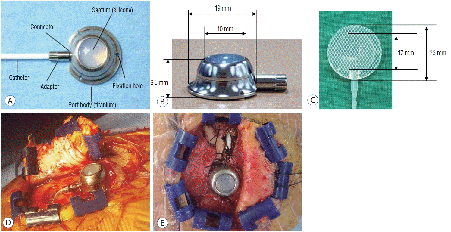
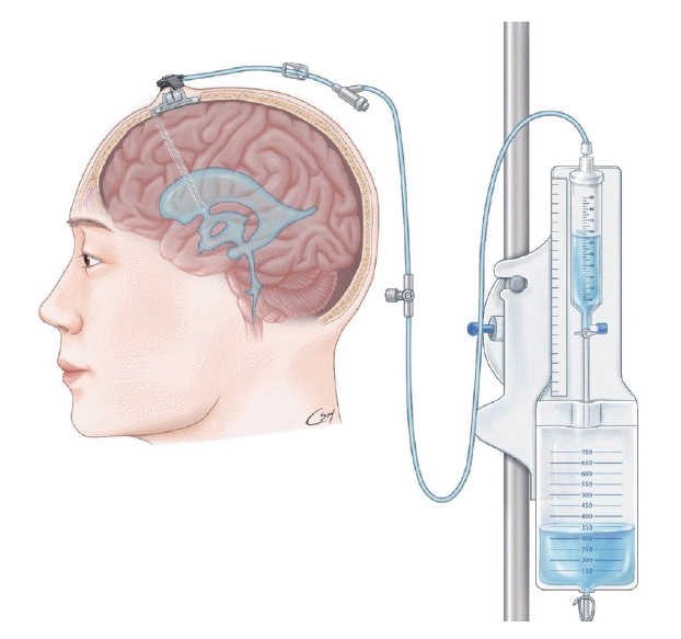
Fig. 1.
Photographs of H-port and installation process. The composition (A) and dimension of H-port (B), and that of Ommaya reservoir for comparison (C), operative side view of burrhole and side outlet port connected to ventricular catheter (D), and photo takne from above of periosteal apron closure (E).
jkns-2023-0178f1.tif
Fig. 2.
Medical illustration of H-port used for continuous extraventricular drainage of cerebrospinal fluid drawn by medical illustrator.
jkns-2023-0178f2.tif
Table 1.
Clinical characteristics of H-port patients
Characteristic H-port (n=152) Ommaya* (n=89) p-value
Gender NS
 Female 103 (67.0) 53 (60.0)
 Male 49 (32.0) 36 (40.0)
Age (years) 56 (5–84) 52 (15–76) NS
Primary cancer
 Lung 79 (52.0) 63 (71.0) 0.004
 Breast 47 (31.0) 18 (20.0)
 Gastric 8 (5.0) 1 (1.0)
 Ovary 4 (3.0) 1 (1.0)
 Other 14 (9.0) 6 (7.0)
KPS 80 (40–100) 60 (40–100) NS
Presenting symptoms and signs
 Headache with N/V 75 (49.0) 73 (82.0) <0.0001
 Altered mentality 25 (16.0) 27 (30.0)
 Cauda equina involvement 41 (27.0) 8 (9.0)
 Cranial neuropathy 23 (15.0) 9 (10.0)
Increased ICP (>15 cm H2O)
 Yes 71 (47.0) 45 (51.0) NS
 No 81 (53.0) 44 (49.0)

Values are presented as median (range) or number (%).

* Historical data from previous literature (see methods for details).

NS : not significant, KPS : Karnofsky performance score, N/V : nausea and vomiting, ICP : intracranial pressure

Table 2.
Adverse events associated with the installation and during the course of intraventricular chemotherapy
H-port (n=152) Ommaya (n=89) p-value
Installation-related NS
 Catheter malposition 2 3 NS
 Intracranial hemorrhage 2 0 NA
Intraventricular chemotherapy-related <0.001
 CSF leakage 3 13 <0.001
 Infection 14 12 NS
 Intracranial hemorrhage 0 4 NA

NS : not significant, NA : not applicable, CSF : cerebrospinal fluid

Table 3.
Increased ICP control rates, according to type of reservoir or method used
ICP control
Control rate (%) p-value
Controlled Uncontrolled
Reservoir type (n=125*) <0.001
 Ommaya 12 46 21.0
 H-port 40 27 60.0
Method (n=69) <0.0001
 Intermittent aspiration 6 50 11.0
 Continuous EVD 33 6 85.0

* In this analysis, patients who had shunt operations for uncontrolled ICP were excluded.

In this analysis, patients, who received additional cerebrospinal fluid drainage for palliation of symptoms from increased ICP control, were included.

ICP : intracranial pressure, EVD : extraventricular drainage

TOOLS
Similar articles