Abstract
Ankylosis of the temporomandibular joint (TMJ) is a condition in which the mandibular condyle fuses with the mandibular fossa through fibrous or bone tissue. It is a debilitating pathology that interferes with chewing, speaking, and oral hygiene. Currently, alloplastic reconstruction is considered the gold standard for treating severely compromised TMJs, such as in ankylosis. The article describes a patient with a history of facial trauma, with bilateral ankylosis of the TMJs, inability to open his mouth, and poor dental condition. Due to a long period of immobilization of approximately 40 years, the initial treatment plan was to remove the ankylosis bilaterally and install customized PMMA (polymethylmethacrylate) spacers. The patient gained mouth opening and improved chewing quality with one year of customized spacer use prior to definitive alloplastic replacement with stock-type TMJ prostheses. Customized joint spacers are a provisional treatment option when definitive alloplastic reconstruction is not indicated. Spacers provide the patient with progressive jaw function and mobility gains.
Ankylosis of the temporomandibular joint (TMJ) is a condition in which the condyle of the mandible fuses with the mandibular fossa through fibrous or bone tissue. It is a debilitating condition that interferes with chewing, speaking, and oral hygiene. Common etiological factors include trauma, inflammatory arthritis, infection, previous TMJ surgery, and congenital deformities, with trauma followed by infection being the most common factors. Multiple surgical modalities are proposed for treating TMJ ankylosis, such as gap arthroplasty, interpositional arthroplasty, and joint replacement. In total joint replacement, use of autogenous tissue such as costochondral grafts poses a risk of excessive growth, resorption, and recurrence1.
Alloplastic replacement is considered the gold standard for treating severely compromised TMJs, such as in cases of ankylosis2. According to Green et al.3, use of polymethylmethacrylate (PMMA) spacers prior to installation of TMJ prostheses leads to an easier and more accurate final surgery and is indicated when immediate installation of the prostheses is contraindicated.
Bone cement or PMMA has been used for decades to cement prostheses and create spacers in the field of orthopedics. The challenge with surgical spacers is that they cannot be made and sterilized prior to surgery4.
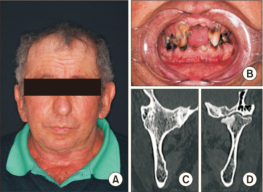
This article presents the case of a 67-year-old male patient who had been unable to open his mouth for over 40 years. He reported trauma during childhood but was unaware of the exact mechanism of facial fractures. On clinical examination, we observed the inability to open the mouth and poor dental condition due to difficulty with oral hygiene. Tomographically, a large ankylotic mass was observed in the right TMJ region, Sawney class IV. On the left side, a degenerated mandibular condyle was observed, leading to the hypothesis of fibrous ankylosis.(Fig. 1)
Due to decades of mandibular immobilization, the decision was made to create customized joint spacers prior to installation of permanent TMJ prostheses. With the help of the Additive Manufacturing and Health Innovation Laboratory of the Federal Institute of Santa Catarina, the joint spacer molds were created from tomographic images of the mandibular TMJ components from patients who underwent alloplastic reconstruction of the TMJ at Hospital Governador Celso Ramos. The same laboratory created a biomodel of the patient’s facial skeleton, which, together with the spacer molds, was sterilized in ethylene oxide.
To perform the surgical procedure, nasotracheal intubation was performed with the aid of a bronchoscope. Bilateral retromandibular and preauricular accesses were performed, and after removing the ankylosis bilaterally (Fig. 2), customized spacers were made from PMMA and fixed to the mandible using long bicortical screws.(Fig. 3)
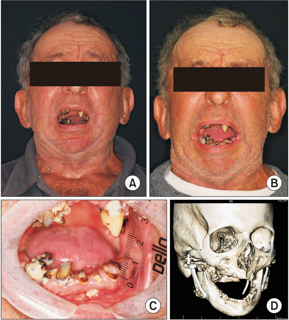
In the immediate postoperative period, a mouth opening of 10 mm, edema, and pain compatible with the surgical procedure were observed. In the 21-day postoperative period, the patient acquired mouth opening of 15 mm, had no clinical signs of infection, and reported significant improvement in chewing quality. At one year postoperatively, the patient had a mouth opening of approximately 21 mm and reestablished masticatory function, despite continued poor dental condition.(Fig. 4) The customized TMJ spacers were replaced with stock-type TMJ prostheses after one year, and multiple extractions were performed by the Service and Residency Program in Oral and Maxillofacial Surgery at Hospital Governador Celso Ramos.
Puricelli5 first proposed biconvex arthroplasty to treat ankylosis of the TMJ, in which two convex PMMA surfaces replace the affected structures. PMMA semi-spheres are molded during the plastic surgery phase and introduced over the condylar remnant and the mandibular fossa. This is a simple, safe, and low-cost technique.
Wolford et al.1 cite two treatment protocols for ankylosis of the TMJ: one single-stage treatment and one two-stage treatment. The two-stage protocol consists first of removing the ankylosis and installing a spacer, which may be made of bone cement, and the second stage involves alloplastic reconstruction. Sinn et al.6 mention the use of silicone balls as temporary spacers in the two-stage alloplastic reconstruction of pediatric patients with ankylosis of the TMJs.
Kong et al.7 carried out a study in which they analyzed the use of bone cement spacers in revision surgeries for infected knee prostheses. In this study, 22 patients were treated using traditional static bone cement spacers, and another 20 patients were treated using customized articulated knee spacers; both groups received antibiotics. In patients who received articulated spacers, less bone loss was observed in the femur and tibia and better satisfaction was reported among patients who received static spacers. Patients who had previously received articulated spacers had better postoperative range of motion after installation of new definitive prostheses than those who received static spacers.
According to Teschke et al.4, in cases of mandibular resection, including those with disarticulation, a surgical spacer is used to maintain symmetry of the hard and soft tissues. The customized surgical spacer offers a safe final mandibular reconstruction, including the TMJ, but spacer infection is the main risk of failure. It is crucial for creation of the customized surgical spacer, virtual surgical planning, and creation of molds to include sterile surgical cement. In cases where the surgical spacer is not inserted, the surgical field will be closed after resection of the pathological bone, and the periosteal tube will be lost, particularly for reconstruction of the TMJ. Second-stage surgery, without insertion of the surgical spacer, will be more difficult due to loss of dissection planes, increasing the risk of injury to the facial nerve.
Teschke et al.4 describe two cases that underwent mandibular resection including the TMJ with installation of a customized PMMA surgical spacer. The patients were treated in a second stage, finishing with removal of the customized spacers and installation of customized TMJ prostheses. The intervals between the first and second surgeries were 16 months and 24 months, respectively. According to Teschke et al.4, a space maintainer must be specifically designed for the patient through virtual planning, must have a suitable format for maintaining the periosteal tube, and must be biocompatible and biomechanically stable, perforable, and low cost. PMMA is an approved material that fulfills the desired properties.
In conclusion, TMJ ankylosis is a debilitating condition that is best treated through total joint replacement with TMJ prostheses. Customized joint spacers are a temporary treatment option when definitive alloplastic reconstruction is not indicated. They provide the patient with progressive jaw function and mobility gains. However, the use of these customized spacers requires second-stage surgery to install permanent prostheses, either stock or customized. PMMA TMJ spacers may be used in future studies for treating advanced joint diseases, especially as an alternative to high-cost joint devices.
Notes
Authors’ Contributions
C.A.M.U., F.B.D.D.S., and L.B.P. participated in data collection and writing the manuscript. C.A.M.U., F.B.D.D.S., and M.C. participated in surgery. M.B.M.B.S. participated in development PMMA condylar device in software. All authors read and approved the final manuscript.
References
1. Wolford L, Movahed R, Teschke M, Fimmers R, Havard D, Schneiderman E. 2016; Temporomandibular joint ankylosis can be successfully treated with TMJ concepts patient-fitted total joint prosthesis and autogenous fat grafts. J Oral Maxillofac Surg. 74:1215–27. https://doi.org/10.1016/j.joms.2016.01.017. DOI: 10.1016/j.joms.2016.01.017. PMID: 26878364.

2. Lotesto A, Miloro M, Mercuri LG, Sukotjo C. 2017; Status of alloplastic total temporomandibular joint replacement procedures performed by members of the American Society of Temporomandibular Joint Surgeons. Int J Oral Maxillofac Surg. 46:93–6. https://doi.org/10.1016/j.ijom.2016.08.002. DOI: 10.1016/j.ijom.2016.08.002. PMID: 27567049.

3. Green JM 3rd, Lawson ST, Liacouras PC, Wise EM, Gentile MA, Grant GT. 2016; Custom anatomical 3D spacer for temporomandibular joint resection and reconstruction. Craniomaxillofac Trauma Reconstr. 9:82–7. https://doi.org/10.1055/s-0035-1546814. DOI: 10.1055/s-0035-1546814. PMID: 26889353. PMCID: PMC4755798.

4. Teschke M, Christensen A, Far F, Reich RH, Naujokat H. 2021; Digitally designed, personalized bone cement spacer for staged TMJ and mandibular reconstruction - introduction of a new technique. J Craniomaxillofac Surg. 49:935–42. https://doi.org/10.1016/j.jcms.2021.05.002. DOI: 10.1016/j.jcms.2021.05.002. PMID: 34238634.

5. Puricelli E. 2022; Puricelli biconvex arthroplasty as an alternative for temporomandibular joint reconstruction: description of the technique and long-term case report. Head Face Med. 18:27. https://doi.org/10.1186/s13005-022-00331-4. DOI: 10.1186/s13005-022-00331-4. PMID: 35906643. PMCID: PMC9335964.

6. Sinn DP, Tandon R, Tiwana PS. 2021; Can alloplastic total temporomandibular joint reconstruction be used in the growing patient? A preliminary report. J Oral Maxillofac Surg. 79:2267.e1–16. https://doi.org/10.1016/j.joms.2021.06.022. DOI: 10.1016/j.joms.2021.06.022. PMID: 34339614.

7. Kong L, Mei J, Ge W, Jin X, Chen X, Zhang X, et al. 2021; Application of 3D printing-assisted articulating spacer in two-stage revision surgery for periprosthetic infection after total knee arthroplasty: a retrospective observational study. Biomed Res Int. 2021:3948638. https://doi.org/10.1155/2021/3948638. DOI: 10.1155/2021/3948638. PMID: 33628779. PMCID: PMC7884112.

Fig. 1
A. 67-year-old patient with both temporomandibular joints (TMJs) affected by ankylosis. B. Poor oral hygiene is observed due to decades of jaw immobilization. C. Coronal view of the right TMJ, in which Sawney class IV ankylosis is observed. D. In the coronal view of the left TMJ, we can observe fibrous ankylosis.

Fig. 2
A. Through a preauricular approach, the large ankylotic mass of the right temporomandibular joint is visualized for osteotomy. B. Initial osteotomy performed using a piezoelectric saw. C. The distal pole of the ankylotic mass is loosened with the aid of osteotomes. D. The deep, medial portion of the ankylotic mass is removed using a titanium screw and steel wire.

Fig. 3
A. Customized spacers manufactured intraoperatively of orthopedic cement, PMMA (polymethylmethacrylate), from molds manufactured by the Additive Manufacturing and Health Innovation Laboratory of the Federal Institute of Santa Catarina. B. Right customized spacer in contact with the mandibular fossa after removal of the ankylotic block. C. Right customized spacer fixed to the mandibular ramus using long titanium screws through the retromandibular approach. D. Customized orthopedic cement spacers installed after removal of the right ankylotic block and left condylectomy of the left condyle with fibrous ankylosis (three-dimensional reconstruction).

Fig. 4
A. Twenty-one days after bilateral arthroplasty with customized spacers. Mouth opening approximately 10 mm. B. One year postoperative, patient achieved mouth opening of approximately 20 mm. There are no painful joint symptoms or chewing complaints. C. Mouth opening after bilateral arthroplasty with installation of PMMA (polymethylmethacrylate) joint spacers (one year postoperatively). Due to personal issues, the patient was unable to receive dental treatment. D. The customized temporomandibular joint (TMJ) spacers were replaced with stock-type TMJ prostheses after one year, and multiple extractions were performed.


 PDF
PDF Citation
 Print
Print


 XML Download