Journal List > J Korean Assoc Oral Maxillofac Surg > v.50(3) > 1516087715

Burman, Lal, Alagarsamy, Kumar, Ankush, Rai, and Yunus: Evidence-based management of isolated dentoalveolar fractures: a systematic review

Abstract

Dentoalveolar (DA) trauma, which can involve tooth, alveolar bone, and surrounding soft tissues, is a significant dentofacial emergency. In emergency settings, physicians might lack comprehensive knowledge of timely procedures, causing delays for specialist referral. This systematic review assesses the literature on isolated DA fractures, emphasizing intervention timing and splinting techniques and duration in both children and adults. This systematic review adhered to PRISMA guidelines and involved a thorough search across PubMed, Google Scholar, Semantic Scholar, and the Cochrane Library from January 1980 to December 2022. Inclusion and exclusion criteria guided study selection, with data extraction and analysis centered on demographics, etiology, injury site, diagnostics, treatment timelines, and outcomes in pediatric (2-12 years) and adult (>12 years) populations. This review analyzed 26 studies, categorized by age into pediatrics (2-12 years) and adults (>12 years). Falls were a common etiology, primarily affecting the anterior maxilla. Immediate management involved replantation, repositioning, and splinting within 24 hours (pediatric) or 48 hours (adult). Composite resin-bonded splints were common. Endodontic treatment was done within a timeframe of 3 days to 12 weeks for children and 2-12 weeks for adults. Tailored management based on patient age, tooth development stage, time elapsed, and resource availability is essential.

I. Introduction

Dentoalveolar (DA) trauma is a common dentofacial injury, with a prevalence of 11%-30% among children and 5%-20% among adults1-5. It involves tooth trauma, alveolar bone fractures, or a combination of the two and constitutes a dental emergency. DA trauma can be due to falls, sports injuries, road traffic accidents, etc.6. Prompt diagnosis and early management of these injuries are crucial for ensuring the best possible outcomes7.
In clinical practice, DA fracture management is a two-stage procedure. The first stage involves the manual reduction of alveolar bone and repositioning or replantation of teeth, if required, followed by splinting for stabilization. Stabilization can be achieved with wire, an arch bar, fiber with composite material, or acrylic capping. The second stage involves whatever restorative, endodontic, or rehabilitative procedures are needed for each case. The timing and type of endodontic treatment depends on the associated injuries, fracture severity, and overall tooth and supporting structure condition8. Various novel and minimally invasive techniques have recently been introduced. The management of DA fractures depends on many factors, such as the site; type and extent of the fracture; associated dental trauma; tooth staging; adjacent available teeth; patient age, medical condition and willingness; patient compliance; presentation timing; choice of splint fixation; available resources; and surgeon preferences and expertise9.
In routine practice, general physicians in emergency settings often encounter these fractures and lack a detailed understanding of the emergency procedures for management, which can lead to delay in treatment and referral to specialists such as maxillofacial surgeons, pediatric dentists, or endodontists for definitive management10. These fractures often occur with other facial bone fractures, and they are usually managed together11. Isolated DA fracture cases receive limited attention in the emergency department and they are generally referred for specialist treatment on an outpatient basis12,13. This systematic review was conducted to summarize the literature on management strategies for and outcomes of isolated DA fractures in pediatric and adult populations.

II. Materials and Methods

The PRISMA (Preferred Reporting Items for Systematic reviews and Meta-Analyses) guidelines were followed to ensure the rigor and transparency of the systematic review process14. The PICO (Population, Intervention, Comparison, and Outcome) criteria were used. The population was individuals (pediatric and adult groups) with isolated DA fractures. The interventions were the various methods used for splinting. No comparisons were considered. The outcomes were basic demographic data, etiology, site involvement, associated dental trauma and its immediate treatment, type and duration of splinting, complications, and timing of endodontic treatment. The inclusion criteria for this systematic review encompassed prospective, retrospective, and comparative studies; case series; and case reports. Studies of DA fractures associated with other facial bone fractures were excluded. When institutions published multiple studies with overlapping patient populations or varying lengths of follow-up, only the most comprehensive reports were included for analysis to prevent the duplication of results. Studies in non-English languages, in vitro and animal studies, reviews, book chapters, and studies with insufficient or missing data were excluded to ensure reliability and minimize publication bias.

1. Search strategy

Electronic searches were conducted in PubMed, Google Scholar, Semantic Scholar, and Cochrane Library for studies published between January 1980 and December 2022 to ensure a comprehensive analysis of the available literature. To achieve maximum sensitivity and ensure the inclusion of all relevant studies, the search was conducted in a staged manner using individual and combined search terms, such as “dentoalveolar fracture,” “dentoalveolar trauma,” “splinting,” “bridle wire,” “wiring,” “arch bar,” “composite splint,” “fiber splinting,” “teeth splinting,” “semi-rigid,” “rigid,” “flexible,” “splint retainer,” and “orthodontic retainer”.(Table 1) Duplicate studies were removed from the combined results of the databases. The reference lists of all final retrieved articles were carefully reviewed to identify other potentially relevant studies that met the inclusion criteria.

2. Data collection

Data collection and analysis involved examining studies that met the specified inclusion and exclusion criteria. Two reviewers (S.B. and B.L.) analyzed the studies independently at the title, abstract, and full-text levels. Any discrepancies or conflicts were resolved through mutual consensus.
The following data were extracted for analysis: the study type, total number of patients, age group (≤12 years and >12 years), sex of patients, mechanism of injury, site of involvement, investigations conducted, time elapsed between injury and splinting, type of splint used, duration of splint placement, late dental management, complications encountered, and follow-up duration.

3. Risk of bias in individual studies

The risk of bias was assessed using the Joanna Brigs University tool15. A high risk of bias was identified when ≤49% of the answers were positive; moderate risk of bias was assumed when 50%-69% of the answers were positive, and a low risk of bias was identified when more than 70% of the answers were positive.

III. Results

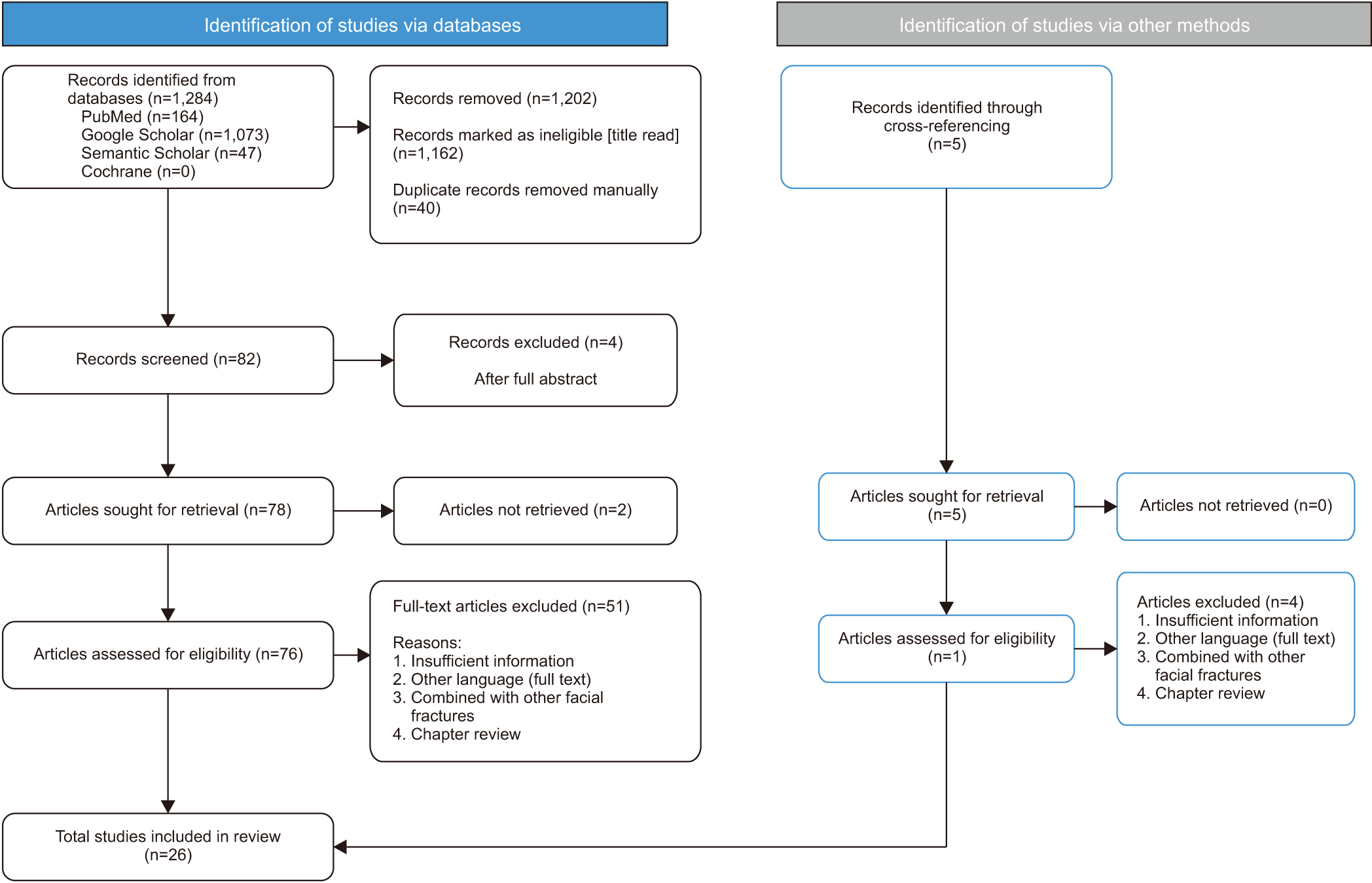
The selection process is depicted in Fig. 1 (PRISMA flowchart). A total of 78 studies were included for full-text reading by the two investigators. Data extraction and analysis were done for 26 studies. For simplicity and better insights, two groups were made based on age: the 2-12 years age group (pediatric) and the older than 12 years age group (adult)16-41.(Tables 2, 3)

1. Pediatric (2-12 years)

Eighteen case reports were analyzed (male 9, female 9). Fall (n=14) was the most common reported etiology. The anterior maxilla (n=10) was the most reported site, followed by the anterior mandible (n=6). Orthopantomogram (OPG) (n=11) was the most frequently conducted radiographic investigation. The reported dental injuries were avulsion (n=9), luxation (n=4), extrusion (n=1), intrusion (n=3), and enamel/crown fracture (n=3). Among the 9 cases of tooth avulsion, replantation was done in 4 permanent teeth, repositioning (n=7) was done in cases of luxation or intrusion and composite restoration (n=3) was done for fractured teeth.
Splinting was done within 24 hours in 7 cases, but the timeframe was not mentioned in the others. The splinting types were a composite resin-bonded splint (n=9), acrylic resin splint (n=5), circummandibular wiring with maxillomandibular fixation (n=1), suture (n=1), titanium mesh (n=1), and vacuum splint (n=1), and splinting was done under either local anesthesia (n=13) or general anesthesia (n=5). The splinting duration was mostly 2-4 weeks (n=13), although it differed among the studies. Endodontic intervention (n=6) was performed between 3 days and 12 weeks after the initial injury. Rehabilitation used removable partial dentures in one case. Complications such as resorption (n=2), abscess (n=1), and gingival recession (n=1) were reported. Follow-up ranged between 1 month and 42 months.

2. Adult (>12 years)

Eight studies were included (male 7, female 3). Fall (n=5) was the most common reported etiology. The anterior maxilla (n=6) was the most reported site, followed by the anterior mandible (n=1). OPG (n=3) was the most conducted radiographic investigation. Dental injuries included avulsion (n=1), luxation (n=1), and enamel/crown fracture (n=3). Replantation was done in one patient, repositioning (n=3) was conducted in cases of luxation, and selective grinding was done in one case.
Splinting was done within 48 hours in 4 patients, and the timeframe was not mentioned in the other studies. Splinting was done with a composite resin-bonded splint (n=4), bone screw stabilization (n=1, 3 cases), and an Erich’s arch bar with wire (n=3) under local anesthesia (n=6, 7 cases) or general anesthesia (n=2). The splinting duration varied between 2-4 weeks (n=4) and 6-8 weeks (n=2). Endodontic intervention (n=4) was performed between 2 and 12 weeks after the initial injury. A porcelain veneer (n=2) was done for fractured teeth, and rehabilitation used removable partial dentures in one study (3 cases). Follow-up ranged between 3 months and 60 months. Comparative results are depicted in Table 4.

3. Risk of bias assessment

Among the included studies (n=26), 22 had a low risk of bias, and four had a moderate risk of bias (Martins and Fávaro31, Al-Hadad et al.40, Nyárády et al.39, Ali et al.36).(Supplementary Table 1)

IV. Discussion

DA fractures involve the alveolar bone, teeth, and associated soft tissue structures42. They can occur in isolation or in association with other facial bone fractures. Cases involving concurrent injuries often present as emergencies and are managed in accordance with established protocols. In contrast, isolated DA cases might not promptly report to an emergency department, and when they do, they might not receive prompt attention. In fact, these cases should be considered sub-acute emergencies because the intervention timing directly influences the prognosis of the affected teeth and bones. In addition, injuries to primary teeth affect the permanent dentition43. Whenever feasible, it is important to expeditiously address such injuries to enhance the long-term outcomes and restoration of normal function and esthetics. Evidence on the epidemiology and management of isolated DA fractures is limited. Therefore, this systematic review was conducted to pool the data on DA management and outcomes in pediatric and adult patients.
The incidence of DA fractures is challenging to estimate because they often occur in conjunction with dental injuries and/or craniofacial trauma. Falls were the most common etiology reported. The pediatric population is more prone to falls during activities such as cycling19,25,26 and playing20,23,24,28,33 and to accidental falls at home16,17,29,31. In contrast, the adult population is susceptible to injuries due to falling under the influence of alcohol, assault, or sports-related activities34,36.
Determining the extent of DA trauma and associated dental injuries through clinical and radiographic examination can facilitate prompt treatment. The present review reveals that alveolar fractures most frequently occur in association with dental trauma. The International Association of Dental Traumatology (IADT) guidelines advise the use of a periapical radiograph and two additional radiographs in angulations to diagnose alveolar fractures, supplemented by panoramic and cone-beam computed tomography (CBCT), if necessary. However, various radiographic investigations were reported in the included studies: intraoral periapical radiographs (IOPA)20,24,25,27,36, OPG16,18-21,23,24,27,29,30,33,35, posteroanterior skull radiographs (PA skull)17, CBCT37,38, and non-contrast computed tomography17,18,29. Among them, OPG was most commonly reported in both age groups. This preference for OPG might be attributed to the emergency settings in which DA fractures typically present, where periapical radiographs are often unavailable, and OPG can reveal a horizontal fracture level above the tooth apices. IOPA helps to diagnose and monitor individual tooth injuries. Computed tomography can help to identify the degree of displacement and make alveolar fractures more apparent. PA skull has a limited role in such injuries. In certain cases, a clinician might perform the initial treatment after clinical examination without radiographic examination to prevent delays.
The anterior maxilla was the most common site in both groups, which can be attributed to its anatomical alignment, which makes it vulnerable. The literature suggests a decrease in the overall incidence of DA injury when improved safety equipment, such as mouth guards and face masks, is used44,45. Among the associated dental injuries, avulsion, luxation, extrusion, and intrusion were more prevalent within the pediatric population, and crown and enamel fractures were more common in the adult group. This might be attributed to differences between pediatric and adult patients in tooth-housing bone. The forces are directly transmitted to the teeth in adults due to their strong bone base.
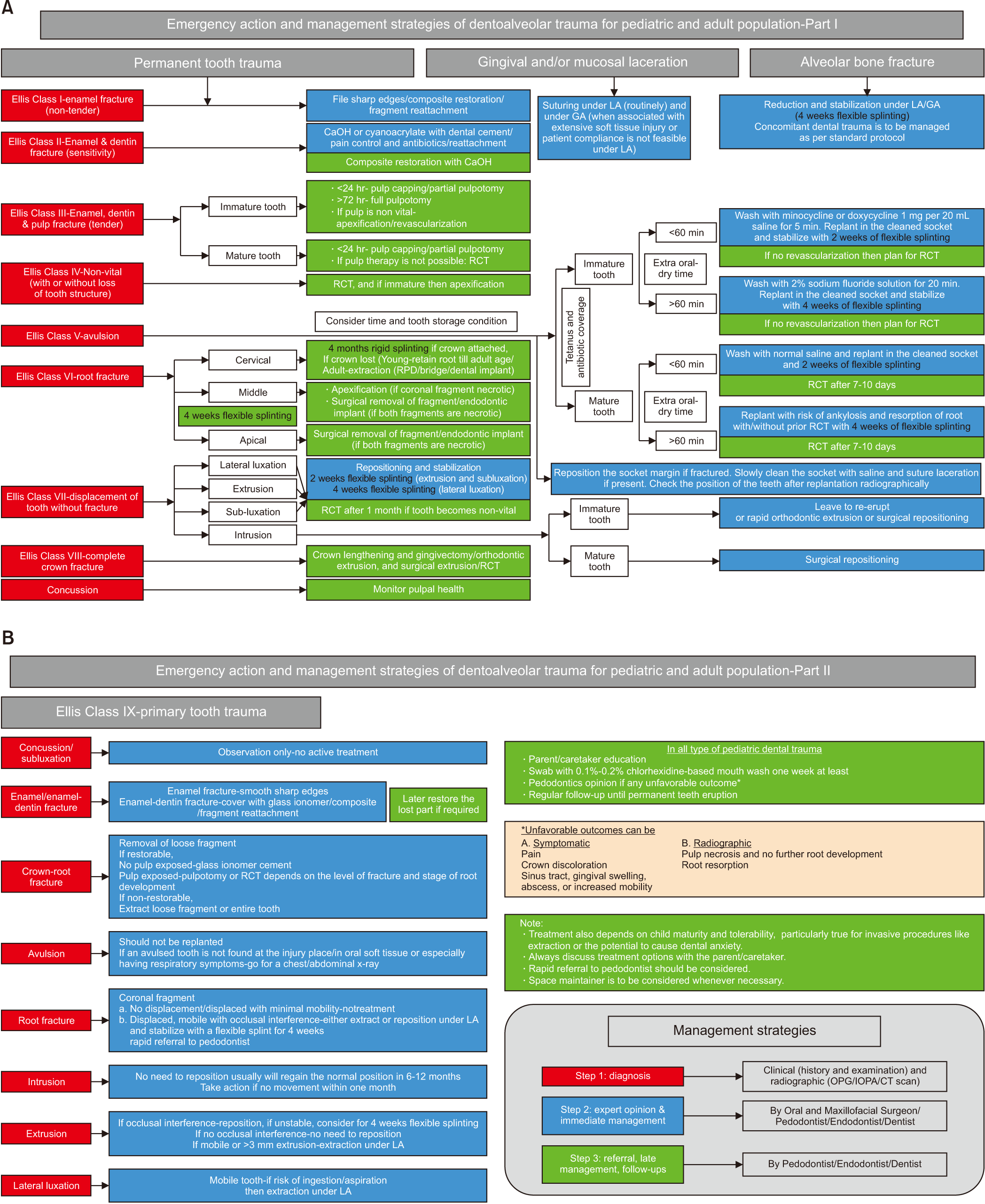
The management of DA fractures and associated dental trauma requires a comprehensive approach tailored to each patient and should be guided by the tooth type and stage of tooth development, which is of greater clinical relevance and significance than chronological age46-48. Repositioning in luxation or intrusion injuries should be performed before the reduction of alveolar bone because they might hinder reduction. However, the replantation of avulsed teeth and repositioning of intruded teeth are not recommended for primary teeth. The flowchart compiled from the literature evidence outlines the recommended actions to be undertaken in emergency settings for strategic management of isolated DA trauma49-57.(Fig. 2) This chart might help clinicians and dental practitioners in emergency settings make prompt decisions for effective management and timely referrals.
Following reduction, stabilization can be achieved by a variety of methods such as composite resin-bonded splints, acrylic resin, suture, titanium mesh, vacuum splints, and circummandibular wires for the pediatric population and Erich arch bars with wires, screw stabilization, titanium mini plates and bonded splints for the adult population. Splinting can aid in the immobilization of both tooth and DA fractures. The choice of a splint depends on patient compliance and the surgeon’s preference. Short-term, non-rigid splints are advised53. Interestingly, composite resin-bonded splints were commonly used in both populations, possibly due to their high patient compliance. Arch bars can pose challenges in hygiene maintenance and can thus affect gingival health. The evidence indicates that the splint type is not significantly related to treatment outcomes58. This review reveals that splinting was typically done for 2-4 weeks using rigid splints in cases of concomitant dental and alveolar fractures, which warrants further research. The IADT guidelines provide distinct recommendations about the duration and type of splinting for alveolar fractures and dental trauma as separate entities. However, scientific evidence is lacking about the best splinting time for combined injuries and the effects on pulpal and periodontal healing. The choice between local and general anesthesia depends on patient factors, such as low compliance, high anxiety, and timing delays that can complicate manipulation under local anesthesia. Both age groups were treated with both types of anesthesia, and general anesthesia was sometimes used for debridement and suturing of associated soft tissue injuries.
Independent of the type of splinting, the timing of splinting is crucial. In the included studies, splinting was done within 24-48 hours, revealing a delay in the management of sub-acute emergencies. The duration of splinting varies between the pediatric and adult populations. A splinting duration of 2-4 weeks was commonly reported in both groups, which contrasts with the IADT recommendation of 4 weeks for all ages. However, bone healing rates and tooth development vary between children and adults. Typically, pediatric cases required shorter splinting durations (2-3 weeks), and adults needed longer splinting periods (3-4 weeks)59. The period of immobilization can also vary on a case-to-case basis depending on the severity of the fracture.
Late dental injury management includes endodontic treatment, definitive restoration, and rehabilitation. Not all cases require additional treatment. The timing and type of endodontic treatment depend on the patient’s age and root development. In pediatric patients with immature teeth, apexification or apexogenesis techniques are used to promote the formation of a natural apical barrier, enabling successful root canal treatment60. In contrast, adults typically have fully formed roots, allowing for standard endodontic procedures. Composite restorations were used to repair tooth, enamel, and crown fractures in both age groups. Veneers or crowns were reported only in the adult group; however, they can also be used in pediatric patients as needed. Rehabilitation procedures such as removable partial dentures were used in cases of permanent tooth loss in both groups, and space maintainers can be used in cases of primary tooth loss61. A detailed algorithm for late treatment is provided in Fig. 2.
Unfavorable outcomes, such as submental abscess17, replacement root resorption23, and gingival recession25, were reported in pediatric patients, but none were reported in adults in the included studies. This emphasizes the need for specialized attention, patient compliance, and follow-up in pediatric populations. Follow-up periods were between one month and five years. Long-term follow-up is crucial because DA structures continue to grow, and achieving functional occlusion is essential for oral function.
This systematic review included isolated DA fractures and their management. Although it offers valuable insights, the limited number of studies and potential for selection bias restrict the generalizability of these results. Persistent efforts and policies are required to raise awareness and thus prevent DA trauma and improve its management in emergency settings.

V. Conclusion

In conclusion, isolated DA trauma should be considered a sub-acute emergency condition that requires immediate attention for optimal outcomes. These injuries are more prevalent among children than adults, and fall is the most common cause for all ages. The upper anterior teeth and maxilla are commonly involved. Staged treatment by specialists and regular follow-up are the keys to overall management, which should be tailored the age of the patient, stage of tooth development, time elapsed since injury, and availability of resources. DA injuries can be prevented by spreading awareness about the importance of safety measures for children during activities such as cycling and playing. Policies should be developed to create awareness and sensitize physicians in emergency settings about the management of DA fractures and prompt referral to specialists.

Supplementary Materials

Supplementary data is available at http://www.jkaoms.org.

Notes

Funding

No funding to declare.

Authors’ Contributions

S.B. and B.L. contributed to formulating the research question, protocol development, literature search, study selection, data extraction, and manuscript writing. R.A. contributed to data synthesis and manuscript writing. J.K., A.A., A.J.R., and M.Y. participated in the study design and coordination and helped to draft the manuscript. All authors read and approved the final manuscript.

Conflict of Interest

No potential conflict of interest relevant to this article was reported.

References

1. Andersson L. 2013; Epidemiology of traumatic dental injuries. J Endod. 39(3 Suppl):S2–5. https://doi.org/10.1016/j.joen.2012.11.021. DOI: 10.1016/j.joen.2012.11.021. PMID: 23439040.
crossref
2. Motamedi MH. 2003; An assessment of maxillofacial fractures: a 5-year study of 237 patients. J Oral Maxillofac Surg. 61:61–4. https://doi.org/10.1053/joms.2003.50049. DOI: 10.1053/joms.2003.50049. PMID: 12524610.
crossref
3. Ravn JJ. 1974; Dental injuries in Copenhagen schoolchildren, school years 1967-1972. Community Dent Oral Epidemiol. 2:231–45. https://doi.org/10.1111/j.1600-0528.1974.tb01658.x. DOI: 10.1111/j.1600-0528.1974.tb01658.x. PMID: 4154162.
crossref
4. Iida S, Matsuya T. 2002; Paediatric maxillofacial fractures: their aetiological characters and fracture patterns. J Craniomaxillofac Surg. 30:237–41. https://doi.org/10.1054/jcms.2002.0295. DOI: 10.1054/jcms.2002.0295. PMID: 12231205.
crossref
5. Andreasen JO. 1970; Fractures of the alveolar process of the jaw. A clinical and radiographic follow-up study. Scand J Dent Res. 78:263–72. https://doi.org/10.1111/j.1600-0722.1970.tb02073.x. DOI: 10.1111/j.1600-0722.1970.tb02073.x. PMID: 5273694.
crossref
6. Glendor U. 2009; Aetiology and risk factors related to traumatic dental injuries--a review of the literature. Dent Traumatol. 25:19–31. https://doi.org/10.1111/j.1600-9657.2008.00694.x. DOI: 10.1111/j.1600-9657.2008.00694.x. PMID: 19208007.
crossref
7. Andreasen JO, Andreasen FM, Skeie A, Hjørting-Hansen E, Schwartz O. 2002; Effect of treatment delay upon pulp and periodontal healing of traumatic dental injuries -- a review article. Dent Traumatol. 18:116–28. https://doi.org/10.1034/j.1600-9657.2002.00079.x. DOI: 10.1034/j.1600-9657.2002.00079.x. PMID: 12110104.
crossref
8. Olynik CR, Gray A, Sinada GG. 2013; Dentoalveolar trauma. Otolaryngol Clin North Am. 46:807–23. https://doi.org/10.1016/j.otc.2013.06.009. DOI: 10.1016/j.otc.2013.06.009. PMID: 24138739.
crossref
9. Goswami M, Eranhikkal A. 2020; Management of traumatic dental injuries using different types of splints: a case series. Int J Clin Pediatr Dent. 13:199–202. https://doi.org/10.5005/jp-journals-10005-1746. DOI: 10.5005/jp-journals-10005-1746. PMID: 32742103. PMCID: PMC7366761.
crossref
10. Abu-Dawoud M, Al-Enezi B, Andersson L. 2007; Knowledge of emergency management of avulsed teeth among young physicians and dentists. Dent Traumatol. 23:348–55. https://doi.org/10.1111/j.1600-9657.2006.00477.x. DOI: 10.1111/j.1600-9657.2006.00477.x. PMID: 17991234.
crossref
11. Lieger O, Zix J, Kruse A, Iizuka T. 2009; Dental injuries in association with facial fractures. J Oral Maxillofac Surg. 67:1680–4. https://doi.org/10.1016/j.joms.2009.03.052. DOI: 10.1016/j.joms.2009.03.052. PMID: 19615582.
crossref
12. Batstone MD, Waters C, Porter SA, Monsour FN. 2004; Treatment delays in paediatric dento-alveolar trauma at a tertiary referral hospital. Aust Dent J. 49:28–32. https://doi.org/10.1111/j.1834-7819.2004.tb00046.x. DOI: 10.1111/j.1834-7819.2004.tb00046.x. PMID: 15104131.
crossref
13. Needleman HL, Stucenski K, Forbes PW, Chen Q, Stack AM. 2013; Massachusetts emergency departments' resources and physicians' knowledge of management of traumatic dental injuries. Dent Traumatol. 29:272–9. https://doi.org/10.1111/j.1600-9657.2012.01170.x. DOI: 10.1111/j.1600-9657.2012.01170.x. PMID: 22804874. PMCID: PMC3955057.
crossref
14. Page MJ, McKenzie JE, Bossuyt PM, Boutron I, Hoffmann TC, Mulrow CD, et al. 2021; The PRISMA 2020 statement: an updated guideline for reporting systematic reviews. BMJ. 372:n71. https://doi.org/10.1136/bmj.n71. DOI: 10.1136/bmj.n71. PMID: 33782057. PMCID: PMC8005924.
crossref
15. Moola S, Munn Z, Tufanaru C, Aromataris E, Sears K, Sfetcu R, et al. Aromataris E, Munn Z, editors. 2020. Systematic reviews of etiology and risk. JBI reviewer's manual. JBI;p. 219–71. DOI: 10.46658/JBIRM-17-06. PMCID: PMC6429893.
16. Khan MH, Singh G, Charul K, Ezhilarasi S. 2020; Management of dentoalveolar fracture of anterior maxilla following traumatic intrusion of permanent maxillary anterior teeth: a review and case report. Traumaxilla. 2:28–31. https://doi.org/10.1177/26323273211072351. DOI: 10.1177/26323273211072351.
crossref
17. Quinto JHS, Bachesk AB, Nogueira LC, Filho LI. 2022; An alternative approach using circummandibular wiring for treatment of dentoalveolar fractures in children: a case report. Craniomaxillofac Trauma Reconstr. 15:169–74. https://doi.org/10.1177/1943387521990283. DOI: 10.1177/1943387521990283. PMID: 35633772. PMCID: PMC9133519.
crossref
18. Thakur A, Thakur S. 2021; Management of a complex dentoalveolar traumatic injury with multiple avulsions. Case Rep Dent. 2021:2373785. https://doi.org/10.1155/2021/2373785. DOI: 10.1155/2021/2373785. PMID: 34258075. PMCID: PMC8261170.
crossref
19. Nilesh K, Mahamuni A, Taur S, Vande AV. 2020; A simple novel technique for the management of a dentoalveolar fracture in a pediatric patient using a vacuum-formed splint. J Dent Res Dent Clin Dent Prospects. 14:68–72. https://doi.org/10.34172/joddd.2020.010. DOI: 10.34172/joddd.2020.010. PMID: 32454961. PMCID: PMC7235701.
crossref
20. Patel C, Bukhari T. 2020; Management of dentoalveolar traumatic injuries in primary dentition: a case report. J Dent Sci. 10:123–7.
21. Vidhya R, Madhu S, Vadakkepurayil K, Anupam KTV. 2017; Management of maxillary dento alveolar fracture with a modified splinting method: a case report. IOSR J Dent Med Sci. 16:51–4.
22. Sreejith VP, Das JR, Anooj PD, Sanal OV. 2015; Management of dentoalveolar fracture in a 2 year old child. Kerala Dent J. 38:27–9.
23. Sangeetha KM, Surendra P, Roshan NM, Subba Reddy VV, Chaur RG, Srinivasa SB. 2014; Management of dentoalveolar fracture with multiple avulsions: a case report with three years follow-up. Int J Case Rep Images. 5:835–8. https://doi.org/10.5348/ijcri-2014144-CR-10455. DOI: 10.5348/ijcri-2014144-CR-10455.
crossref
24. Yonezawa H, Yanamoto S, Hoshino T, Yamada S, Fujiwara T, Umeda M. 2013; Management of maxillary alveolar bone fracture and severely intruded maxillary central incisor: report of a case. Dent Traumatol. 29:416–9. https://doi.org/10.1111/j.1600-9657.2011.01111.x. DOI: 10.1111/j.1600-9657.2011.01111.x. PMID: 22233224.
crossref
25. De Rossi M, De Rossi A, Queiroz AM, Nelson Filho P. 2009; Management of a complex dentoalveolar trauma: a case report. Braz Dent J. 20:259–62. https://doi.org/10.1590/s0103-64402009000300016. DOI: 10.1590/S0103-64402009000300016. PMID: 19784475.
crossref
26. Saito C, Gulinelli J, Cardoso L, Garcia IR Jr, Panzarini S, Poi W, et al. 2009; Severe fracture of the maxillary alveolar process associated with extrusive luxation and tooth avulsion: a case report. J Contemp Dent Pract. 10:91–7. DOI: 10.5005/jcdp-10-1-91. PMID: 19142261.
crossref
27. Koyuturk AE, Kusgoz A. 2008; Multiple dentoalveolar traumatic injury: a case report (3 years follow up). Dent Traumatol. 24:e16–9. https://doi.org/10.1111/j.1600-9657.2008.00579.x. DOI: 10.1111/j.1600-9657.2008.00579.x. PMID: 18557754.
crossref
28. Sahin S, Saygun NI, Kaya Y, Ozdemir A. 2008; Treatment of complex dentoalveolar injury--avulsion and loss of periodontal tissue: a case report. Dent Traumatol. 24:581–4. https://doi.org/10.1111/j.1600-9657.2008.00607.x. DOI: 10.1111/j.1600-9657.2008.00607.x. PMID: 18821969.
crossref
29. Das UM, Viswanath D, Subramanian V, Agarwal M. 2007; Management of dentoalveolar injuries in children: a case report. J Indian Soc Pedod Prev Dent. 25:183–6. https://doi.org/10.4103/0970-4388.37015. DOI: 10.4103/0970-4388.37015. PMID: 18007105.
crossref
30. Prabhakar AR, Tauro DP, Shubha AB. 2006; Management of an unusual maxillary dentoalveolar fracture: a case report. J Dent Child (Chic). 73:112–5.
31. Martins WD, Fávaro DM. 2005; Fracture of the alveolar process in a 2-year-old child: a report of an unconventional immobilization. J Contemp Dent Pract. 6:134–8. DOI: 10.5005/jcdp-6-3-134. PMID: 16127482.
crossref
32. Martins WD, Westphalen FH, Westphalen VP, Souza PH. 2004; Multiple dentoalveolar traumatic lesions: report of a case and proposition of dental polytrauma as a new term. J Contemp Dent Pract. 5:139–47. DOI: 10.5005/jcdp-5-4-139. PMID: 15558099.
crossref
33. Sheroan MM, Roberts MW. 2004; Management of a complex dentoalveolar trauma with multiple avulsions: a case report. Dent Traumatol. 20:222–5. https://doi.org/10.1111/j.1600-9657.2004.00226.x. DOI: 10.1111/j.1600-9657.2004.00226.x. PMID: 15245521.
crossref
34. Dhusia AH, Sonawane H, Verma RS, Uchale P, Jagdale H. 2021; Emergency management of maxillary dentoalveolar fracture: a case report. J Dent Spec. 9:76–9. https://doi.org/10.18231/j.jds.2021.019. DOI: 10.18231/j.jds.2021.019.
crossref
35. Firstyananda W, Sjamsudin E. 2018; Management of dentoalveolar fracture by using rigid wire and composite splint: a case report. Intisari Sains Medis. 9:85–8. https://doi.org/10.15562/ism.v9i2.266. DOI: 10.15562/ism.v9i2.266.
crossref
36. Ali FM, Khan MMA, Faqihi AAMA, Mutawwam FA. 2017; Maxillary anterior region contact sport trauma in a patient having history of proclined maxillary anterior teeth. Saudi J Sports Med. 17:115–7. https://doi.org/10.4103/1319-6308.207571. DOI: 10.4103/1319-6308.207571.
crossref
37. Serra-Pastor B, Penarrocha-Diago M, Penarrocha-Diago M, Agustín-Panadero R. 2016; Treatment and restoration of adult dentoalveolar trauma: a clinical case report. J Clin Exp Dent. 8:e634–7. https://doi.org/10.4317/jced.52990. DOI: 10.4317/jced.52990. PMID: 27957283. PMCID: PMC5149104.
crossref
38. Faus-Matoses V, Martínez-Viñarta M, Alegre-Domingo T, Faus-Matoses I, Faus-Llácer VJ. 2014; Treatment of multiple traumatized anterior teeth associated with an alveolar bone fracture in a 20-year-old patient: a 3-year follow up. J Clin Exp Dent. 6:e425–9. https://doi.org/10.4317/jced.51374. DOI: 10.4317/jced.51374. PMID: 25593668. PMCID: PMC4282913.
crossref
39. Nyárády Z, Orsi E, Nagy K, Olasz L, Nyárády J. 2010; Transgingival lag-screw osteosynthesis of alveolar process fracture. Int J Oral Maxillofac Surg. 39:779–82. https://doi.org/10.1016/j.ijom.2010.01.022. DOI: 10.1016/j.ijom.2010.01.022. PMID: 20452745.
crossref
40. Al-Hadad I, Burke GA, Webster K. 2009; Dentoalveolar fracture of the posterior maxilla. Br J Oral Maxillofac Surg. 47:165. https://doi.org/10.1016/j.bjoms.2008.08.004. DOI: 10.1016/j.bjoms.2008.08.004. PMID: 18818006.
crossref
41. Finucane D, Fleming P, Smith O. 2004; Dentoalveolar trauma in a patient with chronic idiopathic thrombocytopenic purpura: a case report. Pediatr Dent. 26:352–4.
42. Dale RA. 2000; Dentoalveolar trauma. Emerg Med Clin North Am. 18:521–38. https://doi.org/10.1016/s0733-8627(05)70141-3. DOI: 10.1016/S0733-8627(05)70141-3. PMID: 10967737.
crossref
43. Tewari N, Mathur VP, Singh N, Singh S, Pandey RK. 2018; Long-term effects of traumatic dental injuries of primary dentition on permanent successors: a retrospective study of 596 teeth. Dent Traumatol. 34:129–34. https://doi.org/10.1111/edt.12391. DOI: 10.1111/edt.12391. PMID: 29495106.
crossref
44. Maeda Y, Kumamoto D, Yagi K, Ikebe K. 2009; Effectiveness and fabrication of mouthguards. Dent Traumatol. 25:556–64. https://doi.org/10.1111/j.1600-9657.2009.00822.x. DOI: 10.1111/j.1600-9657.2009.00822.x. PMID: 19788425.
crossref
45. Farrington T, Onambele-Pearson G, Taylor RL, Earl P, Winwood K. 2012; A review of facial protective equipment use in sport and the impact on injury incidence. Br J Oral Maxillofac Surg. 50:233–8. https://doi.org/10.1016/j.bjoms.2010.11.020. DOI: 10.1016/j.bjoms.2010.11.020. PMID: 21295384.
crossref
46. Turkistani J, Hanno A. 2011; Recent trends in the management of dentoalveolar traumatic injuries to primary and young permanent teeth. Dent Traumatol. 27:46–54. https://doi.org/10.1111/j.1600-9657.2010.00950.x. DOI: 10.1111/j.1600-9657.2010.00950.x. PMID: 21244628.
crossref
47. Muñante-Cárdenas JL, Olate S, Asprino L, de Albergaria Barbosa JR, de Moraes M, Moreira RW. 2011; Pattern and treatment of facial trauma in pediatric and adolescent patients. J Craniofac Surg. 22:1251–5. https://doi.org/10.1097/scs.0b013e31821c696c. DOI: 10.1097/SCS.0b013e31821c696c. PMID: 21772206.
crossref
48. Andrade NN, Choradia S, Sriram SG. 2015; An institutional experience in the management of pediatric mandibular fractures: a study of 74 cases. J Craniomaxillofac Surg. 43:995–9. https://doi.org/10.1016/j.jcms.2015.03.020. DOI: 10.1016/j.jcms.2015.03.020. PMID: 26027869.
crossref
49. Day PF, Flores MT, O'Connell AC, Abbott PV, Tsilingaridis G, Fouad AF, et al. 2020; International Association of Dental Traumatology guidelines for the management of traumatic dental injuries: 3. Injuries in the primary dentition. Dent Traumatol. 36:343–59. https://doi.org/10.1111/edt.12576. DOI: 10.1111/edt.12576. PMID: 32458553.
crossref
50. Tewari N, Bansal K, Mathur VP. 2019; Dental trauma in children: a quick overview on management. Indian J Pediatr. 86:1043–7. https://doi.org/10.1007/s12098-019-02984-7. DOI: 10.1007/s12098-019-02984-7. PMID: 31197645.
crossref
51. Bourguignon C, Cohenca N, Lauridsen E, Flores MT, O'Connell AC, Day PF, et al. 2020; International Association of Dental Traumatology guidelines for the management of traumatic dental injuries: 1. Fractures and luxations. Dent Traumatol. 36:314–30. https://doi.org/10.1111/edt.12578. DOI: 10.1111/edt.12578. PMID: 32475015.
crossref
52. Levin L, Day PF, Hicks L, O'Connell A, Fouad AF, Bourguignon C, et al. 2020; International Association of Dental Traumatology guidelines for the management of traumatic dental injuries: general introduction. Dent Traumatol. 36:309–13. https://doi.org/10.1111/edt.12574. DOI: 10.1111/edt.12574. PMID: 32472740.
crossref
53. Diangelis AJ, Andreasen JO, Ebeleseder KA, Kenny DJ, Trope M, Sigurdsson A, et al. 2012; ; International Association of Dental Traumatology. International Association of Dental Traumatology guidelines for the management of traumatic dental injuries: 1. Fractures and luxations of permanent teeth. Dent Traumatol. 28:2–12. https://doi.org/10.1111/j.1600-9657.2011.01103.x. DOI: 10.1111/j.1600-9657.2011.01103.x.
crossref
54. Fouad AF, Abbott PV, Tsilingaridis G, Cohenca N, Lauridsen E, Bourguignon C, et al. 2020; International Association of Dental Traumatology guidelines for the management of traumatic dental injuries: 2. Avulsion of permanent teeth. Dent Traumatol. 36:331–42. https://doi.org/10.1111/edt.12573. DOI: 10.1111/edt.12573. PMID: 32460393.
crossref
55. Saikia A, Patil SS, Ms M, Cv D, Sabarish R, Pandian S, et al. 2023; Systematic review of clinical practice guidelines for traumatic dental injuries. Dent Traumatol. 39:371–80. https://doi.org/10.1111/edt.12838. DOI: 10.1111/edt.12838. PMID: 36920339.
crossref
56. Zaror C, Seiffert A, Deana NF, Espinoza-Espinoza G, Atala-Acevedo C, Diaz R, et al. 2023; Emergency and sequalae management of traumatic dental injuries: a systematic survey of clinical practice guidelines. BMC Oral Health. 23:704. https://doi.org/10.1186/s12903-023-03409-w. DOI: 10.1186/s12903-023-03409-w. PMID: 37777731. PMCID: PMC10543333.
crossref
57. Moule A, Cohenca N. 2016; Emergency assessment and treatment planning for traumatic dental injuries. Aust Dent J. 61 Suppl 1:21–38. https://doi.org/10.1111/adj.12396. DOI: 10.1111/adj.12396. PMID: 26923446.
crossref
58. Kahler B, Heithersay GS. 2008; An evidence-based appraisal of splinting luxated, avulsed and root-fractured teeth. Dent Traumatol. 24:2–10. https://doi.org/10.1111/j.1600-9657.2006.00480.x. DOI: 10.1111/j.1600-9657.2006.00480.x. PMID: 18173657.
crossref
59. Theologie-Lygidakis N, Schoinohoriti OK, Leventis M, Iatrou I. 2017; Evaluation of dentoalveolar trauma in children and adolescents: a modified classification system and surgical treatment strategies for its management. J Craniofac Surg. 28:e383–7. https://doi.org/10.1097/scs.0000000000003720. DOI: 10.1097/SCS.0000000000003720. PMID: 28328609.
crossref
60. Garcia-Godoy F, Murray PE. 2012; Recommendations for using regenerative endodontic procedures in permanent immature traumatized teeth. Dent Traumatol. 28:33–41. https://doi.org/10.1111/j.1600-9657.2011.01044.x. DOI: 10.1111/j.1600-9657.2011.01044.x. PMID: 21794081.
crossref
61. Law CS. 2013; Management of premature primary tooth loss in the child patient. J Calif Dent Assoc. 41:612–8. https://doi.org/10.1080/19424396.2013.12222343. DOI: 10.1080/19424396.2013.12222343. PMID: 24073500.
crossref

Fig. 1
PRISMA (Preferred Reporting Items for Systematic reviews and Meta-Analyses) flow chart.
jkaoms-50-3-123-f1.tif
Fig. 2
A. Flowchart outlining the recommended actions to be undertaken in emergency settings for the strategic management of isolated dentoalveolar trauma-Part I. B. Flowchart outlining the recommended actions to be undertaken in emergency settings for the strategic management of isolated dentoalveolar trauma-Part II. (LA: local anesthesia, GA: general anesthesia, CaOH: calcium hydroxide, RCT: root canal treatment, RPD: removable partial denture, OPG: orthopantomogram, IOPA: intra oral periapical, CT: computed tomography)
jkaoms-50-3-123-f2.tif
Table 1
Search strategy
PubMed Google Scholar Semantic Scholar
“Dentoalveolar trauma” OR “Dentoalveolar fracture” “splinting” OR “bridle wire” OR “wiring” OR “arch bar” OR “composite splint” OR “fiber splinting” OR “teeth splinting” OR “semi rigid” OR “rigid” OR “flexible” OR “splint retainer” OR “orthodontic retainer” OR “dentoalveolar fracture” “Dentoalveolar trauma” and “splinting”
“splinting” OR “bridle wire” OR “wiring” OR “arch bar” OR “composite splint” OR “fiber splinting” OR “teeth splinting” OR “semi rigid” OR “rigid” OR “flexible” OR “splint retainer” OR “orthodontic retainer” “dentoalveolar trauma”
Table 2
Characteristics of the included studies (pediatric group: 2-12 years)
No. Study No. of cases Age (yr) Sex (M/F) Study type Etiology Jaw site Dental trauma Immediate dental management Radiographs Injury to splinting time (hr) Type of splint Type of anesthesia Duration of splinting (wk) Late dental management (since trauma) Unfavorable outcome Follow-up duration (mo)
1 Khan et al.16 (2020) 1 9 M CR Fall (first floor) Upper anterior Intrusion (11, 21, 22) Reposition OPG 12 Composite resin-bonded (semi-rigid) LA 4 Not done None 2
Avulsion (12) No replantation (loss)
2 Quinto et al.17 (2022) 1 10 M CR Fall (own height) Lower anterior NM NM NCCT & PA skull NM Circummandibular cerclage with steel wire (rigid) GA 12 RCT, 90 days Abscess (submental) 42
3 Thakur and Thakur18 (2021) 1 11 M CR RTA Lower anterior Avulsion (11, 12, 21, 22, 23, 24, 65, 41) No replantation (alveolar sockets damage) NCCT & OPG 24 Composite resin bonded (semi-rigid) LA 4 RPD after 42 days NM NM
Luxation (31, 42) Reposition
4 Nilesh et al.19 (2020) 1 12 M CR Fall (bicycle) Lower anterior NM NM OPG NM Vacuum-formed (rigid) LA 3 NM NM 1
5 Patel and Bukhari20 (2020) 1 4 M CR Fall (during playing) Lower anterior None NM OPG, IOPA NM Interdental composite (rigid) LA 4 NM None 6
6 Vidhya R et al.21 (2017) 1 7 F CR RTA Upper anterior NM NM OPG NM Open-cap acrylic splint cemented (rigid) LA 3 NM NM NM
7 Sreejith et al.22 (2015) 1 2 F CR Fall (window height) Lower anterior NM NM Occlusal radiograph NM Acrylic splint fixation (rigid) GA (associated soft injury) 3 NM NM <1
8 Sangeetha et al.23 (2014) 1 10 M CR Fall (during cricket playing) Upper posterior fracture of interdental bone between 25 and 26 Avulsion (21, 24) Replantation OPG 2 Modified acrylic cap (rigid) LA 2 RCT, 60 days Replacement resorption of root 36
Luxation (22) Reposition
Extrusion (25, 26, 31, 32, 41) Fiber splint
9 Yonezawa et al.24 (2013) 1 11 F CR Fall (while playing) Upper anterior Intrusion (11) Reposition OPG & IOPA NM Open reduction and internal fixation using titanium micromesh plate (rigid) LA 20 RCT, 3 days None 12
10 De Rossi et al.25 (2009) 1 12 M CR Fall (bicycle) Upper anterior Enamel fracture (11) Composite restoration. IOPA & lateral radiograph 10 Composite resin with orthodontic wire (semi-rigid) LA 36 (patient failed to keep up with treatment plan) RCT, 15 days Loss of labial alveolar bone and gingival recession 24
11 Saito et al.26 (2009) 1 12 F CR Fall (bicycle) Upper anterior Luxation (11, 21) Reposition NM NM Stainless steel orthodontic wire, with cyanoacrylate ester (semi-rigid) LA 4 RCT, 15 days None 24
Avulsion (12, 22) No replantation
12 Koyuturk et al.27 (2008) 1 12 F CR Fall (slip in bathroom) Upper anterior Crown fracture (11, 21) Composite restoration OPG & IOPA <2 Composite resin bonded (semi-rigid) LA 4 RCT, 28 days NM 36
Intrusion (11, 12, 13), Luxation (11, 12, 13) Reposition
Avulsion (14) Replantation
13 Sahin et al.28 (2008) 1 9 F CR Fall (during playing) Upper anterior Avulsion (11, 21) Replantation NM 0.75 Composite resin bonded (semi-rigid) LA 6 RCT, 150 days None 5
14 Das et al.29 (2007) 1 11 M CR Fall (first floor) Lower anterior Avulsion (21, 22, 63) No replantation, RPD after healing NCCT, PNS & OPG NM Composite resin bonded (semi-rigid) LA 3 NM NM NM
Enamel fracture (12, 32, 41) Composite restoration
15 Prabhakar et al.30 (2006) 1 8 M CR Blast (scooter silencer) Upper anterior NM NM OPG NM Acrylic splint with wiring (open-cap) (rigid) GA (associated soft injury) 3 NM None 2
16 Martins and Fávaro31 (2005) 1 2 F CR Fall (bed) Upper anterior NM NM NM NM Immobilization with suturing (2-0 chromic gut suture) (semi-rigid) GA 2 Not required None 12
17 Martins et al.32 (2004) 1 8 F CR During swimming Upper anterior Avulsion (21, 53) No Replantation IOPA 7 Acrylic resin (rigid) LA 4 NM Resorption of tooth 36
Luxation (11, 12) Reposition
18 Sheroan and Roberts33 (2004) 1 9 F CR Fall (during basket playing) Upper anterior and posterior Avulsion (16, 55, 54, 12, 13, 21, 22, 64, 65, 24, 25, 26) Replantation (12, 22) OPG NM Stabilized with silk suture material and composite resin bonded (semi-rigid) GA (associated soft injury) NM RCT, 21 days One extraction 9

(M: male, F: female, CR: case report, RTA: road traffic accident, NM: not mentioned, RPD: removable partial denture, OPG: orthopantomogram, NCCT: non-contrast computed tomography, PA: posterior-anterior, IOPA: intra oral periapical, PNS: paranasal sinus, LA: local anesthesia, GA: general anesthesia, RCT: root canal treatment)

Table 3
Characteristics of included studies (adult group: >12 years)
No. Study No. of cases Age (yr) Sex (M/F) Study type Etiology Jaw site Dental trauma Immediate dental management Radiographs Injury to splinting time (hr) Type of splint Type of anesthesia Duration of splinting (wk) Late dental management (since trauma) Unfavorable outcome Follow-up duration (mo)
1 Dhusia et al.34 (2021) 1 29 M CR Fall (under influence of alcohol) Upper anterior Avulsion (21, 22) Replantation None Immediate Erich’s arch bar with wire (rigid) LA 6 RCT, 6 wk NM 6
2 Firstyananda and Sjamsudin 35 (2018) 1 67 F CR Fall (slip in bathroom) Upper anterior Enamel fracture (11, 21, 22) Selective grinding OPG NM Composite resin bonded (rigid) LA 6-7 NM NM NM
3 Ali et al.36 (2017) 1 20 M CR Sports Upper anterior Extrusion (11, 21) Repositioning IOPA 24 Erich’s arch bar with wire (rigid) LA NM NM NM NM
4 Serra-Pastor et al.37 (2016) 1 25 F CR Fall (NM) Upper anterior Crown fracture (11, 12, 21, 22) IOPA & CBCT NM Composite resin bonded (semi-rigid) LA 3 RCT & ceramic crown, 3 wk None 24
Luxation (21) Repositioning of luxated teeth
5 Faus-Matoses et al.38 (2014) 1 20 M CR Fall Upper anterior Intrusions (12, 11, 21) Repositioning CBCT, OPG 4 Wire-composite (semi-rigid) LA 4 RCT, 3 wk NM 60
Crown fractures (11, 21) and crown-root fracture (22) Ceramic crown
6 Nyárády et al.39 (2010) 6 (3 included) 28-71 M 2, F 1 CS NM Upper anterior NM OPG NM Bone screw stabilization (rigid) GA 1, LA 2 12 RPD (3 cases) None NM
7 Al-Hadad et al.40 (2009) 1 21 M CR Assault Posterior maxilla NM NM NM NM Custom-made arch bars and intermaxillary fixation (rigid) NM 4 NM NM NM
8 Finucane et al.41 (2004) 1 13 M CR Fall (NM) Lower anterior NM NM NM 48 Composite resin bonded (rigid) GA 2 RCT, 12 wk None 3

(M: male, F: female, CR: case report, CS: case series, NM: not mentioned, RCT: root canal treatment, RPD: removable partial denture, OPG: orthopantomogram, IOPA: intra oral periapical, CBCT: cone-beam computed tomography, LA: local anesthesia, GA: general anesthesia)

Table 4
Outcome comparison of both age groups
Parameter Pediatric (2-12 years) Adult (>12 years)
Year 2004-2022 2004-2021
Total studies 18 8
Type of studies CR CS 1, CR 7
Total cases 18 10
Sex, male/female 9/9 7/3
Etiology Fall 14 (sport 5, bicycle 3, fall from height 5, bathroom skid 1), blast 1, RTA 2, swimming 1 Fall 5, sport 1, NM 1, assault 1
Radiographic investigations OPG 11 and PNS 1, IOPA 5, NCCT 3, lateral radiograph 1, occlusal radiograph 1, PA skull 1, NM 3 OPG 3, IOPA 2, CBCT 2, none 1, NM 2
Jaw type & regions Maxilla (anterior) 10, mandible (anterior) 6, maxilla posterior 1, maxilla (anterior-posterior) 1 Maxilla (anterior) 6, mandible (anterior) 1, maxillary posterior 1
Dental trauma components Avulsion 9, luxation 4, intrusion 3, extrusion 1, crown/enamel fracture 3, NM 6 Avulsion 1, luxation 1, intrusion 1, extrusion 1 crown/enamel fracture 3, NM 3
Immediate dental injury management Repositioning 7, replantation 4, no reimplantation 5, composite restoration 3
NM 5
Repositioning 3, replantation 1, selective grinding 1, NM 2
Injury to splinting time Within 24 hours: 7
NM 11
Immediate to 48 hours: 4
NM 4
Type of splint Composite resin bonded 9, acrylic resin splint 5, suture 1, titanium mesh 1, vacuum splint 1, circummandibular wires with MMF 1 (rigid 9, semi-rigid 9) Composite resin bonded 4, bone screw stabilization 1, Erich’s arch bar with wire/custom made bar 3 (rigid 6 and semi-rigid 2)
Type of anesthesia (case) LA 13, GA 5 LA 6 (7 cases), GA 2, NM 1
Duration of splinting 2-4 weeks: 13
6-12 weeks: 2
5-9 months: 2
NM 1
2-4 weeks: 4
6-8 weeks: 2
3 months: 1 (3 cases)
NM 1
Late dental management Endodontics (3 days-12 weeks: 6, >12 weeks: 2, not required/not done: 2, RPD: 1, NM: 7)
RPD 1
Endodontics (2-12 weeks: 4, NM: 3, not required: 1 [3 cases])
Veneers/crowns 2, RPD 1 (3 cases)
Complications Resorption 2, abscess 1, recession 1, extraction 1, none 7, NM 6 None 3, NM 5
Follow-up duration 1-42 months 3-60 months

(CR: case report, CS: case series, RTA: road traffic accident, NM: not mentioned, OPG: orthopantomogram, IOPA: intra oral periapical, CBCT: cone-beam computed tomography, NCCT: non-contrast computed tomography, PNS: paranasal sinus view, PA: posterior-anterior, MMF: maxillomandibular fixation, LA: local anesthesia, GA: general anesthesia, RPD: removable partial denture)

TOOLS
Similar articles